|
Parietal Operculum
Parietal (literally: "pertaining or relating to walls") is an adjective used predominantly for the parietal lobe and other relevant anatomy Parietal may also refer to: Human anatomy Brain *The parietal lobe is found in all mammals. The human brain has a number of connected, related, and proximal suborgans and bones which contain the "parietal" in their names. **Inferior parietal lobule, below the horizontal portion of the intraparietal sulcus and behind the lower part of the postcentral sulcus ** Parietal operculum, portion of the parietal lobe on the outside surface of the brain ** Parietal pericardium, double-walled sac that contains the heart and the roots of the great vessel **Posterior parietal cortex, portion of parietal neocortex posterior to the primary somatosensory cortex **Superior parietal lobule, bounded in front by the upper part of the postcentral sulcus ** Parietal branch of superficial temporal artery, curves upward and backward on the side of the head * Parie ... [...More Info...] [...Related Items...] OR: [Wikipedia] [Google] [Baidu] |
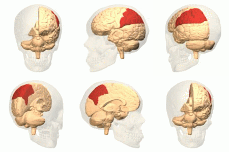
Parietal Lobe
The parietal lobe is one of the four major lobes of the cerebral cortex in the brain of mammals. The parietal lobe is positioned above the temporal lobe and behind the frontal lobe and central sulcus. The parietal lobe integrates sensory information among various modalities, including spatial sense and navigation (proprioception), the main sensory receptive area for the sense of touch in the somatosensory cortex which is just posterior to the central sulcus in the postcentral gyrus, and the dorsal stream of the visual system. The major sensory inputs from the skin ( touch, temperature, and pain receptors), relay through the thalamus to the parietal lobe. Several areas of the parietal lobe are important in language processing. The somatosensory cortex can be illustrated as a distorted figure – the cortical homunculus (Latin: "little man") in which the body parts are rendered according to how much of the somatosensory cortex is devoted to them. The superior parietal lobule a ... [...More Info...] [...Related Items...] OR: [Wikipedia] [Google] [Baidu] |
Parietal Eminence
The parietal eminence (parietal tuber, parietal tuberosity) is a convex, smooth eminence on the external surface of the parietal bone of the skull. It is the site where intramembranous ossification of the parietal bone begins during embryological development. It tends to be slightly more prominent in women than in men A man is an adult male human. Prior to adulthood, a male human is referred to as a boy (a male child or adolescent). Like most other male mammals, a man's genome usually inherits an X chromosome from the mother and a Y chro ..., so may be used to help to identify the sex of a skull. Additional images File:Parietal eminence - animation01.gif, Parietal eminence shown in red File:Braus 1921 362.png, Skull showing parietal eminence as Tuber parietale References External links * Bones of the head and neck {{musculoskeletal-stub ... [...More Info...] [...Related Items...] OR: [Wikipedia] [Google] [Baidu] |
Parietal Wall
In the shell of gastropod mollusks (a snail shell), the lip is the free margin of the peristome (synonym: peritreme) or aperture (the opening) of the gastropod shell. In dextral (right-handed) shells (most snail shells are right-handed), the right side or outer side of the aperture is known as the outer lip (''labrum''). The left side of the aperture is known as the inner lip or columellar lip (''labium'') if there is a pronounced lip there. In those species where there is no pronounced lip, the part of the body whorl that adjoins the aperture is known as the parietal wall. The outer lip is usually thin and sharp in immature shells, and in some adults (e.g. the land snails '' Helicella'' and ''Bulimulus''). However, in some other land snails and in many marine species the outer lip is ''thickened'' (also called ''callused''), or ''reflected'' (turned outwards). In some other marine species it is curled inwards (''inflected''), as in the cowries such as '' Cypraea''. It can also be ... [...More Info...] [...Related Items...] OR: [Wikipedia] [Google] [Baidu] |
Parietal Scales
Parietal scale refers to the scales of a snake which are on the head of the snake and are connected to the frontals towards the posterior. These scales are analogous to and take their name from the parietal bone which forms the roof and sides of the cranium in humans. See also * Parietal bone * Snake scales * Anatomical terms of location Standard anatomical terms of location are used to unambiguously describe the anatomy of animals, including humans. The terms, typically derived from Latin or Greek roots, describe something in its standard anatomical position. This position p ... Snake scales {{Snake-stub They are also great for protecting themselves ... [...More Info...] [...Related Items...] OR: [Wikipedia] [Google] [Baidu] |
Parietal Eye
A parietal eye, also known as a third eye or pineal eye, is a part of the epithalamus present in some vertebrates. The eye is located at the top of the head, is photoreceptive and is associated with the pineal gland, regulating circadian rhythmicity and hormone production for thermoregulation. The hole in the head which contains the eye is known as a pineal foramen or parietal foramen, since it is often enclosed by the parietal bones. Presence in various animals The parietal eye is found in the tuatara, most lizards, frogs, salamanders, certain bony fish, sharks, and lampreys. It is absent in mammals, but was present in their closest extinct relatives, the therapsids, suggesting it was lost during the course of the mammalian evolution due to it being useless in endothermic animals. It is also absent in the ancestrally endothermic ("warm-blooded") archosaurs such as birds. The parietal eye is also lost in ectothermic ("cold-blooded") archosaurs like crocodilians, and in turtles, ... [...More Info...] [...Related Items...] OR: [Wikipedia] [Google] [Baidu] |
Parietal Callus
A parietal callus is a feature of the shell anatomy of some groups of snails, i.e. gastropods. It is a thickened calcareous deposit which may be present on the parietal wall of the aperture of the adult shell. The parietal wall is the margin of the aperture and part of the wall of the body whorl that is closest to the columella. The callus is often smooth and glossy, but can also be decorated with raised ribs or wrinkles. This feature is found in such marine families as Ranellidae (pictured), Cassidae, Nassariidae, Ringiculidae and others. It is also found in some families of land snails, terrestrial pulmonates. See also * Gastropod shell * Aperture (mollusc) The aperture is an opening in certain kinds of mollusc shells: it is the main opening of the shell, where the head-foot part of the body of the animal emerges for locomotion, feeding, etc. The term ''aperture'' is used for the main opening in ... References Gastropod anatomy Mollusc shells {{gastropod-st ... [...More Info...] [...Related Items...] OR: [Wikipedia] [Google] [Baidu] |
Parietal Pleura
The pulmonary pleurae (''sing.'' pleura) are the two opposing layers of serous membrane overlying the lungs and the inside of the surrounding chest walls. The inner pleura, called the visceral pleura, covers the surface of each lung and dips between the lobes of the lung as ''fissures'', and is formed by the invagination of lung buds into each thoracic sac during embryonic development. The outer layer, called the parietal pleura, lines the inner surfaces of the thoracic cavity on each side of the mediastinum, and can be subdivided into ''mediastinal'' (covering the side surfaces of the fibrous pericardium, oesophagus and thoracic aorta), ''diaphragmatic'' (covering the upper surface of the diaphragm), ''costal'' (covering the inside of rib cage) and cervical (covering the underside of the suprapleural membrane) pleurae. The visceral and the mediastinal parietal pleurae are connected at the root of the lung ("hilum") through a smooth fold known as ''pleural reflections ... [...More Info...] [...Related Items...] OR: [Wikipedia] [Google] [Baidu] |
Parietal Placentation
Placentation refers to the formation, type and structure, or arrangement of the placenta. The function of placentation is to transfer nutrients, respiratory gases, and water from maternal tissue to a growing embryo, and in some instances to remove waste from the embryo. Placentation is best known in live-bearing mammals (theria), but also occurs in some fish, reptiles, amphibians, a diversity of invertebrates, and flowering plants. In vertebrates, placentas have evolved more than 100 times independently, with the majority of these instances occurring in squamate reptiles. The placenta can be defined as an organ formed by the sustained apposition or fusion of fetal membranes and parental tissue for physiological exchange. This definition is modified from the original Mossman (1937) definition, which constrained placentation in animals to only those instances where it occurred in the uterus. In mammals In live bearing mammals, the placenta forms after the embryo implants into the ... [...More Info...] [...Related Items...] OR: [Wikipedia] [Google] [Baidu] |
Parietal Cell
Parietal cells (also known as oxyntic cells) are epithelial cells in the stomach that secrete hydrochloric acid (HCl) and intrinsic factor. These cells are located in the gastric glands found in the lining of the fundus and body regions of the stomach. They contain an extensive secretory network of canaliculi from which the HCl is secreted by active transport into the stomach. The enzyme hydrogen potassium ATPase (H+/K+ ATPase) is unique to the parietal cells and transports the H+ against a concentration gradient of about 3 million to 1, which is the steepest ion gradient formed in the human body. Parietal cells are primarily regulated via histamine, acetylcholine and gastrin signalling from both central and local modulators. Structure Canaliculus A canaliculus is an adaptation found on gastric parietal cells. It is a deep infolding, or little channel, which serves to increase the surface area, e.g. for secretion. The parietal cell membrane is dynamic; the numbers of canalic ... [...More Info...] [...Related Items...] OR: [Wikipedia] [Google] [Baidu] |
Parietal Foramen (other) , paired openings in the parietal bones of humans, which host the emissary veins
{{Disambiguation ...
"Parietal foramen" may refer to: * Pineal foramen, a midline hole in the skull roof which hosts the parietal eye in many vertebrate species * Parietal foramina A parietal foramen is an opening in the skull for the parietal emissary vein, which drains into the superior sagittal sinus. Occasionally, a small branch of the occipital artery can also pass through it. It is located at the back part of the par ... [...More Info...] [...Related Items...] OR: [Wikipedia] [Google] [Baidu] |
Inferior Parietal Lobule
The inferior parietal lobule (subparietal district) lies below the horizontal portion of the intraparietal sulcus, and behind the lower part of the postcentral sulcus. Also known as Geschwind's territory after Norman Geschwind, an American neurologist, who in the early 1960s recognised its importance. It is a part of the parietal lobe. Structure It is divided from rostral to caudal into two gyri: * One, the supramarginal gyrus, arches over the upturned end of the lateral fissure; it is continuous in front with the postcentral gyrus, and behind with the superior temporal gyrus. * The second, the angular gyrus, arches over the posterior end of the superior temporal sulcus, behind which it is continuous with the middle temporal gyrus. In macaque neuroanatomy, this region is often divided into caudal and rostral portions, cIPL and rIPL, respectively. The cIPL is further divided into areas Opt and PG whereas rIPL is divided into PFG and PF areas. Function Inferior parietal lobul ... [...More Info...] [...Related Items...] OR: [Wikipedia] [Google] [Baidu] |
Parietal Bone
The parietal bones () are two bones in the skull which, when joined at a fibrous joint, form the sides and roof of the cranium. In humans, each bone is roughly quadrilateral in form, and has two surfaces, four borders, and four angles. It is named from the Latin ''paries'' (''-ietis''), wall. Surfaces External The external surface ig. 1is convex, smooth, and marked near the center by an eminence, the parietal eminence (''tuber parietale''), which indicates the point where ossification commenced. Crossing the middle of the bone in an arched direction are two curved lines, the superior and inferior temporal lines; the former gives attachment to the temporal fascia, and the latter indicates the upper limit of the muscular origin of the temporal muscle. Above these lines the bone is covered by a tough layer of fibrous tissue – the epicranial aponeurosis; below them it forms part of the temporal fossa, and affords attachment to the temporal muscle. At the back part an ... [...More Info...] [...Related Items...] OR: [Wikipedia] [Google] [Baidu] |


