|
Parafovea
Parafovea or the parafoveal belt is a region in the retina that circumscribes the fovea and is part of the macula lutea. It is circumscribed by the perifovea. Effect on reading In reading, information within 1° (approximately 6–8 characters) of the point of fixation is processed in foveal vision, while information up to 6° of visual angle benefits from parafoveal preview. Studies have shown that people can tell the difference in the letters of a word in the fovea and near-parafovea (the part of the parafovea closest to the fovea), but not in the outer edges of the parafovea. In languages that read from left to right, the word immediately to the right of the fixated word is known as the parafoveal word. Information present in the parafovea can interact with information present in the fovea. The benefit the parafoveal preview has is also mediated by how common the word in the parafovea is, with less common words providing less of a reduction in fixation duration when they reach ... [...More Info...] [...Related Items...] OR: [Wikipedia] [Google] [Baidu] |
Retina
The retina (from la, rete "net") is the innermost, light-sensitive layer of tissue of the eye of most vertebrates and some molluscs. The optics of the eye create a focused two-dimensional image of the visual world on the retina, which then processes that image within the retina and sends nerve impulses along the optic nerve to the visual cortex to create visual perception. The retina serves a function which is in many ways analogous to that of the film or image sensor in a camera. The neural retina consists of several layers of neurons interconnected by synapses and is supported by an outer layer of pigmented epithelial cells. The primary light-sensing cells in the retina are the photoreceptor cells, which are of two types: rods and cones. Rods function mainly in dim light and provide monochromatic vision. Cones function in well-lit conditions and are responsible for the perception of colour through the use of a range of opsins, as well as high-acuity vision used for task ... [...More Info...] [...Related Items...] OR: [Wikipedia] [Google] [Baidu] |
Fovea Centralis
The fovea centralis is a small, central pit composed of closely packed cones in the eye. It is located in the center of the macula lutea of the retina. The fovea is responsible for sharp central vision (also called foveal vision), which is necessary in humans for activities for which visual detail is of primary importance, such as reading and driving. The fovea is surrounded by the ''parafovea'' belt and the ''perifovea'' outer region. The parafovea is the intermediate belt, where the ganglion cell layer is composed of more than five layers of cells, as well as the highest density of cones; the perifovea is the outermost region where the ganglion cell layer contains two to four layers of cells, and is where visual acuity is below the optimum. The perifovea contains an even more diminished density of cones, having 12 per 100 micrometres versus 50 per 100 micrometres in the most central fovea. That, in turn, is surrounded by a larger peripheral area, which delivers highly compres ... [...More Info...] [...Related Items...] OR: [Wikipedia] [Google] [Baidu] |
Macula Lutea
The macula (/ˈmakjʊlə/) or macula lutea is an oval-shaped pigmented area in the center of the retina of the human eye and in other animals. The macula in humans has a diameter of around and is subdivided into the umbo, foveola, foveal avascular zone, fovea, parafovea, and perifovea areas. The anatomical macula at a size of is much larger than the clinical macula which, at a size of , corresponds to the anatomical fovea. The macula is responsible for the central, high-resolution, color vision that is possible in good light; and this kind of vision is impaired if the macula is damaged, for example in macular degeneration. The clinical macula is seen when viewed from the pupil, as in ophthalmoscopy or retinal photography. The term macula lutea comes from Latin ''macula'', "spot", and ''lutea'', "yellow". Structure The macula is an oval-shaped pigmented area in the center of the retina of the human eye and other animal eyes. Its center is shifted slightly away from the ... [...More Info...] [...Related Items...] OR: [Wikipedia] [Google] [Baidu] |
Perifovea
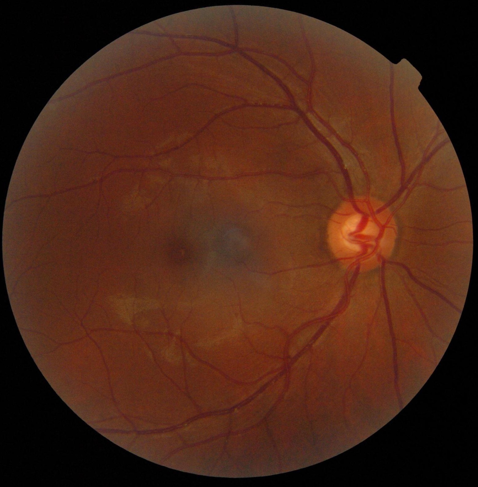
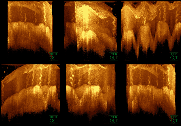
Perifovea is a region in the retina that circumscribes the parafovea and fovea and is a part of the macula lutea. The perifovea is a belt that covers a 10° radius around the fovea and is 1.5 mm wide. The perifovea ends when the Henle's fiber layer disappears and the ganglion cells are one-layered. Additional images File:Macula lutea.svg, Schematic diagram of the macula lutea of the retina, showing perifovea, parafovea, fovea, and clinical macula File:Retina-OCT800.png, Time-Domain OCT of the macular area of a retina at 800 nm, axial resolution 3 µm File:SD-OCT Macula Cross-Section.png, Spectral-Domain OCT macula cross-section scan. File:Macula Histology OCT.jpg, alt=macula histology (OCT), macula histology (OCT) File:Retinography.jpg, A fundus photograph showing the macula as a spot to the left. The optic disc is the area on the right where blood vessels converge. The grey, more diffuse spot in the centre is a shadow artifact. See also * Eye movements in reading * Fixation (v ... [...More Info...] [...Related Items...] OR: [Wikipedia] [Google] [Baidu] |
Macula
The macula (/ˈmakjʊlə/) or macula lutea is an oval-shaped pigmented area in the center of the retina of the human eye and in other animals. The macula in humans has a diameter of around and is subdivided into the umbo, foveola, foveal avascular zone, fovea, parafovea, and perifovea areas. The anatomical macula at a size of is much larger than the clinical macula which, at a size of , corresponds to the anatomical fovea. The macula is responsible for the central, high-resolution, color vision that is possible in good light; and this kind of vision is impaired if the macula is damaged, for example in macular degeneration. The clinical macula is seen when viewed from the pupil, as in ophthalmoscopy or retinal photography. The term macula lutea comes from Latin ''macula'', "spot", and ''lutea'', "yellow". Structure The macula is an oval-shaped pigmented area in the center of the retina of the human eye and other animal eyes. Its center is shifted slightly away from the ... [...More Info...] [...Related Items...] OR: [Wikipedia] [Google] [Baidu] |
Fundus Photograph
Fundus photography involves photographing the rear of an eye, also known as the fundus. Specialized fundus cameras consisting of an intricate microscope attached to a flash enabled camera are used in fundus photography. The main structures that can be visualized on a fundus photo are the central and peripheral retina, optic disc and macula. Fundus photography can be performed with colored filters, or with specialized dyes including fluorescein and indocyanine green. The models and technology of fundus photography have advanced and evolved rapidly over the last century. Since the equipment is sophisticated and challenging to manufacture to clinical standards, only a few manufacturers/brands are available in the market: Welch Allyn, Digisight, Volk, Topcon, Zeiss, Canon, Nidek, Kowa, CSO, CenterVue, Ezer and Optos are some example of fundus camera manufacturers. History The concept of fundus photography was first introduced in the mid 19th century, after the introduction of p ... [...More Info...] [...Related Items...] OR: [Wikipedia] [Google] [Baidu] |
Visual Artifact
Visual artifacts (also artefacts) are artifact (error), anomalies apparent during visual representation as in digital graphics and other forms of imagery, especially photography and microscopy. In digital graphics * Image quality#Image quality factors, Image quality factors, different types of visual artifacts * Compression artifacts * Digital artifacts, visual artifacts resulting from digital image processing * Image noise, Noise * Screen-door effect, also known as fixed-pattern noise (FPN), a visual artifact of digital projection technology *Ghosting (television) *Screen burn-in * Distortion * Silk screen effect * Rainbow effect * Screen tearing * Moiré pattern * Color banding In video entertainment Many people who use their computers as a hobby experience artifacting due to a hardware or software malfunction. The cases can differ but the usual causes are: * Temperature issues, such as failure of cooling fan. * Unsuited video card (graphics card) drivers. * Drivers that have ... [...More Info...] [...Related Items...] OR: [Wikipedia] [Google] [Baidu] |
Eye Movement In Reading
Eye movement in reading involves the visual processing of written text. This was described by the French ophthalmologist Louis Émile Javal in the late 19th century. He reported that eyes do not move continuously along a line of text, but make short, rapid movements (saccades) intermingled with short stops ( fixations). Javal's observations were characterised by a reliance on naked-eye observation of eye movement in the absence of technology. From the late 19th to the mid-20th century, investigators used early tracking technologies to assist their observation, in a research climate that emphasised the measurement of human behaviour and skill for educational ends. Most basic knowledge about eye movement was obtained during this period. Since the mid-20th century, there have been three major changes: the development of non-invasive eye-movement tracking equipment; the introduction of computer technology to enhance the power of this equipment to pick up, record, and process the huge vo ... [...More Info...] [...Related Items...] OR: [Wikipedia] [Google] [Baidu] |
Eye Movement In Music Reading
Eye movement in music reading is the scanning of a musical score by a musician's eyes. This usually occurs as the music is read during performance, although musicians sometimes scan music silently to study it. The phenomenon has been studied by researchers from a range of backgrounds, including cognitive psychology and music education. These studies have typically reflected a curiosity among performing musicians about a central process in their craft, and a hope that investigating eye movement might help in the development of more effective methods of training musicians' sight reading skills. A central aspect of music reading is the sequence of alternating saccades and fixations, as it is for most oculomotor tasks. Saccades are the rapid ‘flicks’ that move the eyes from location to location over a music score. Saccades are separated from each other by fixations, during which the eyes are relatively stationary on the page. It is well established that the perception of visual i ... [...More Info...] [...Related Items...] OR: [Wikipedia] [Google] [Baidu] |
Fixation (visual)
Fixation or visual fixation is the maintaining of the gaze on a single location. An animal can exhibit visual fixation if it possess a fovea in the anatomy of their eye. The fovea is typically located at the center of the retina and is the point of clearest vision. The species in which fixational eye movement has been verified thus far include humans, primates, cats, rabbits, turtles, salamanders, and owls. Regular eye movement alternates between saccades and visual fixations, the notable exception being in smooth pursuit, controlled by a different neural substrate that appears to have developed for hunting prey. The term "fixation" can either be used to refer to the point in time and space of focus or the act of fixating. Fixation, in the act of fixating, is the point between any two saccades, during which the eyes are relatively stationary and virtually all visual input occurs. In the absence of retinal jitter, a laboratory condition known as retinal stabilization, perceptions ... [...More Info...] [...Related Items...] OR: [Wikipedia] [Google] [Baidu] |
Optical Coherence Tomography
Optical coherence tomography (OCT) is an imaging technique that uses low-coherence light to capture micrometer-resolution, two- and three-dimensional images from within optical scattering media (e.g., biological tissue). It is used for medical imaging and industrial nondestructive testing (NDT). Optical coherence tomography is based on low-coherence interferometry, typically employing near-infrared light. The use of relatively long wavelength light allows it to penetrate into the scattering medium. Confocal microscopy, another optical technique, typically penetrates less deeply into the sample but with higher resolution. Depending on the properties of the light source ( superluminescent diodes, ultrashort pulsed lasers, and supercontinuum lasers have been employed), optical coherence tomography has achieved sub-micrometer resolution (with very wide-spectrum sources emitting over a ~100 nm wavelength range). Optical coherence tomography is one of a class of optical tom ... [...More Info...] [...Related Items...] OR: [Wikipedia] [Google] [Baidu] |
Human Eye Anatomy
Humans (''Homo sapiens'') are the most abundant and widespread species of primate, characterized by bipedalism and exceptional cognitive skills due to a large and complex brain. This has enabled the development of advanced tools, culture, and language. Humans are highly social and tend to live in complex social structures composed of many cooperating and competing groups, from families and kinship networks to political states. Social interactions between humans have established a wide variety of values, social norms, and rituals, which bolster human society. Its intelligence and its desire to understand and influence the environment and to explain and manipulate phenomena have motivated humanity's development of science, philosophy, mythology, religion, and other fields of study. Although some scientists equate the term ''humans'' with all members of the genus ''Homo'', in common usage, it generally refers to ''Homo sapiens'', the only extant member. Anatomically modern huma ... [...More Info...] [...Related Items...] OR: [Wikipedia] [Google] [Baidu] |


