|
Paracolic Gutter
The paracolic gutters (paracolic sulci, paracolic recesses) are peritoneal recesses – spaces between the colon and the abdominal wall. Structure There are two paracolic gutters: * The right lateral paracolic gutter. * The left medial paracolic gutter. The right and left paracolic gutters are peritoneal recesses on the posterior abdominal wall lying alongside the ascending and descending colon. The main paracolic gutter lies lateral to the colon on each side. A less obvious medial paracolic gutter may be formed, especially on the right side, if the colon possesses a short mesentery for part of its length. The right (lateral) paracolic gutter runs from the superiolateral aspect of the hepatic flexure of the colon, down the lateral aspect of the ascending colon, and around the cecum. It is continuous with the peritoneum as it descends into the pelvis over the pelvic brim. Superiorly, it is continuous with the peritoneum which lines the hepatorenal pouch and, through the epiploic fo ... [...More Info...] [...Related Items...] OR: [Wikipedia] [Google] [Baidu] |
Peritoneal Recesses
Peritoneal recesses (or peritoneal gutters) are the spaces formed by peritoneum draping over viscera. The term refers mainly to four spaces in the abdominal cavity; the two paracolic gutters and the two paramesenteric gutters. There are other smaller recesses including those around the duodenojejunal flexure, cecum, and the sigmoid colon. These gutters are clinically important because they allow a passage for infectious fluids from different compartments of the abdomen. For example; fluid from an infected Vermiform appendix, appendix can track up the right paracolic gutter to the hepatorenal recess. The four peritoneal recesses are: * The left and right paracolic gutters. * The left and right paramesenteric gutters. See also * Hepatorenal recess References External links * — "Abdominal Cavity: Peritoneal Gutters" page 1 * — "Abdominal Cavity: Peritoneal Gutters" page 2 * — "Abdominal Cavity: Peritoneal Gutters" page 3 Abdomen {{Anatomy-stub ... [...More Info...] [...Related Items...] OR: [Wikipedia] [Google] [Baidu] |
Lesser Sac
The lesser sac, also known as the omental bursa, is a part of the peritoneal cavity that is formed by the lesser and greater omentum. Usually found in mammals, it is connected with the greater sac via the omental foramen or ''Foramen of Winslow''. In mammals, it is common for the lesser sac to contain considerable amounts of fat. Anatomic margins ;Anterior margin: listed from the top-to-bottom margin: Quadrate lobe of the liver, lesser omentum, stomach, gastrocolic ligament ;Lateral margin: listed from the most anterior to the most posterior margin: Gastrosplenic ligament, spleen, phrenicosplenic ligament ;Posterior margin: Left kidney and adrenal gland, pancreas ;Inferior margin: Greater omentum ;Superior margin: Liver If any of the marginal structures rupture their contents could leak into the lesser sac. If the stomach were to rupture on its anterior side though the leak would collect in the greater sac. The lesser sac is formed during embryogenesis from an infolding of ... [...More Info...] [...Related Items...] OR: [Wikipedia] [Google] [Baidu] |
Peritoneal Recesses
Peritoneal recesses (or peritoneal gutters) are the spaces formed by peritoneum draping over viscera. The term refers mainly to four spaces in the abdominal cavity; the two paracolic gutters and the two paramesenteric gutters. There are other smaller recesses including those around the duodenojejunal flexure, cecum, and the sigmoid colon. These gutters are clinically important because they allow a passage for infectious fluids from different compartments of the abdomen. For example; fluid from an infected Vermiform appendix, appendix can track up the right paracolic gutter to the hepatorenal recess. The four peritoneal recesses are: * The left and right paracolic gutters. * The left and right paramesenteric gutters. See also * Hepatorenal recess References External links * — "Abdominal Cavity: Peritoneal Gutters" page 1 * — "Abdominal Cavity: Peritoneal Gutters" page 2 * — "Abdominal Cavity: Peritoneal Gutters" page 3 Abdomen {{Anatomy-stub ... [...More Info...] [...Related Items...] OR: [Wikipedia] [Google] [Baidu] |
Abscess
An abscess is a collection of pus that has built up within the tissue of the body. Signs and symptoms of abscesses include redness, pain, warmth, and swelling. The swelling may feel fluid-filled when pressed. The area of redness often extends beyond the swelling. Carbuncles and boils are types of abscess that often involve hair follicles, with carbuncles being larger. They are usually caused by a bacterial infection. Often many different types of bacteria are involved in a single infection. In many areas of the world, the most common bacteria present is ''methicillin-resistant Staphylococcus aureus''. Rarely, parasites can cause abscesses; this is more common in the developing world. Diagnosis of a skin abscess is usually made based on what it looks like and is confirmed by cutting it open. Ultrasound imaging may be useful in cases in which the diagnosis is not clear. In abscesses around the anus, computer tomography (CT) may be important to look for deeper infection. Standa ... [...More Info...] [...Related Items...] OR: [Wikipedia] [Google] [Baidu] |
Appendicitis
Appendicitis is inflammation of the appendix. Symptoms commonly include right lower abdominal pain, nausea, vomiting, and decreased appetite. However, approximately 40% of people do not have these typical symptoms. Severe complications of a ruptured appendix include widespread, painful inflammation of the inner lining of the abdominal wall and sepsis. Appendicitis is caused by a blockage of the hollow portion of the appendix. This is most commonly due to a calcified "stone" made of feces. Inflamed lymphoid tissue from a viral infection, parasites, gallstone, or tumors may also cause the blockage. This blockage leads to increased pressures in the appendix, decreased blood flow to the tissues of the appendix, and bacterial growth inside the appendix causing inflammation. The combination of inflammation, reduced blood flow to the appendix and distention of the appendix causes tissue injury and tissue death. If this process is left untreated, the appendix may burst, releasing ba ... [...More Info...] [...Related Items...] OR: [Wikipedia] [Google] [Baidu] |
Pelvis
The pelvis (plural pelves or pelvises) is the lower part of the trunk, between the abdomen and the thighs (sometimes also called pelvic region), together with its embedded skeleton (sometimes also called bony pelvis, or pelvic skeleton). The pelvic region of the trunk includes the bony pelvis, the pelvic cavity (the space enclosed by the bony pelvis), the pelvic floor, below the pelvic cavity, and the perineum, below the pelvic floor. The pelvic skeleton is formed in the area of the back, by the sacrum and the coccyx and anteriorly and to the left and right sides, by a pair of hip bones. The two hip bones connect the spine with the lower limbs. They are attached to the sacrum posteriorly, connected to each other anteriorly, and joined with the two femurs at the hip joints. The gap enclosed by the bony pelvis, called the pelvic cavity, is the section of the body underneath the abdomen and mainly consists of the reproductive organs (sex organs) and the rectum, while the pelvic f ... [...More Info...] [...Related Items...] OR: [Wikipedia] [Google] [Baidu] |
Gallbladder
In vertebrates, the gallbladder, also known as the cholecyst, is a small hollow organ where bile is stored and concentrated before it is released into the small intestine. In humans, the pear-shaped gallbladder lies beneath the liver, although the structure and position of the gallbladder can vary significantly among animal species. It receives and stores bile, produced by the liver, via the common hepatic duct, and releases it via the common bile duct into the duodenum, where the bile helps in the digestion of fats. The gallbladder can be affected by gallstones, formed by material that cannot be dissolved – usually cholesterol or bilirubin, a product of haemoglobin breakdown. These may cause significant pain, particularly in the upper-right corner of the abdomen, and are often treated with removal of the gallbladder (called a cholecystectomy). Cholecystitis, inflammation of the gallbladder, has a wide range of causes, including result from the impaction of gallstones, inf ... [...More Info...] [...Related Items...] OR: [Wikipedia] [Google] [Baidu] |
Duodenum
The duodenum is the first section of the small intestine in most higher vertebrates, including mammals, reptiles, and birds. In fish, the divisions of the small intestine are not as clear, and the terms anterior intestine or proximal intestine may be used instead of duodenum. In mammals the duodenum may be the principal site for iron absorption. The duodenum precedes the jejunum and ileum and is the shortest part of the small intestine. In humans, the duodenum is a hollow jointed tube about 25–38 cm (10–15 inches) long connecting the stomach to the middle part of the small intestine. It begins with the duodenal bulb and ends at the suspensory muscle of duodenum. Duodenum can be divided into four parts: the first (superior), the second (descending), the third (horizontal) and the fourth (ascending) parts. Structure The duodenum is a C-shaped structure lying adjacent to the stomach. It is divided anatomically into four sections. The first part of the duodenum lies ... [...More Info...] [...Related Items...] OR: [Wikipedia] [Google] [Baidu] |
Stomach
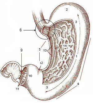
The stomach is a muscular, hollow organ in the gastrointestinal tract of humans and many other animals, including several invertebrates. The stomach has a dilated structure and functions as a vital organ in the digestive system. The stomach is involved in the gastric phase of digestion, following chewing. It performs a chemical breakdown by means of enzymes and hydrochloric acid. In humans and many other animals, the stomach is located between the oesophagus and the small intestine. The stomach secretes digestive enzymes and gastric acid to aid in food digestion. The pyloric sphincter controls the passage of partially digested food ( chyme) from the stomach into the duodenum, where peristalsis takes over to move this through the rest of intestines. Structure In the human digestive system, the stomach lies between the oesophagus and the duodenum (the first part of the small intestine). It is in the left upper quadrant of the abdominal cavity. The top of the stomach lies ag ... [...More Info...] [...Related Items...] OR: [Wikipedia] [Google] [Baidu] |
Abdomen
The abdomen (colloquially called the belly, tummy, midriff, tucky or stomach) is the part of the body between the thorax (chest) and pelvis, in humans and in other vertebrates. The abdomen is the front part of the abdominal segment of the torso. The area occupied by the abdomen is called the abdominal cavity. In arthropods it is the posterior (anatomy), posterior tagma (biology), tagma of the body; it follows the thorax or cephalothorax. In humans, the abdomen stretches from the thorax at the thoracic diaphragm to the pelvis at the pelvic brim. The pelvic brim stretches from the lumbosacral joint (the intervertebral disc between Lumbar vertebrae, L5 and Vertebra#Sacrum, S1) to the pubic symphysis and is the edge of the pelvic inlet. The space above this inlet and under the thoracic diaphragm is termed the abdominal cavity. The boundary of the abdominal cavity is the abdominal wall in the front and the peritoneal surface at the rear. In vertebrates, the abdomen is a large body c ... [...More Info...] [...Related Items...] OR: [Wikipedia] [Google] [Baidu] |
Large Intestine
The large intestine, also known as the large bowel, is the last part of the gastrointestinal tract and of the digestive system in tetrapods. Water is absorbed here and the remaining waste material is stored in the rectum as feces before being removed by defecation. The colon is the longest portion of the large intestine, and the terms are often used interchangeably but most sources define the large intestine as the combination of the cecum, colon, rectum, and anal canal. Some other sources exclude the anal canal. In humans, the large intestine begins in the right iliac region of the pelvis, just at or below the waist, where it is joined to the end of the small intestine at the cecum, via the ileocecal valve. It then continues as the colon ascending the abdomen, across the width of the abdominal cavity as the transverse colon, and then descending to the rectum and its endpoint at the anal canal. Overall, in humans, the large intestine is about long, which is about one-fi ... [...More Info...] [...Related Items...] OR: [Wikipedia] [Google] [Baidu] |
Supine
In grammar, a supine is a form of verbal noun used in some languages. The term is most often used for Latin, where it is one of the four principal parts of a verb. The word refers to a position of lying on one's back (as opposed to 'prone', lying face downward), but there exists no widely accepted etymology that explains why or how the term came to be used to also describe this form of a verb. Latin There are two supines, I (first) and II (second). They are originally the accusativeFortson, §5.59. and dative or ablative forms of a verbal noun in the fourth declension, respectively. First supine The first supine ends in ''-tum''. It has two uses. The first supine comes with verbs of motion. In one usage, it indicates purpose: * 'Mater pompam me ''spectatum'' duxit' is 'Mother took me ''to watch'' the procession'. * 'Legati ad Caesarem ''gratulatum'' convenerunt' is 'The ambassadors came to Caesar ''to congratulate'' him'. The translation of this first usage of the f ... [...More Info...] [...Related Items...] OR: [Wikipedia] [Google] [Baidu] |


