|
Paraaortic Lymph Nodes
The periaortic lymph nodes (also known as lumbar) are a group of lymph nodes that lie in front of the lumbar vertebrae near the aorta. These lymph nodes receive drainage from the gastrointestinal tract and the abdominal organs. The periaortic lymph nodes are different from the paraaortic lymph nodes. The periaortic group is the general group, that is subdivided into: preaortic, paraaortic, and retroaortic groups. The paraaortic group is synonymous with the lateral aortic group. Divisions The periaortic lymph node group is divided into three subgroups: preaortic, paraaortic, and retroaortic: * The preaortic group drains the gastrointestinal viscera. They can be subdivided into three groups: the celiac nodes, the superior mesenteric nodes, and the inferior mesenteric nodes. *The paraaortic group (also known as lateral aortic group) drains the iliac nodes, the ovaries, the testes and other pelvic organs. The lateral group nodes are located adjacent to the aorta, anterior to the ... [...More Info...] [...Related Items...] OR: [Wikipedia] [Google] [Baidu] |
Lymph Node
A lymph node, or lymph gland, is a kidney-shaped organ of the lymphatic system and the adaptive immune system. A large number of lymph nodes are linked throughout the body by the lymphatic vessels. They are major sites of lymphocytes that include B and T cells. Lymph nodes are important for the proper functioning of the immune system, acting as filters for foreign particles including cancer cells, but have no detoxification function. In the lymphatic system a lymph node is a secondary lymphoid organ. A lymph node is enclosed in a fibrous capsule and is made up of an outer cortex and an inner medulla. Lymph nodes become inflamed or enlarged in various diseases, which may range from trivial throat infections to life-threatening cancers. The condition of lymph nodes is very important in cancer staging, which decides the treatment to be used and determines the prognosis. Lymphadenopathy refers to glands that are enlarged or swollen. When inflamed or enlarged, lymph nodes can be ... [...More Info...] [...Related Items...] OR: [Wikipedia] [Google] [Baidu] |
Renal Vein
The renal veins are large-calibre veins that drain blood filtered by the kidneys into the inferior vena cava. There is one renal vein draining each kidney. Because the inferior vena cava is on the right half of the body, the left renal vein is longer than the right one. Structure One renal vein drains each kidney. A renal vein is situated anterior to its corresponding accompanying renal artery. The renal veins empty into the inferior vena cava, entering it at nearly a 90° angle. Due to the right-ward displacement of the inferior vena cava from the midline, the left renal vein is some 3 times longer than the right one (~7.5 cm and ~2.5 cm, respectively). The renal vein divides into 4 divisions upon entering the kidney: * the anterior branch which receives blood from the anterior portion of the kidney and, * the posterior branch which receives blood from the posterior portion. Tributaries Because the tributaries of the inferior vena cava are not bilaterally symmetrical, the l ... [...More Info...] [...Related Items...] OR: [Wikipedia] [Google] [Baidu] |
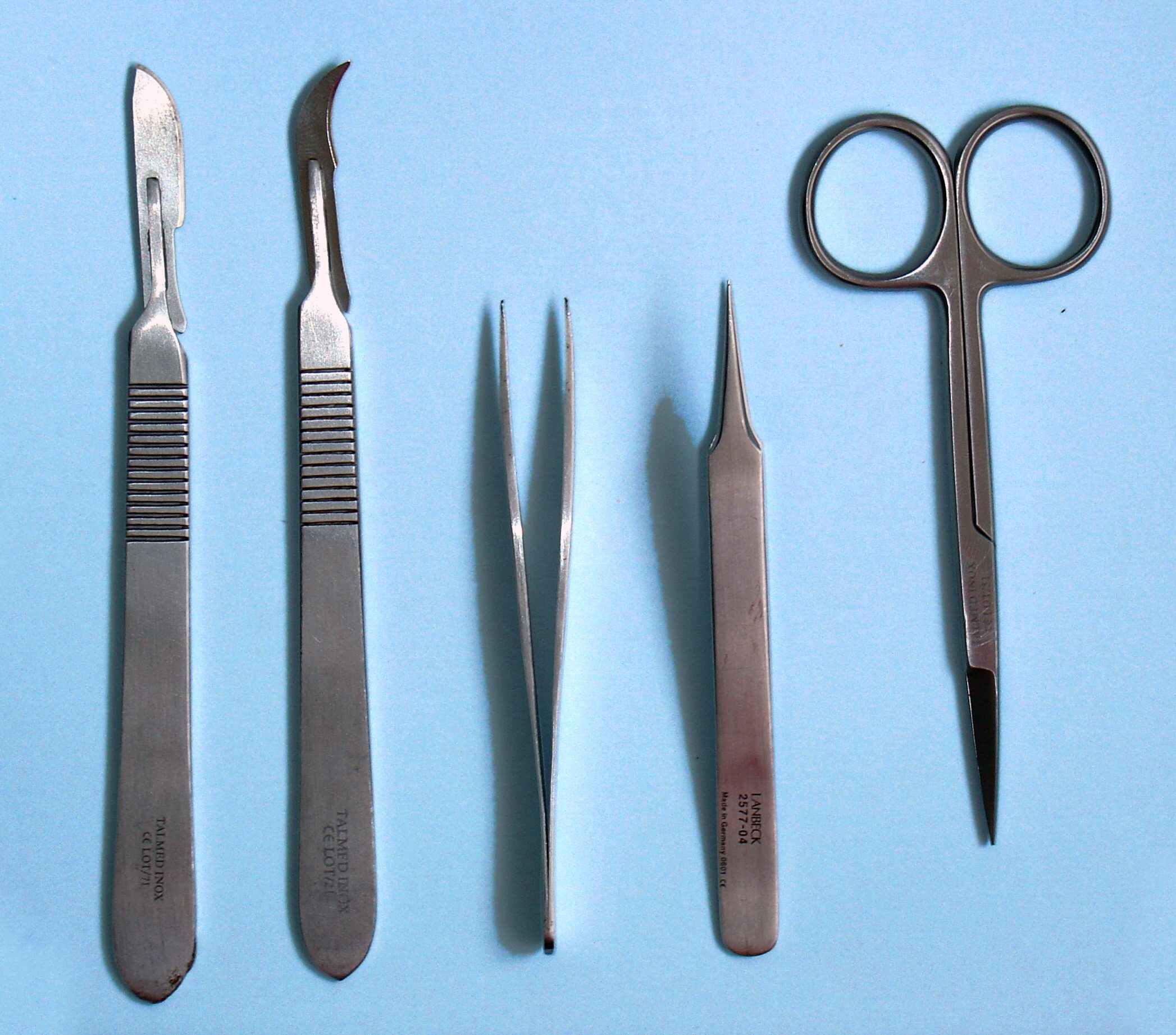
Dissection
Dissection (from Latin ' "to cut to pieces"; also called anatomization) is the dismembering of the body of a deceased animal or plant to study its anatomical structure. Autopsy is used in pathology and forensic medicine to determine the cause of death in humans. Less extensive dissection of plants and smaller animals preserved in a formaldehyde solution is typically carried out or demonstrated in biology and natural science classes in middle school and high school, while extensive dissections of cadavers of adults and children, both fresh and preserved are carried out by medical students in medical schools as a part of the teaching in subjects such as anatomy, pathology and forensic medicine. Consequently, dissection is typically conducted in a morgue or in an anatomy lab. Dissection has been used for centuries to explore anatomy. Objections to the use of cadavers have led to the use of alternatives including virtual dissection of computer models. Overview Plant and animal b ... [...More Info...] [...Related Items...] OR: [Wikipedia] [Google] [Baidu] |
Thoracic Duct
In human anatomy, the thoracic duct is the larger of the two lymph ducts of the lymphatic system. It is also known as the ''left lymphatic duct'', ''alimentary duct'', ''chyliferous duct'', and ''Van Hoorne's canal''. The other duct is the right lymphatic duct. The thoracic duct carries chyle, a liquid containing both lymph and emulsified fats, rather than pure lymph. It also collects most of the lymph in the body other than from the right thorax, arm, head, and neck (which are drained by the right lymphatic duct). The thoracic duct usually starts from the level of the twelfth thoracic vertebra (T12) and extends to the root of the neck. It drains into the systemic (blood) circulation at the junction of the left subclavian and internal jugular veins, at the commencement of the brachiocephalic vein. When the duct ruptures, the resulting flood of liquid into the pleural cavity is known as chylothorax. Structure In adults, the thoracic duct is typically 38–45 cm in length an ... [...More Info...] [...Related Items...] OR: [Wikipedia] [Google] [Baidu] |
Cisterna Chyli
The cisterna chyli (or cysterna chyli, and etymologically more correct, receptaculum chyli) is a dilated sac at the lower end of the thoracic duct in most mammals into which lymph from the intestinal trunk and two lumbar lymphatic trunks flow. It receives fatty chyle from the intestines and thus acts as a conduit for the lipid products of digestion. It is the most common drainage trunk of most of the body's lymphatics. The cisterna chyli is a retro-peritoneal structure. Structure In humans, the cisterna chyli is located posterior to the abdominal aorta on the anterior aspect of the bodies of the first and second lumbar vertebrae (L1 and L2). There it forms the beginning of the primary lymph vessel, the thoracic duct, which transports lymph and chyle from the abdomen via the aortic opening of the diaphragm up to the junction of left subclavian vein and internal jugular veins. Other animals In dogs, the cisterna chyli is located to the left and often ventral to the aorta; in cat ... [...More Info...] [...Related Items...] OR: [Wikipedia] [Google] [Baidu] |
Lumbar Trunks
The lumbar trunks are formed by the union of the efferent vessels from the lateral aortic lymph nodes. They receive the lymph from the lower limbs, from the walls and viscera of the pelvis, from the kidneys and suprarenal glands and the deep lymphatics of the greater part of the abdominal wall. Ultimately, the lumbar trunks empty into the cisterna chyli, a dilatation at the beginning of the thoracic duct In human anatomy, the thoracic duct is the larger of the two lymph ducts of the lymphatic system. It is also known as the ''left lymphatic duct'', ''alimentary duct'', ''chyliferous duct'', and ''Van Hoorne's canal''. The other duct is the righ .... References External links Overview at uams.edu Lymphatics of the torso {{lymphatic-stub ... [...More Info...] [...Related Items...] OR: [Wikipedia] [Google] [Baidu] |
Lumbar Veins
The lumbar veins are four pairs of veins running along the inside of the posterior abdominal wall, and drain venous blood from parts of the abdominal wall. Each lumbar vein accompanies a single lumbar artery. The lower two pairs of lumbar veins all drain directly into the inferior vena cava, whereas the fate of the upper two pairs is more variable. Lumbar veins are the lumbar equivalent of the posterior intercostal veins of the thorax. Structure A lumbar vein accompanies each of the four lumbar arteries on each side of the body. Distribution and tributaries Collectively, the lumbar veins drain blood from the territories supplied by the corresponding lumbar arteries (the posterior, lateral, and anterior abdominal wall). The lumbar veins drain the anterior spinal veins. Fate The 3rd and 4th lumbar veins drain into the inferior vena cava. The fate of the superior two lumbar veins us far more variable, and may drain into either the inferior vena cava, ascending lumbar vei ... [...More Info...] [...Related Items...] OR: [Wikipedia] [Google] [Baidu] |
Suprarenal Gland
The adrenal glands (also known as suprarenal glands) are endocrine glands that produce a variety of hormones including adrenaline and the steroids aldosterone and cortisol. They are found above the kidneys. Each gland has an outer cortex which produces steroid hormones and an inner medulla. The adrenal cortex itself is divided into three main zones: the zona glomerulosa, the zona fasciculata and the zona reticularis. The adrenal cortex produces three main types of steroid hormones: mineralocorticoids, glucocorticoids, and androgens. Mineralocorticoids (such as aldosterone) produced in the zona glomerulosa help in the regulation of blood pressure and electrolyte balance. The glucocorticoids cortisol and cortisone are synthesized in the zona fasciculata; their functions include the regulation of metabolism and immune system suppression. The innermost layer of the cortex, the zona reticularis, produces androgens that are converted to fully functional sex hormones in the gonads and ... [...More Info...] [...Related Items...] OR: [Wikipedia] [Google] [Baidu] |
Kidney
The kidneys are two reddish-brown bean-shaped organs found in vertebrates. They are located on the left and right in the retroperitoneal space, and in adult humans are about in length. They receive blood from the paired renal arteries; blood exits into the paired renal veins. Each kidney is attached to a ureter, a tube that carries excreted urine to the bladder. The kidney participates in the control of the volume of various body fluids, fluid osmolality, acid–base balance, various electrolyte concentrations, and removal of toxins. Filtration occurs in the glomerulus: one-fifth of the blood volume that enters the kidneys is filtered. Examples of substances reabsorbed are solute-free water, sodium, bicarbonate, glucose, and amino acids. Examples of substances secreted are hydrogen, ammonium, potassium and uric acid. The nephron is the structural and functional unit of the kidney. Each adult human kidney contains around 1 million nephrons, while a mouse kidney contains on ... [...More Info...] [...Related Items...] OR: [Wikipedia] [Google] [Baidu] |
Uterus
The uterus (from Latin ''uterus'', plural ''uteri'') or womb () is the organ in the reproductive system of most female mammals, including humans that accommodates the embryonic and fetal development of one or more embryos until birth. The uterus is a hormone-responsive sex organ that contains glands in its lining that secrete uterine milk for embryonic nourishment. In the human, the lower end of the uterus, is a narrow part known as the isthmus that connects to the cervix, leading to the vagina. The upper end, the body of the uterus, is connected to the fallopian tubes, at the uterine horns, and the rounded part above the openings to the fallopian tubes is the fundus. The connection of the uterine cavity with a fallopian tube is called the uterotubal junction. The fertilized egg is carried to the uterus along the fallopian tube. It will have divided on its journey to form a blastocyst that will implant itself into the lining of the uterus – the endometrium, where it will ... [...More Info...] [...Related Items...] OR: [Wikipedia] [Google] [Baidu] |
Uterine Tube
The uterus (from Latin ''uterus'', plural ''uteri'') or womb () is the organ in the reproductive system of most female mammals, including humans that accommodates the embryonic and fetal development of one or more embryos until birth. The uterus is a hormone-responsive sex organ that contains glands in its lining that secrete uterine milk for embryonic nourishment. In the human, the lower end of the uterus, is a narrow part known as the isthmus that connects to the cervix, leading to the vagina. The upper end, the body of the uterus, is connected to the fallopian tubes, at the uterine horns, and the rounded part above the openings to the fallopian tubes is the fundus. The connection of the uterine cavity with a fallopian tube is called the uterotubal junction. The fertilized egg is carried to the uterus along the fallopian tube. It will have divided on its journey to form a blastocyst that will implant itself into the lining of the uterus – the endometrium, where it will re ... [...More Info...] [...Related Items...] OR: [Wikipedia] [Google] [Baidu] |
Testis
A testicle or testis (plural testes) is the male reproductive gland or gonad in all bilaterians, including humans. It is homologous to the female ovary. The functions of the testes are to produce both sperm and androgens, primarily testosterone. Testosterone release is controlled by the anterior pituitary luteinizing hormone, whereas sperm production is controlled both by the anterior pituitary follicle-stimulating hormone and gonadal testosterone. Structure Appearance Males have two testicles of similar size contained within the scrotum, which is an extension of the abdominal wall. Scrotal asymmetry, in which one testicle extends farther down into the scrotum than the other, is common. This is because of the differences in the vasculature's anatomy. For 85% of men, the right testis hangs lower than the left one. Measurement and volume The volume of the testicle can be estimated by palpating it and comparing it to ellipsoids of known sizes. Another method is to use caliper ... [...More Info...] [...Related Items...] OR: [Wikipedia] [Google] [Baidu] |
.jpg)


