|
Papillary Muscle
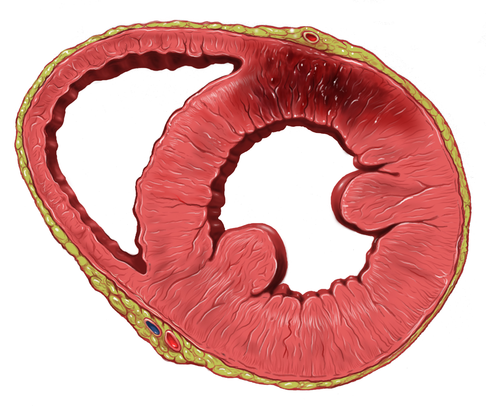
The papillary muscles are muscles located in the ventricles of the heart. They attach to the cusps of the atrioventricular valves (also known as the mitral and tricuspid valves) via the chordae tendineae and contract to prevent inversion or prolapse of these valves on systole (or ventricular contraction). The papillary muscles constitute about 10% of the total heart mass. Structure There are five total papillary muscles in the heart; three in the right ventricle and two in the left. The anterior, posterior, and septal papillary muscles of the right ventricle each attach via chordae tendineae to the tricuspid valve. The anterolateral and posteromedial papillary muscles of the left ventricle attach via chordae tendineae to the mitral valve.Netter's Atlas of Human Anatomy, plates 216B and 217A Blood supply The mitral valve papillary muscles in the left ventricle are called the anterolateral and posteromedial muscles. * Anterolateral muscle blood supply: left anterior descending a ... [...More Info...] [...Related Items...] OR: [Wikipedia] [Google] [Baidu] |
Heart
The heart is a muscular organ in most animals. This organ pumps blood through the blood vessels of the circulatory system. The pumped blood carries oxygen and nutrients to the body, while carrying metabolic waste such as carbon dioxide to the lungs. In humans, the heart is approximately the size of a closed fist and is located between the lungs, in the middle compartment of the chest. In humans, other mammals, and birds, the heart is divided into four chambers: upper left and right atria and lower left and right ventricles. Commonly the right atrium and ventricle are referred together as the right heart and their left counterparts as the left heart. Fish, in contrast, have two chambers, an atrium and a ventricle, while most reptiles have three chambers. In a healthy heart blood flows one way through the heart due to heart valves, which prevent backflow. The heart is enclosed in a protective sac, the pericardium, which also contains a small amount of fluid. The wall ... [...More Info...] [...Related Items...] OR: [Wikipedia] [Google] [Baidu] |
Muscle Contraction
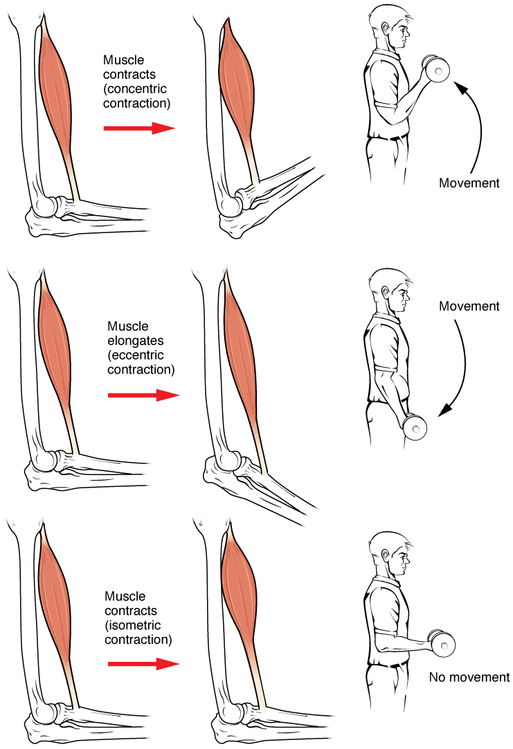
Muscle contraction is the activation of tension-generating sites within muscle cells. In physiology, muscle contraction does not necessarily mean muscle shortening because muscle tension can be produced without changes in muscle length, such as when holding something heavy in the same position. The termination of muscle contraction is followed by muscle relaxation, which is a return of the muscle fibers to their low tension-generating state. For the contractions to happen, the muscle cells must rely on the interaction of two types of filaments which are the thin and thick filaments. Thin filaments are two strands of actin coiled around each, and thick filaments consist of mostly elongated proteins called myosin. Together, these two filaments form myofibrils which are important organelles in the skeletal muscle system. Muscle contraction can also be described based on two variables: length and tension. A muscle contraction is described as isometric if the muscle tension changes ... [...More Info...] [...Related Items...] OR: [Wikipedia] [Google] [Baidu] |
Mitral Regurgitation
Mitral regurgitation (MR), also known as mitral insufficiency or mitral incompetence, is a form of valvular heart disease in which the mitral valve is insufficient and does not close properly when the heart pumps out blood.Mitral valve regurgitation at Mount Sinai Hospital It is the abnormal leaking of blood backwards – regurgitation from the , through the mitral valve, into the |
Ischemia
Ischemia or ischaemia is a restriction in blood supply to any tissue, muscle group, or organ of the body, causing a shortage of oxygen that is needed for cellular metabolism (to keep tissue alive). Ischemia is generally caused by problems with blood vessels, with resultant damage to or dysfunction of tissue i.e. hypoxia and microvascular dysfunction. It also implies local hypoxia in a part of a body resulting from constriction (such as vasoconstriction, thrombosis, or embolism). Ischemia causes not only insufficiency of oxygen, but also reduced availability of nutrients and inadequate removal of metabolic wastes. Ischemia can be partial (poor perfusion) or total blockage. The inadequate delivery of oxygenated blood to the organs must be resolved either by treating the cause of the inadequate delivery or reducing the oxygen demand of the system that needs it. For example, patients with myocardial ischemia have a decreased blood flow to the heart and are prescribed with medi ... [...More Info...] [...Related Items...] OR: [Wikipedia] [Google] [Baidu] |
Myocardial Infarct
A myocardial infarction (MI), commonly known as a heart attack, occurs when blood flow decreases or stops to the coronary artery of the heart, causing damage to the heart muscle. The most common symptom is chest pain or discomfort which may travel into the shoulder, arm, back, neck or jaw. Often it occurs in the center or left side of the chest and lasts for more than a few minutes. The discomfort may occasionally feel like heartburn. Other symptoms may include shortness of breath, nausea, feeling faint, a cold sweat or feeling tired. About 30% of people have atypical symptoms. Women more often present without chest pain and instead have neck pain, arm pain or feel tired. Among those over 75 years old, about 5% have had an MI with little or no history of symptoms. An MI may cause heart failure, an irregular heartbeat, cardiogenic shock or cardiac arrest. Most MIs occur due to coronary artery disease. Risk factors include high blood pressure, smoking, diabetes, lack of exercis ... [...More Info...] [...Related Items...] OR: [Wikipedia] [Google] [Baidu] |
Ventricular Systole
The cardiac cycle is the performance of the human heart from the beginning of one heartbeat to the beginning of the next. It consists of two periods: one during which the heart muscle relaxes and refills with blood, called diastole, following a period of robust contraction and pumping of blood, called systole. After emptying, the heart immediately relaxes and expands to receive another influx of blood returning from the lungs and other systems of the body, before again contracting to pump blood to the lungs and those systems. A normally performing heart must be fully expanded before it can efficiently pump again. Assuming a healthy heart and a typical rate of 70 to 75 beats per minute, each cardiac cycle, or heartbeat, takes about 0.8 second to complete the cycle. There are two atrial and two ventricle chambers of the heart; they are paired as the left heart and the right heart—that is, the left atrium with the left ventricle, the right atrium with the right ventricle—and t ... [...More Info...] [...Related Items...] OR: [Wikipedia] [Google] [Baidu] |
Posterior Interventricular Artery
In the coronary circulation, the posterior interventricular artery (PIV, PIA, or PIVA), most often called the posterior descending artery (PDA), is an artery running in the posterior interventricular sulcus to the apex of the heart where it meets with the anterior interventricular artery or also known as Left Anterior Descending artery. It supplies the posterior third of the interventricular septum. The remaining anterior two-thirds is supplied by the anterior interventricular artery which is a septal branch of the left anterior descending artery, which is a branch of left coronary artery. It is typically a branch of the right coronary artery (70%, known as right dominance). Alternately, the PIV can be a branch of the circumflex coronary artery The circumflex branch of left coronary artery, or left circumflex artery or circumflex artery, is a branch of the left coronary artery. Description The left circumflex artery follows the left part of the coronary sulcus, running firs ... [...More Info...] [...Related Items...] OR: [Wikipedia] [Google] [Baidu] |
Right Coronary Artery
In the blood supply of the heart, the right coronary artery (RCA) is an artery originating above the right cusp of the aortic valve, at the right aortic sinus in the heart. It travels down the right coronary sulcus, towards the crux of the heart. It supplies the right side of the heart, and the interventricular septum. Structure The right coronary artery originates above the right aortic sinus above the aortic valve. It passes through the right coronary sulcus (right atrioventricular groove), towards the crux of the heart. It gives off many branches, including the posterior interventricular artery, the right marginal artery, the conus artery, and the sinoatrial nodal artery. Segments * Proximal: starting at RCA origin, spanning half the distance to the acute margin * Middle: from proximal segment to the acute margin * Distal: from middle segment to origination point of the posterior interventricular artery, where the posterior interventricular sulcus meets the atrioven ... [...More Info...] [...Related Items...] OR: [Wikipedia] [Google] [Baidu] |
Obtuse Marginal Branch
The left marginal artery (or obtuse marginal artery) is a branch of the circumflex artery, originating at the left atrioventricular sulcus, traveling along the left margin of heart towards the apex of the heart. See also * Right marginal branch of right coronary artery The right marginal branch of right coronary artery (or right marginal artery) is the largest marginal branch of the right coronary artery. It follows the acute margin of the heart. It supplies blood to both surfaces of the right ventricle. Stru ... Additional images External links * - "Heart: The Left Coronary Artery and its Branches" * - "Posterior view of the heart." * Image at texheartsurgeons.com Arteries of the thorax {{circulatory-stub ... [...More Info...] [...Related Items...] OR: [Wikipedia] [Google] [Baidu] |
Left Circumflex Artery
The circumflex branch of left coronary artery, or left circumflex artery or circumflex artery, is a branch of the left coronary artery. Description The left circumflex artery follows the left part of the coronary sulcus, running first to the left and then to the right, reaching nearly as far as the posterior longitudinal sulcus. There have been multiple anomalies described, for example the left circumflex having an aberrant course from the right coronary artery. Branches The circumflex artery curves to the left around the heart within the coronary sulcus, giving rise to one or more left marginal arteries (also called obtuse marginal branches) as it curves toward the posterior surface of the heart. It helps form the posterior left ''ventricular branch'' or posterolateral artery. The circumflex artery ends at the point where it joins to form to the posterior interventricular artery in 15% of all cases, which lies in the posterior interventricular sulcus. In the other 85% of all ... [...More Info...] [...Related Items...] OR: [Wikipedia] [Google] [Baidu] |
Left Anterior Descending Artery
The left anterior descending artery (also LAD, anterior interventricular branch of left coronary artery, or anterior descending branch) is a branch of the left coronary artery. Blockage of this artery is often called the ''widow-maker infarction'' due to a high death risk. Structure It passes at first behind the pulmonary artery and then comes forward between that vessel and the left atrium to reach the anterior interventricular sulcus, along which it descends to the notch of cardiac apex. Although rare, multiple anomalous courses of the LAD have been described. These include the origin of the artery from the right aortic sinus. In 78% of cases, it reaches the apex of the heart. Branches The LAD gives off two types of branches: ''septals'' and ''diagonals''. * Septals originate from the LAD at 90 degrees to the surface of the heart, perforating and supplying the anterior 2/3 of the interventricular septum. * Diagonals run along the surface of the heart and supply the lateral ... [...More Info...] [...Related Items...] OR: [Wikipedia] [Google] [Baidu] |
Systole
Systole ( ) is the part of the cardiac cycle during which some chambers of the heart contract after refilling with blood. The term originates, via New Latin, from Ancient Greek (''sustolē''), from (''sustéllein'' 'to contract'; from ''sun'' 'together' + ''stéllein'' 'to send'), and is similar to the use of the English term ''to squeeze''. The mammalian heart has four chambers: the left atrium above the left ventricle (lighter pink, see graphic), which two are connected through the mitral (or bicuspid) valve; and the right atrium above the right ventricle (lighter blue), connected through the tricuspid valve. The atria are the receiving blood chambers for the circulation of blood and the ventricles are the discharging chambers. In late ventricular diastole, the atrial chambers contract and send blood to the larger, lower ventricle chambers. This flow fills the ventricles with blood, and the resulting pressure closes the valves to the atria. The ventricles now perform i ... [...More Info...] [...Related Items...] OR: [Wikipedia] [Google] [Baidu] |


