|
Palatine Process Of Maxilla
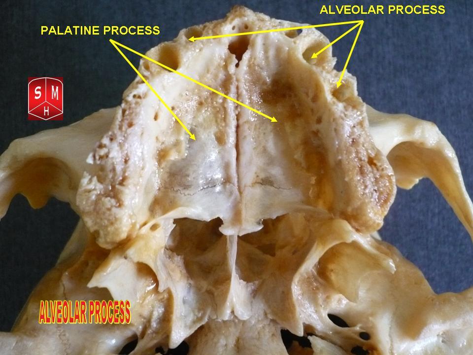
In human anatomy of the mouth, the palatine process of maxilla (palatal process), is a thick, horizontal process of the maxilla. It forms the anterior three quarters of the hard palate, the horizontal plate of the palatine bone making up the rest. Structure It is perforated by numerous foramina for the passage of the nutrient vessels; is channelled at the back part of its lateral border by a groove, sometimes a canal, for the transmission of the descending palatine vessels and the anterior palatine nerve from the spheno-palatine ganglion; and presents little depressions for the lodgement of the palatine glands. When the two maxillae are articulated, a funnel-shaped opening, the incisive foramen, is seen in the middle line, immediately behind the incisor teeth. In this opening the orifices of two lateral canals are visible; they are named the incisive canals or foramina of Stenson; through each of them passes the terminal branch of the descending palatine artery and the nasopala ... [...More Info...] [...Related Items...] OR: [Wikipedia] [Google] [Baidu] |
Maxilla
The maxilla (plural: ''maxillae'' ) in vertebrates is the upper fixed (not fixed in Neopterygii) bone of the jaw formed from the fusion of two maxillary bones. In humans, the upper jaw includes the hard palate in the front of the mouth. The two maxillary bones are fused at the intermaxillary suture, forming the anterior nasal spine. This is similar to the mandible (lower jaw), which is also a fusion of two mandibular bones at the mandibular symphysis. The mandible is the movable part of the jaw. Structure In humans, the maxilla consists of: * The body of the maxilla * Four processes ** the zygomatic process ** the frontal process of maxilla ** the alveolar process ** the palatine process * three surfaces – anterior, posterior, medial * the Infraorbital foramen * the maxillary sinus * the incisive foramen Articulations Each maxilla articulates with nine bones: * two of the cranium: the frontal and ethmoid * seven of the face: the nasal, zygomatic, lacrimal, ... [...More Info...] [...Related Items...] OR: [Wikipedia] [Google] [Baidu] |
Descending Palatine Artery
The descending palatine artery is a branch of the third part of the maxillary artery supplying the hard and soft palate. Course It descends through the greater palatine canal with the greater and lesser palatine branches of the pterygopalatine ganglion, and, emerging from the greater palatine foramen, runs forward in a groove on the medial side of the alveolar border of the hard palate to the incisive canal; the terminal branch of the artery passes upward through this canal to anastomosis, anastomose with the sphenopalatine artery. Branches Branches are distributed to the gums, the palatine glands, and the mucous membrane of the roof of the mouth; while in the Greater palatine canal, pterygopalatine canal it gives off twigs which descend in the lesser palatine canals to supply the soft palate and palatine tonsil, anastomosing with the ascending palatine artery. According to Terminologia Anatomica, the descending palatine artery branches into the greater palatine artery and lesser ... [...More Info...] [...Related Items...] OR: [Wikipedia] [Google] [Baidu] |
Posterior Canal
The semicircular canals or semicircular ducts are three semicircular, interconnected tubes located in the innermost part of each ear, the inner ear. The three canals are the horizontal, superior and posterior semicircular canals. Structure The semicircular canals are a component of the bony labyrinth that are at right angles from each other. At one end of each of the semicircular canals is a dilated sac called an osseous ampulla, which is more than twice the diameter of the canal. Each ampulla contains an ampullary crest, the crista ampullaris which consists of a thick gelatinous cap called a cupula and many hair cells. The superior and posterior semicircular canals are oriented vertically at right angles to each other. The lateral semicircular canal is about a 30-degree angle from the horizontal plane. The orientations of the canals cause a different canal to be stimulated by movement of the head in different planes, and more than one canal is stimulated at once if the movement ... [...More Info...] [...Related Items...] OR: [Wikipedia] [Google] [Baidu] |
Nasopalatine Nerves
The nasopalatine nerve (long sphenopalatine nerve) is a nerve of the head. It is a branch of the pterygopalatine ganglion, a continuation from the maxillary nerve (V2). It supplies parts of the palate and nasal septum. Structure The nasopalatine nerve communicates with the corresponding nerve of the opposite side and with the greater palatine nerve. The medial superior posterior nasal branches of the maxillary nerve usually branch from the nasopalatine nerve. Origin The nasopalatine nerve is a branch of the pterygopalatine ganglion, a continuation from the maxillary nerve (V2), itself a branch of the trigeminal nerve. It enters the nasal cavity through the sphenopalatine foramen. Course It passes across the roof of the nasal cavity below the orifice of the sphenoidal sinus to reach the nasal septum. It then runs obliquely downward and forward between the periosteum and mucous membrane of the lower part of the nasal septum. It descends to the roof of the mouth ... [...More Info...] [...Related Items...] OR: [Wikipedia] [Google] [Baidu] |
Foramina Of Scarpa
In the maxilla, occasionally two additional canals are present in the middle line of the palatine process; they are termed the foramina of Scarpa, and when present transmit the nasopalatine nerves, the left passing through the anterior, and the right through the posterior canal. See also * Antonio Scarpa – anatomist References External links * Foramina of the skull {{Portal bar, Anatomy ... [...More Info...] [...Related Items...] OR: [Wikipedia] [Google] [Baidu] |
Anterior Nasal Spine
The anterior nasal spine, or anterior nasal spine of maxilla, is a bony projection in the skull that serves as a cephalometric landmark. The anterior nasal spine is the projection formed by the fusion of the two maxillary bones at the intermaxillary suture. It is placed at the level of the nostrils, at the uppermost part of the philtrum and rarely fractures. Additional images File:Anterior nasal spine of maxilla - animation02.gif, Animation. Anterior nasal spine shown in red. File:Anterior nasal spine of maxilla - animation00.gif, Left maxilla. Anterior nasal spine shown in red. File:Anterior nasal spine of maxilla - skull - anterior view.png, Skull. Anterior view. Anterior nasal spine shown in red. File:Slide12hhhh.JPG, Right maxilla. Anterior nasal spine labeled at center left. See also * Posterior nasal spine The posterior nasal spine is part of the horizontal plate of the palatine bone of the skull. It is found at the medial end of its posterior border. It is paired ... [...More Info...] [...Related Items...] OR: [Wikipedia] [Google] [Baidu] |
Incisor Crest
Incisors (from Latin ''incidere'', "to cut") are the front teeth present in most mammals. They are located in the premaxilla above and on the mandible below. Humans have a total of eight (two on each side, top and bottom). Opossums have 18, whereas armadillos have none. Structure Adult humans normally have eight incisors, two of each type. The types of incisor are: * maxillary central incisor (upper jaw, closest to the center of the lips) * maxillary lateral incisor (upper jaw, beside the maxillary central incisor) * mandibular central incisor (lower jaw, closest to the center of the lips) * mandibular lateral incisor (lower jaw, beside the mandibular central incisor) Children with a full set of deciduous teeth (primary teeth) also have eight incisors, named the same way as in permanent teeth. Young children may have from zero to eight incisors depending on the stage of their tooth eruption and tooth development. Typically, the mandibular central incisors erupt first, followe ... [...More Info...] [...Related Items...] OR: [Wikipedia] [Google] [Baidu] |
Vomer
The vomer (; lat, vomer, lit=ploughshare) is one of the unpaired facial bones of the skull. It is located in the midsagittal line, and articulates with the sphenoid, the ethmoid, the left and right palatine bones, and the left and right maxillary bones. The vomer forms the inferior part of the nasal septum in humans, with the superior part formed by the perpendicular plate of the ethmoid bone. The name is derived from the Latin word for a ploughshare and the shape of the bone. In humans The vomer is situated in the median plane, but its anterior portion is frequently bent to one side. It is thin, somewhat quadrilateral in shape, and forms the hinder and lower part of the nasal septum; it has two surfaces and four borders. The surfaces are marked by small furrows for blood vessels, and on each is the nasopalatine groove, which runs obliquely downward and forward, and lodges the nasopalatine nerve and vessels. Borders The ''superior border'', the thickest, presents a d ... [...More Info...] [...Related Items...] OR: [Wikipedia] [Google] [Baidu] |
Nasal Crest
Nasal is an adjective referring to the nose, part of human or animal anatomy. It may also be shorthand for the following uses in combination: * With reference to the human nose: ** Nasal administration, a method of pharmaceutical drug delivery ** Nasal emission, the abnormal passing of oral air through a palatal cleft, or from some other type of pharyngeal inadequacy ** Nasal hair, the hair in the nose * With reference to phonetics: ** Nasalization, the production of a sound with a lowered velum, allowing some of the air to escape through the nose; the resulting being either: *** a nasal consonant, or *** a nasal vowel * With reference to the nose of humans or other animals: ** Nasal bone, two small oblong bones placed side by side at the middle and upper part of the face, and form, by their junction, "the bridge" of the nose ** Nasal cavity, a large air filled space above and behind the nose in the middle of the face ** Nasal concha, a long, narrow and curled bone shelf which pro ... [...More Info...] [...Related Items...] OR: [Wikipedia] [Google] [Baidu] |
Dental Alveolus
Dental alveoli (singular ''alveolus'') are sockets in the jaws in which the roots of teeth are held in the alveolar process with the periodontal ligament. The lay term for dental alveoli is tooth sockets. A joint that connects the roots of the teeth and the alveolus is called '' gomphosis'' (plural ''gomphoses''). Alveolar bone is the bone that surrounds the roots of the teeth forming bone sockets. In mammals, tooth sockets are found in the maxilla, the premaxilla, and the mandible. Etymology 1706, "a hollow," especially "the socket of a tooth," from Latin alveolus "a tray, trough, basin; bed of a small river; small hollow or cavity," diminutive of alvus "belly, stomach, paunch, bowels; hold of a ship," from PIE root *aulo- "hole, cavity" (source also of Greek aulos "flute, tube, pipe;" Serbo-Croatian, Polish, Russian ulica "street," originally "narrow opening;" Old Church Slavonic uliji, Lithuanian aulys "beehive" (hollow trunk), Armenian yli "pregnant"). The word was extende ... [...More Info...] [...Related Items...] OR: [Wikipedia] [Google] [Baidu] |
Canine Tooth
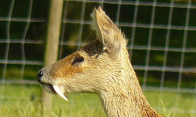
In mammalian oral anatomy, the canine teeth, also called cuspids, dog teeth, or (in the context of the upper jaw) fangs, eye teeth, vampire teeth, or vampire fangs, are the relatively long, pointed teeth. They can appear more flattened however, causing them to resemble incisors and leading them to be called ''incisiform''. They developed and are used primarily for firmly holding food in order to tear it apart, and occasionally as weapons. They are often the largest teeth in a mammal's mouth. Individuals of most species that develop them normally have four, two in the upper jaw and two in the lower, separated within each jaw by incisors; humans and dogs are examples. In most species, canines are the anterior-most teeth in the maxillary bone. The four canines in humans are the two maxillary canines and the two mandibular canines. Details There are generally four canine teeth: two in the upper (maxillary) and two in the lower (mandibular) arch. A canine is placed laterall ... [...More Info...] [...Related Items...] OR: [Wikipedia] [Google] [Baidu] |
Nasopalatine Nerve
The nasopalatine nerve (long sphenopalatine nerve) is a nerve of the head. It is a branch of the pterygopalatine ganglion, a continuation from the maxillary nerve (V2). It supplies parts of the palate and nasal septum. Structure The nasopalatine nerve communicates with the corresponding nerve of the opposite side and with the greater palatine nerve. The medial superior posterior nasal branches of the maxillary nerve usually branch from the nasopalatine nerve. Origin The nasopalatine nerve is a branch of the pterygopalatine ganglion, a continuation from the maxillary nerve (V2), itself a branch of the trigeminal nerve. It enters the nasal cavity through the sphenopalatine foramen. Course It passes across the roof of the nasal cavity below the orifice of the sphenoidal sinus to reach the nasal septum. It then runs obliquely downward and forward between the periosteum and mucous membrane of the lower part of the nasal septum. It descends to the roof of the mouth ... [...More Info...] [...Related Items...] OR: [Wikipedia] [Google] [Baidu] |


