|
Pachychoroid Spectrum
Pachychoroid disorders of the macula represent a group of diseases affecting the central part of the retina of the eye, the macula. Due to thickening and congestion of the highly vascularized layer underneath the macula, the choroid, damage to the retinal pigment epithelium and the retinal photoreceptor cells ensues. This leads to impaired vision. The best known representative of the pachychoroid disease spectrum, central serous chorioretinopathy, is the fourth most common cause of irreversible damage to the macula:. The term "pachychoroid" was first introduced in 2013 by David Warrow, Quan Hoang and K. Bailey Freund. Disease mechanism The disease mechanisms are not completely understood. All pachychoroid disorders of the macula show choroidal thickening and congestion with increased blood vessel diameter, especially in the deep choroid (the so-called Haller's layer). This results in increased pressure from the deep choroid against the superficial choroid close to the retina, d ... [...More Info...] [...Related Items...] OR: [Wikipedia] [Google] [Baidu] |
Central Serous Chorioretinopathy With Increased Choroidal Thickness
Central is an adjective usually referring to being in the center of some place or (mathematical) object. Central may also refer to: Directions and generalised locations * Central Africa, a region in the centre of Africa continent, also known as Middle Africa * Central America, a region in the centre of America continent * Central Asia, a region in the centre of Eurasian continent * Central Australia, a region of the Australian continent * Central Belt, an area in the centre of Scotland * Central Europe, a region of the European continent * Central London, the centre of London * Central Region (other) * Central United States, a region of the United States of America Specific locations Countries * Central African Republic, a country in Africa States and provinces * Blue Nile (state) or Central, a state in Sudan * Central Department, Paraguay * Central Province (Kenya) * Central Province (Papua New Guinea) * Central Province (Solomon Islands) * Central Province, Sri Lank ... [...More Info...] [...Related Items...] OR: [Wikipedia] [Google] [Baidu] |
Retinal Pigment Epithelium
The pigmented layer of retina or retinal pigment epithelium (RPE) is the pigmented cell layer just outside the neurosensory retina that nourishes retinal visual cells, and is firmly attached to the underlying choroid and overlying retinal visual cells. History The RPE was known in the 18th and 19th centuries as the pigmentum nigrum, referring to the observation that the RPE is dark (black in many animals, brown in humans); and as the tapetum nigrum, referring to the observation that in animals with a tapetum lucidum, in the region of the tapetum lucidum the RPE is not pigmented. Anatomy The RPE is composed of a single layer of hexagonal cells that are densely packed with pigment granules. When viewed from the outer surface, these cells are smooth and hexagonal in shape. When seen in section, each cell consists of an outer non-pigmented part containing a large oval nucleus and an inner pigmented portion which extends as a series of straight thread-like processes between the rods, ... [...More Info...] [...Related Items...] OR: [Wikipedia] [Google] [Baidu] |
Focal Choroidal Excavation
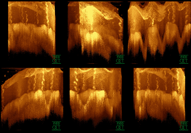
Focal choroidal excavation (FCE) is a concavity in the choroidal layer of the eye that can be detected by optical coherence tomography. The disease is usually unilateral and not associated with any accompanying systemic diseases. Pathophysiology Focal choroidal excavation (FCE) is a concavity in the choroidal layer of the eye without posterior staphyloma or scleral ectasia, that can be detected by optical coherence tomography. The concavity is commonly seen in the macular region. The disease is usually unilateral and not associated with any accompanying systemic diseases. Choroidal vascular disorders which cause visual symptoms, including central serous chorioretinopathy (CSCR), choroidal neovascularization (CNV), and polypoidal choroidal vasculopathy (PCV) may also present with focal choroidal excavation. Etiology The exact etiology of FCE is still (as of 2022) unknown. It was previously considered a congenital disease, but later it was suggested that FCEs can also occur with ... [...More Info...] [...Related Items...] OR: [Wikipedia] [Google] [Baidu] |
Photodynamic Therapy
Photodynamic therapy (PDT) is a form of phototherapy involving light and a photosensitizing chemical substance, used in conjunction with molecular oxygen to elicit cell death (phototoxicity). PDT is popularly used in treating acne. It is used clinically to treat a wide range of medical conditions, including wet age-related macular degeneration, psoriasis, atherosclerosis and has shown some efficacy in anti-viral treatments, including herpes. It also treats malignant cancers including head and neck, lung, bladder and particular skin. The technology has also been tested for treatment of prostate cancer, both in a dog model and in human prostate cancer patients. It is recognised as a treatment strategy that is both minimally invasive and minimally toxic. Other light-based and laser therapies such as laser wound healing and rejuvenation, or intense pulsed light hair removal do not require a photosensitizer. Photosensitisers have been employed to sterilise blood plasma and wate ... [...More Info...] [...Related Items...] OR: [Wikipedia] [Google] [Baidu] |
Polypoidal Choroidal Vasculopathy
Polypoidal choroidal vasculopathy (PCV) is an eye disease primarily affecting the choroid. It may cause sudden blurring of vision or a scotoma in the central field of vision. Since Indocyanine green angiography gives better imaging of choroidal structures, it is more preferred in diagnosing PCV. Treatment options of PCV include careful observation, photodynamic therapy, Gas dynamic laser, thermal laser, intravitreal injection of Anti–vascular endothelial growth factor therapy, anti-VEGF therapy, or combination therapy. Pathophysiology PCV is an ocular disease characterised by abnormally shaped vessels in the choroid. It is described as an exudative maculopathy, characterised by multiple recurrent serosanguineous Retinal pigment epithelium, retinal pigment epithelial detachments. Elevated reddish to orange lesions on fundus examination, dilated inner choroidal vessels, and polypoidal vascular structures beneath the retinal detachment are other features of PCV. Etiology PCV is bel ... [...More Info...] [...Related Items...] OR: [Wikipedia] [Google] [Baidu] |
Aneurysm
An aneurysm is an outward bulging, likened to a bubble or balloon, caused by a localized, abnormal, weak spot on a blood vessel wall. Aneurysms may be a result of a hereditary condition or an acquired disease. Aneurysms can also be a nidus (starting point) for clot formation (thrombosis) and embolization. As an aneurysm increases in size, the risk of rupture, which leads to uncontrolled bleeding, increases. Although they may occur in any blood vessel, particularly lethal examples include aneurysms of the Circle of Willis in the brain, aortic aneurysms affecting the thoracic aorta, and abdominal aortic aneurysms. Aneurysms can arise in the heart itself following a heart attack, including both ventricular and atrial septal aneurysms. There are congenital atrial septal aneurysms, a rare heart defect. Etymology The word is from Greek: ἀνεύρυσμα, aneurysma, "dilation", from ἀνευρύνειν, aneurynein, "to dilate". Classification Aneurysms are classified by type, ... [...More Info...] [...Related Items...] OR: [Wikipedia] [Google] [Baidu] |
Anti–vascular Endothelial Growth Factor Therapy
Anti–vascular endothelial growth factor therapy, also known as anti-VEGF () therapy or medication, is the use of medications that block vascular endothelial growth factor. This is done in the treatment of certain cancers and in age-related macular degeneration. They can involve monoclonal antibodies such as bevacizumab, antibody derivatives such as ranibizumab (Lucentis), or orally-available small molecules that inhibit the tyrosine kinases stimulated by VEGF: sunitinib, sorafenib, axitinib, and pazopanib (some of these therapies target VEGF receptors rather than the VEGFs). Both antibody-based compounds and the first three orally available compounds are commercialized. The latter two, axitinib and pazopanib, are in clinical trials. Bergers and Hanahan concluded in 2008 that anti-VEGF drugs can show therapeutic efficacy in mouse models of cancer and in an increasing number of human cancers. But, "the benefits are at best transitory and are followed by a restoration of tumour ... [...More Info...] [...Related Items...] OR: [Wikipedia] [Google] [Baidu] |
Choroidal Neovascularization
Choroidal neovascularization (CNV) is the creation of new blood vessels in the choroid layer of the eye. Choroidal neovascularization is a common cause of neovascular degenerative maculopathy (i.e. 'wet' macular degeneration) commonly exacerbated by extreme myopia, malignant myopic degeneration, or age-related developments. Causes CNV can occur rapidly in individuals with defects in Bruch's membrane, the innermost layer of the choroid. It is also associated with excessive amounts of vascular endothelial growth factor (VEGF). As well as in wet macular degeneration, CNV can also occur frequently with the rare genetic disease pseudoxanthoma elasticum and rarely with the more common optic disc drusen. CNV has also been associated with extreme myopia or malignant myopic degeneration, where in choroidal neovascularization occurs primarily in the presence of cracks within the retinal (specifically) macular tissue known as lacquer cracks. Symptoms CNV can create a sudden deterioration o ... [...More Info...] [...Related Items...] OR: [Wikipedia] [Google] [Baidu] |
Optical Coherence Tomography
Optical coherence tomography (OCT) is an imaging technique that uses low-coherence light to capture micrometer-resolution, two- and three-dimensional images from within optical scattering media (e.g., biological tissue). It is used for medical imaging and industrial nondestructive testing (NDT). Optical coherence tomography is based on low-coherence interferometry, typically employing near-infrared light. The use of relatively long wavelength light allows it to penetrate into the scattering medium. Confocal microscopy, another optical technique, typically penetrates less deeply into the sample but with higher resolution. Depending on the properties of the light source ( superluminescent diodes, ultrashort pulsed lasers, and supercontinuum lasers have been employed), optical coherence tomography has achieved sub-micrometer resolution (with very wide-spectrum sources emitting over a ~100 nm wavelength range). Optical coherence tomography is one of a class of optical tom ... [...More Info...] [...Related Items...] OR: [Wikipedia] [Google] [Baidu] |
Funduscopy
Ophthalmoscopy, also called funduscopy, is a test that allows a health professional to see inside the fundus of the eye and other structures using an ophthalmoscope (or funduscope). It is done as part of an eye examination and may be done as part of a routine physical examination. It is crucial in determining the health of the retina, optic disc, and vitreous humor. The pupil is a hole through which the eye's interior will be viewed. Opening the pupil wider (dilating it) is a simple and effective way to better see the structures behind it. Therefore, dilation of the pupil (mydriasis) is often accomplished with medicated eye drops before funduscopy. However, although dilated fundus examination is ideal, undilated examination is more convenient and is also helpful (albeit not as comprehensive), and it is the most common type in primary care. An alternative or complement to ophthalmoscopy is to perform a fundus photography, where the image can be analysed later by a professional. T ... [...More Info...] [...Related Items...] OR: [Wikipedia] [Google] [Baidu] |
Bruch's Membrane
Bruch's membrane is the innermost layer of the choroid of the eye. It is also called the ''vitreous lamina'' or ''Membrane vitriae'', because of its glassy microscopic appearance. It is 2–4 μm thick. Layers Bruch's membrane consists of five layers (from inside to outside): #the basement membrane of the retinal pigment epithelium #the inner collagenous zone #a central band of elastic fibers #the outer collagenous zone #the basement membrane of the choriocapillaris The retinal pigment epithelium transports metabolic waste from the photoreceptors across Bruch's membrane to the choroid. Embryology Bruch's membrane is present by midterm in fetal development as an elastic sheet. Pathology Bruch's membrane thickens with age, slowing the transport of metabolites. This may lead to the formation of drusen in age-related macular degeneration. There is also a buildup of deposits (Basal Linear Deposits or BLinD and Basal Lamellar Deposits BLamD) on and within the membrane, primarily co ... [...More Info...] [...Related Items...] OR: [Wikipedia] [Google] [Baidu] |
Retina
The retina (from la, rete "net") is the innermost, light-sensitive layer of tissue of the eye of most vertebrates and some molluscs. The optics of the eye create a focused two-dimensional image of the visual world on the retina, which then processes that image within the retina and sends nerve impulses along the optic nerve to the visual cortex to create visual perception. The retina serves a function which is in many ways analogous to that of the film or image sensor in a camera. The neural retina consists of several layers of neurons interconnected by synapses and is supported by an outer layer of pigmented epithelial cells. The primary light-sensing cells in the retina are the photoreceptor cells, which are of two types: rods and cones. Rods function mainly in dim light and provide monochromatic vision. Cones function in well-lit conditions and are responsible for the perception of colour through the use of a range of opsins, as well as high-acuity vision used for task ... [...More Info...] [...Related Items...] OR: [Wikipedia] [Google] [Baidu] |


