|
Pyloromyotomy
Pyloromyotomy is a surgical procedure in which a portion of the muscle fibers of the pyloric muscle are cut. This is typically done in cases where the contents from the stomach are inappropriately stopped by the pyloric muscle, causing the stomach contents to build up in the stomach and unable to be appropriately digested. The procedure is typically performed in cases of " hypertrophic pyloric stenosis" in young children. In most cases, the procedure can be performed with either an open approach or a laparoscopic approach and the patients typically have good outcomes with minimal complications. History and development The development of the procedure has attributed to Dr. Conrad Ramstedt in 1911, who originally named the procedure Ramstedt's Operation. However, the procedure was truly performed about 17 months earlier by Sir Harold Stiles in 1910 at the Royal Hospital for sick children. In 1991, the first laparoscopic pyloromyotomy was performed by Dr. Alain and Dr. Grousse ... [...More Info...] [...Related Items...] OR: [Wikipedia] [Google] [Baidu] |
Conrad Ramstedt
Wilhelm Conrad Ramstedt (1 February 1867 – 7 February 1963) was a German surgeon remembered for describing pyloromyotomy, Ramstedt's operation. Biography Conrad Ramstedt was born in 1867 in Hamersleben, Saxony-Anhalt, the son of physician Constantin Ramstedt. He was educated at the Gymnasium (school), gymnasium in Magdeburg before studying medicine at Heidelberg University Faculty of Medicine, Heidelberg, Humboldt University of Berlin, Berlin and finally University of Halle-Wittenberg, Halle from where he qualified in 1894. He became assistant in the University Surgical Clinic in Halle under Fritz Gustav von Bramann from 1895 to 1901. In 1901 he joined the German Army (German Empire), German Army as a medical officer in the 4th (Westphalian) Cuirassiers "von Driesen", Westphalian Cuirassiers, serving until 1909. On his discharge from the Army, he became chief surgeon to the RafaelKlinik in Münster, a position he held until 1947. During World War I he served as Oberstabsar ... [...More Info...] [...Related Items...] OR: [Wikipedia] [Google] [Baidu] |
Pyloric Stenosis
Pyloric stenosis is a narrowing of the opening from the stomach to the first part of the small intestine (the pylorus). Symptoms include projectile vomiting without the presence of bile. This most often occurs after the baby is fed. The typical age that symptoms become obvious is two to twelve weeks old. The cause of pyloric stenosis is unclear. Risk factors in babies include birth by cesarean section, preterm birth, bottle feeding, and being first born. The diagnosis may be made by feeling an olive-shaped mass in the baby's abdomen. This is often confirmed with ultrasound. Treatment initially begins by correcting dehydration and electrolyte problems. This is then typically followed by surgery, although some treat the condition without surgery by using atropine. Results are generally good both in the short term and in the long term. About one to two per 1,000 babies are affected, and males are affected about four times more often than females. The condition is very rare in ... [...More Info...] [...Related Items...] OR: [Wikipedia] [Google] [Baidu] |
Pylorus
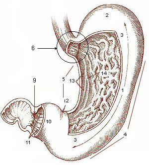
The pylorus ( or ), or pyloric part, connects the stomach to the duodenum. The pylorus is considered as having two parts, the ''pyloric antrum'' (opening to the body of the stomach) and the ''pyloric canal'' (opening to the duodenum). The ''pyloric canal'' ends as the ''pyloric orifice'', which marks the junction between the stomach and the duodenum. The orifice is surrounded by a sphincter, a band of muscle, called the ''pyloric sphincter''. The word ''pylorus'' comes from Greek πυλωρός, via Latin. The word ''pylorus'' in Greek means "gatekeeper", related to "gate" ( el, pyle) and is thus linguistically related to the word " pylon". Structure The pylorus is the furthest part of the stomach that connects to the duodenum. It is divided into two parts, the ''antrum'', which connects to the body of the stomach, and the ''pyloric canal'', which connects to the duodenum. Antrum The ''pyloric antrum'' is the initial portion of the pylorus. It is near the bottom of the stomach, ... [...More Info...] [...Related Items...] OR: [Wikipedia] [Google] [Baidu] |
Harold Stiles
Sir Harold Jalland Stiles (21 March 1863 – 19 April 1946) was an English surgeon who was known for his research into cancer and tuberculosis and for treatment of nerve injuries. Early years Harold Stiles was born in Spalding, Lincolnshire in 1863 the son of Henry Tournay Stiles MD and his wife, Elizabeth Ellen Jalland. He came from a family of doctors. He studied Medicine at the University of Edinburgh, graduating MB ChB in 1885. He earned the Ettles scholarship for the most distinguished graduate of the year. For two years he then taught anatomy at Edinburgh. He was House Surgeon to Professor John Chiene FRSE, Demonstrator in the University Department of Anatomy under Sir William Turner, and Assistant in Charge of Pathology in the university's surgical laboratory. In 1889 Stiles was admitted as a Fellow of the Royal College of Surgeons of Edinburgh. He was then living at 5 Castle Terrace, south of Edinburgh Castle. He trained for six months under Professor Theodore ... [...More Info...] [...Related Items...] OR: [Wikipedia] [Google] [Baidu] |
Laparoscopy
Laparoscopy () is an operation performed in the abdomen or pelvis using small incisions (usually 0.5–1.5 cm) with the aid of a camera. The laparoscope aids diagnosis or therapeutic interventions with a few small cuts in the abdomen.MedlinePlus > Laparoscopy Update Date: 21 August 2009. Updated by: James Lee, MD // No longer valid Laparoscopic surgery, also called minimally invasive procedure, bandaid surgery, or keyhole surgery, is a modern surgical technique. There are a number of advantages to the patient with laparoscopic surgery versus an exploratory laparotomy. These include reduced pain due to smaller incisions, reduced hemorrhaging, and shorter recovery time. The key element is the use of a laparoscope, a long fiber optic cable system that allows viewing of the affected area by snaking the cable from a more distant, but more easily accessible location. Laparoscopic surgery includes operations within the abdominal or pelvic cavities, whereas keyhole surgery perform ... [...More Info...] [...Related Items...] OR: [Wikipedia] [Google] [Baidu] |
Surgical Suture
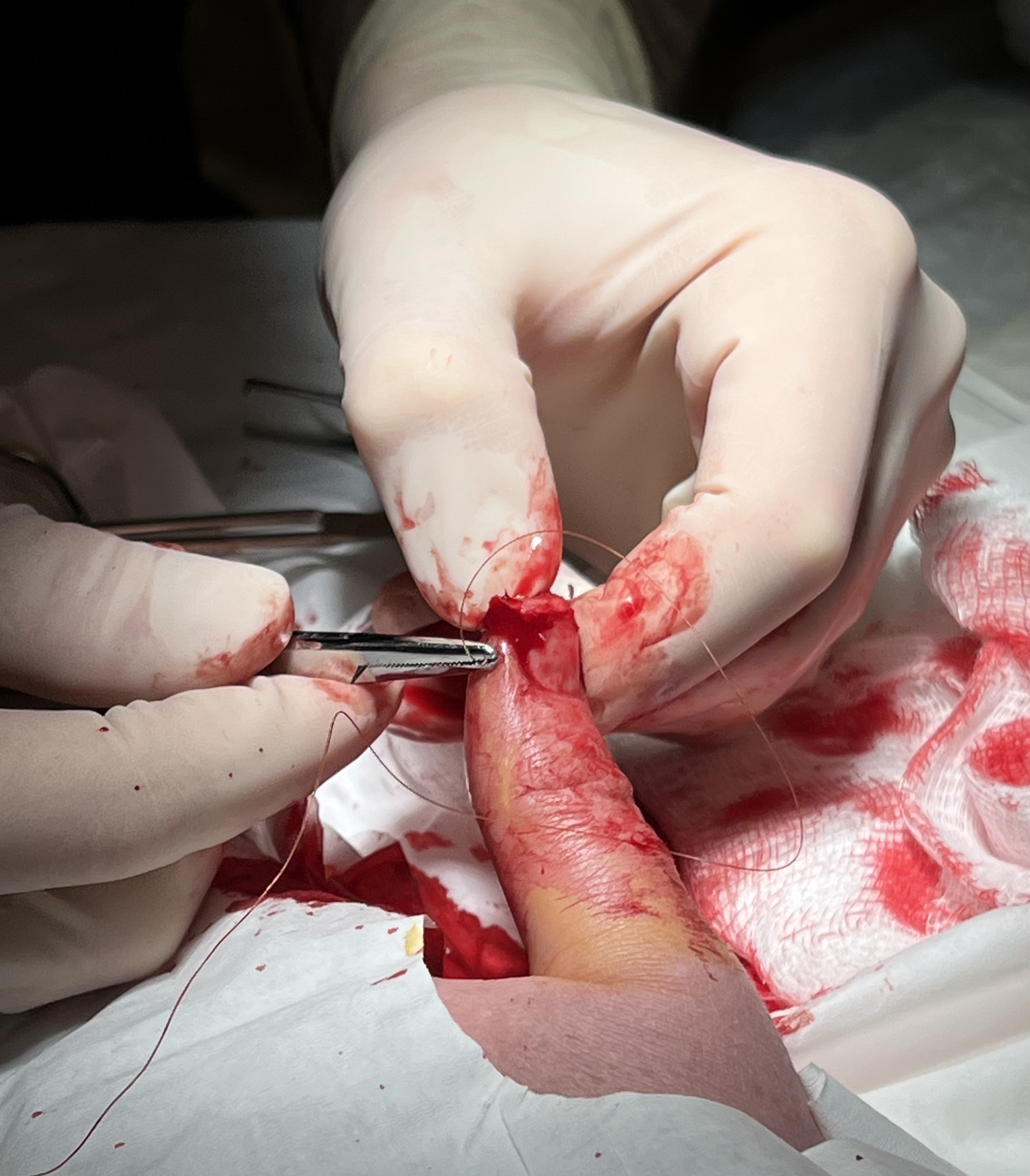
A surgical suture, also known as a stitch or stitches, is a medical device used to hold body tissues together and approximate wound edges after an injury or surgery. Application generally involves using a needle with an attached length of thread. There are numerous types of suture which differ by needle shape and size as well as thread material and characteristics. Selection of surgical suture should be determined by the characteristics and location of the wound or the specific body tissues being approximated. In selecting the needle, thread, and suturing technique to use for a specific patient, a medical care provider must consider the tensile strength of the specific suture thread needed to efficiently hold the tissues together depending on the mechanical and shear forces acting on the wound as well as the thickness of the tissue being approximated. One must also consider the elasticity of the thread and ability to adapt to different tissues, as well as the memory of the threa ... [...More Info...] [...Related Items...] OR: [Wikipedia] [Google] [Baidu] |
Incisional Hernia
An incisional hernia is a type of hernia caused by an incompletely-healed surgical wound. Since median incisions in the abdomen are frequent for abdominal exploratory surgery, ventral incisional hernias are often also classified as ventral hernias due to their location. Not all ventral hernias are from incisions, as some may be caused by other trauma or congenital problems. Signs and symptoms Clinically, incisional hernias present as a bulge or protrusion at or near the area of a surgical incision. Virtually any prior abdominal operation can develop an incisional hernia at the scar area (provided adequate healing does not occur due to infection), including large abdominal procedures such as intestinal or vascular surgery, and small incisions, such as ( appendix removal or abdominal exploratory surgery). While incisional hernias can occur at any incision, they tend to occur more commonly along a straight line from the xiphoid process of the sternum straight down to the pubis, ... [...More Info...] [...Related Items...] OR: [Wikipedia] [Google] [Baidu] |
Wound Dehiscence
Wound dehiscence is a surgical complication in which a wound ruptures along a surgical incision. Risk factors include age, collagen disorder such as Ehlers–Danlos syndrome, diabetes, obesity, poor knotting or grabbing of stitches, and trauma to the wound after surgery. Signs Signs of dehiscence can include bleeding, pain, inflammation, fever, or the wound opening spontaneously. An internal surgical wound dehiscence can occur internally, as a consequence of hysterectomy, at the site of the vaginal cuff. Cause A primary cause of wound dehiscence is sub-acute infection, resulting from inadequate or imperfect aseptic technique. Coated suture, such as Vicryl, generally breaks down at a rate predicted to correspond with tissue healing, but is hastened in the presence of bacteria. In the absence of other known metabolic factors which inhibit healing and may have contributed to suture dehiscence, subacute infection should be suspected, and the protocol for obtaining wound culture ... [...More Info...] [...Related Items...] OR: [Wikipedia] [Google] [Baidu] |
Fascia
A fascia (; plural fasciae or fascias; adjective fascial; from Latin: "band") is a band or sheet of connective tissue, primarily collagen, beneath the skin that attaches to, stabilizes, encloses, and separates muscles and other internal organs. Fascia is classified by layer, as superficial fascia, deep fascia, and ''visceral'' or ''parietal'' fascia, or by its function and anatomical location. Like ligaments, aponeuroses, and tendons, fascia is made up of fibrous connective tissue containing closely packed bundles of collagen fibers oriented in a wavy pattern parallel to the direction of pull. Fascia is consequently flexible and able to resist great unidirectional tension forces until the wavy pattern of fibers has been straightened out by the pulling force. These collagen fibers are produced by fibroblasts located within the fascia. Fasciae are similar to ligaments and tendons as they have collagen as their major component. They differ in their location and function: ligament ... [...More Info...] [...Related Items...] OR: [Wikipedia] [Google] [Baidu] |
Mucosa
A mucous membrane or mucosa is a membrane that lines various cavities in the body of an organism and covers the surface of internal organs. It consists of one or more layers of epithelial cells overlying a layer of loose connective tissue. It is mostly of endodermal origin and is continuous with the skin at body openings such as the eyes, eyelids, ears, inside the nose, inside the mouth, lips, the genital areas, the urethral opening and the anus. Some mucous membranes secrete mucus, a thick protective fluid. The function of the membrane is to stop pathogens and dirt from entering the body and to prevent bodily tissues from becoming dehydrated. Structure The mucosa is composed of one or more layers of epithelial cells that secrete mucus, and an underlying lamina propria of loose connective tissue. The type of cells and type of mucus secreted vary from organ to organ and each can differ along a given tract. Mucous membranes line the digestive, respiratory and reproductive trac ... [...More Info...] [...Related Items...] OR: [Wikipedia] [Google] [Baidu] |


