|
Pupillary Dilatation
Mydriasis is the Pupillary dilation, dilation of the pupil, usually having a non-physiological cause, or sometimes a physiological pupillary response. Non-physiological causes of mydriasis include disease, Physical trauma, trauma, or the use of certain types of drugs. Normally, as part of the pupillary light reflex, the pupil dilates in the dark and miosis, constricts in the light to respectively improve vividity at night and to protect the retina from sunlight damage during the day. A ''mydriatic'' pupil will remain excessively large even in a bright environment. The excitation of the radial fibres of the iris which increases the pupillary aperture is referred to as a mydriasis. More generally, mydriasis also refers to the natural dilation of pupils, for instance in low light conditions or under sympathetic stimulation. Fixed, unilateral mydriasis could be a symptom of raised intracranial pressure. The opposite, constriction of the pupil, is referred to as miosis. Both mydriasis ... [...More Info...] [...Related Items...] OR: [Wikipedia] [Google] [Baidu] |
Sympathetic System
The sympathetic nervous system (SNS) is one of the three divisions of the autonomic nervous system, the others being the parasympathetic nervous system and the enteric nervous system. The enteric nervous system is sometimes considered part of the autonomic nervous system, and sometimes considered an independent system. The autonomic nervous system functions to regulate the body's unconscious actions. The sympathetic nervous system's primary process is to stimulate the body's fight or flight response. It is, however, constantly active at a basic level to maintain homeostasis. The sympathetic nervous system is described as being antagonistic to the parasympathetic nervous system which stimulates the body to "feed and breed" and to (then) "rest-and-digest". Structure There are two kinds of neurons involved in the transmission of any signal through the sympathetic system: pre-ganglionic and post-ganglionic. The shorter preganglionic neurons originate in the thoracolumbar division of ... [...More Info...] [...Related Items...] OR: [Wikipedia] [Google] [Baidu] |
Iris (anatomy)
In humans and most mammals and birds, the iris (plural: ''irides'' or ''irises'') is a thin, annular structure in the eye, responsible for controlling the diameter and size of the pupil, and thus the amount of light reaching the retina. Eye color is defined by the iris. In optical terms, the pupil is the eye's aperture, while the iris is the diaphragm. Structure The iris consists of two layers: the front pigmented fibrovascular layer known as a stroma and, beneath the stroma, pigmented epithelial cells. The stroma is connected to a sphincter muscle (sphincter pupillae), which contracts the pupil in a circular motion, and a set of dilator muscles ( dilator pupillae), which pull the iris radially to enlarge the pupil, pulling it in folds. The sphincter pupillae is the opposing muscle of the dilator pupillae. The pupil's diameter, and thus the inner border of the iris, changes size when constricting or dilating. The outer border of the iris does not change size. The constricti ... [...More Info...] [...Related Items...] OR: [Wikipedia] [Google] [Baidu] |
Photophobia
Photophobia is a medical symptom of abnormal intolerance to visual perception of light. As a medical symptom photophobia is not a morbid fear or phobia, but an experience of discomfort or pain to the eyes due to light exposure or by presence of actual physical sensitivity of the eyes, though the term is sometimes additionally applied to abnormal or irrational fear of light such as heliophobia. The term ''photophobia'' comes from the Greek language, Greek φῶς (''phōs''), meaning "light", and φόβος (''phóbos''), meaning "fear". Causes Patients may develop photophobia as a result of several different medical conditions, related to the human eye, eye, the nervous system, genetic, or other causes. Photophobia may manifest itself in an increased response to light starting at any step in the visual system, such as: *Too much light entering the eye. Too much light can enter the eye if it is damaged, such as with corneal abrasion and retinal damage, or if its pupil(s) is unabl ... [...More Info...] [...Related Items...] OR: [Wikipedia] [Google] [Baidu] |
Cycloplegia
Cycloplegia is paralysis of the ciliary muscle of the eye, resulting in a loss of accommodation. Because of the paralysis of the ciliary muscle, the curvature of the lens can no longer be adjusted to focus on nearby objects. This results in similar problems as those caused by presbyopia, in which the lens has lost elasticity and can also no longer focus on close-by objects. Cycloplegia with accompanying mydriasis (dilation of pupil) is usually due to topical application of muscarinic antagonists such as atropine and cyclopentolate. Belladonna alkaloids are used for testing the error of refraction and examination of eye. Management Cycloplegic drugs are generally muscarinic receptor blockers. These include atropine, cyclopentolate, homatropine, scopolamine and tropicamide. They are indicated for use in cycloplegic refraction (to paralyze the ciliary muscle in order to determine the true refractive error of the eye) and the treatment of uveitis. All cycloplegics are also mydriat ... [...More Info...] [...Related Items...] OR: [Wikipedia] [Google] [Baidu] |
Spasm Of Accommodation
A spasm of accommodation (also known as a ciliary spasm, an accommodation, or accommodative spasm) is a condition in which the ciliary muscle of the eye remains in a constant state of contraction. Normal accommodation allows the eye to "accommodate" for near-vision. However, in a state of perpetual contraction, the ciliary muscle cannot relax when viewing distant objects. This causes vision to blur when attempting to view objects from a distance. This may cause pseudomyopia or latent hyperopia. Although antimuscarinic drops (homoatropine 5%) can be applied topically to relax the muscle, this leaves the individual without any accommodation and, depending on refractive error, unable to see well at near distances. Also, excessive pupil dilation may occur as an unwanted side effect. This dilation may pose a problem since a larger pupil is less efficient at focusing light (see pupil, aperture, and optical aberration for more.) Patients who have accommodative spasm may benefit from ... [...More Info...] [...Related Items...] OR: [Wikipedia] [Google] [Baidu] |
Medicine
Medicine is the science and practice of caring for a patient, managing the diagnosis, prognosis, prevention, treatment, palliation of their injury or disease, and promoting their health. Medicine encompasses a variety of health care practices evolved to maintain and restore health by the prevention and treatment of illness. Contemporary medicine applies biomedical sciences, biomedical research, genetics, and medical technology to diagnose, treat, and prevent injury and disease, typically through pharmaceuticals or surgery, but also through therapies as diverse as psychotherapy, external splints and traction, medical devices, biologics, and ionizing radiation, amongst others. Medicine has been practiced since prehistoric times, and for most of this time it was an art (an area of skill and knowledge), frequently having connections to the religious and philosophical beliefs of local culture. For example, a medicine man would apply herbs and say prayers for healing, o ... [...More Info...] [...Related Items...] OR: [Wikipedia] [Google] [Baidu] |
Tropicamide
Tropicamide, sold under the brand name Mydriacyl among others, is a medication used to dilate the pupil and help with examination of the eye. Specifically it is used to help examine the back of the eye. It is applied as eye drops. Effects occur within 40 minutes and last for up to a day. Common side effects include blurry vision, increased intraocular pressure, and sensitivity to light. Another rare but severe side effect is psychosis, particularly in children. It is unclear if use during pregnancy is safe for the fetus. Tropicamide is in the antimuscarinic part of the anticholinergic family of medications. It works by making the muscles within the eye unable to respond to nerve signals. Tropicamide was approved for medical use in the United States in 1960. It is on the World Health Organization's List of Essential Medicines. Medical use Tropicamide is an antimuscarinic drug that produces short acting mydriasis (dilation of the pupil) and cycloplegia when applied as eye ... [...More Info...] [...Related Items...] OR: [Wikipedia] [Google] [Baidu] |
Parasympathetic Nerve
The parasympathetic nervous system (PSNS) is one of the three divisions of the autonomic nervous system, the others being the sympathetic nervous system and the enteric nervous system. The enteric nervous system is sometimes considered part of the autonomic nervous system, and sometimes considered an independent system. The autonomic nervous system is responsible for regulating the body's unconscious actions. The parasympathetic system is responsible for stimulation of "rest-and-digest" or "feed and breed" activities that occur when the body is at rest, especially after eating, including sexual arousal, salivation, lacrimation (tears), urination, digestion, and defecation. Its action is described as being complementary to that of the sympathetic nervous system, which is responsible for stimulating activities associated with the fight-or-flight response. Nerve fibres of the parasympathetic nervous system arise from the central nervous system. Specific nerves include several crani ... [...More Info...] [...Related Items...] OR: [Wikipedia] [Google] [Baidu] |
Radial Muscle
The iris dilator muscle (pupil dilator muscle, pupillary dilator, radial muscle of iris, radiating fibers), is a smooth muscle of the eye, running radially in the iris and therefore fit as a dilator. The pupillary dilator consists of a spokelike arrangement of modified contractile cells called myoepithelial cells. These cells are stimulated by the sympathetic nervous system. When stimulated, the cells contract, widening the pupil and allowing more light to enter the eye. Structure Innervation It is innervated by the sympathetic system, which acts by releasing noradrenaline, which acts on α1-receptors. page 163 Thus, when presented with a threatening stimulus that activates the fight-or-flight response, this innervation contracts the muscle and dilates the pupil, thus temporarily letting more light reach the retina. The dilator muscle is innervated more specifically by postganglionic sympathetic nerves arising from the superior cervical ganglion as the sympathetic root of cilia ... [...More Info...] [...Related Items...] OR: [Wikipedia] [Google] [Baidu] |
Muscle Contraction
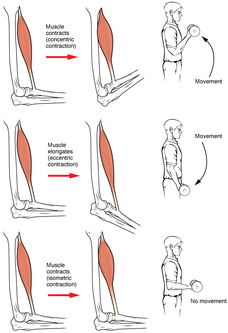
Muscle contraction is the activation of tension-generating sites within muscle cells. In physiology, muscle contraction does not necessarily mean muscle shortening because muscle tension can be produced without changes in muscle length, such as when holding something heavy in the same position. The termination of muscle contraction is followed by muscle relaxation, which is a return of the muscle fibers to their low tension-generating state. For the contractions to happen, the muscle cells must rely on the interaction of two types of filaments which are the thin and thick filaments. Thin filaments are two strands of actin coiled around each, and thick filaments consist of mostly elongated proteins called myosin. Together, these two filaments form myofibrils which are important organelles in the skeletal muscle system. Muscle contraction can also be described based on two variables: length and tension. A muscle contraction is described as isometric if the muscle tension changes ... [...More Info...] [...Related Items...] OR: [Wikipedia] [Google] [Baidu] |
Adrenergic Receptors
The adrenergic receptors or adrenoceptors are a class of G protein-coupled receptors that are targets of many catecholamines like norepinephrine (noradrenaline) and epinephrine (adrenaline) produced by the body, but also many medications like beta blockers, beta-2 (β2) agonists and alpha-2 (α2) agonists, which are used to treat high blood pressure and asthma, for example. Many cells have these receptors, and the binding of a catecholamine to the receptor will generally stimulate the sympathetic nervous system (SNS). The SNS is responsible for the fight-or-flight response, which is triggered by experiences such as exercise or fear-causing situations. This response dilates pupils, increases heart rate, mobilizes energy, and diverts blood flow from non-essential organs to skeletal muscle. These effects together tend to increase physical performance momentarily. History By the turn of the 19th century, it was agreed that the stimulation of sympathetic nerves could cause different ... [...More Info...] [...Related Items...] OR: [Wikipedia] [Google] [Baidu] |
Sympathetic Nervous System
The sympathetic nervous system (SNS) is one of the three divisions of the autonomic nervous system, the others being the parasympathetic nervous system and the enteric nervous system. The enteric nervous system is sometimes considered part of the autonomic nervous system, and sometimes considered an independent system. The autonomic nervous system functions to regulate the body's unconscious actions. The sympathetic nervous system's primary process is to stimulate the body's fight or flight response. It is, however, constantly active at a basic level to maintain homeostasis. The sympathetic nervous system is described as being antagonistic to the parasympathetic nervous system which stimulates the body to "feed and breed" and to (then) "rest-and-digest". Structure There are two kinds of neurons involved in the transmission of any signal through the sympathetic system: pre-ganglionic and post-ganglionic. The shorter preganglionic neurons originate in the thoracolumbar division o ... [...More Info...] [...Related Items...] OR: [Wikipedia] [Google] [Baidu] |


