|
Pulmonary Angiography
Pulmonary angiography (or pulmonary arteriography,conventional pulmonary angiography, selective pulmonary angiography) is a medical fluoroscopic procedure used to visualize the pulmonary arteries and much less frequently, the pulmonary veins. It is a minimally invasive procedure performed most frequently by an interventional radiologist or interventional cardiologist to visualise the arteries of the lungs. Uses Pulmonary angiography is useful as the confirmation test where the non-invasive imaging such as CT pulmonary angiography is inconclusive on determining the presence of pulmonary embolism. The accuracy of pulmonary angiography may be higher than clinical examination, arterial blood gas results, and ventilation/perfusion scan. Pulmonary angiography is also used to confirm chronic thromboembolic pulmonary hypertension (CTEPH) and provides a platform for balloon pulmonary angioplasty to treat the disease. CT pulmonary angiography has nearly entirely replaced conventional pul ... [...More Info...] [...Related Items...] OR: [Wikipedia] [Google] [Baidu] |
Pulmonary Embolism Selective Angiogram
The lungs are the primary organs of the respiratory system in humans and most other animals, including some snails and a small number of fish. In mammals and most other vertebrates, two lungs are located near the backbone on either side of the heart. Their function in the respiratory system is to extract oxygen from the air and transfer it into the bloodstream, and to release carbon dioxide from the bloodstream into the atmosphere, in a process of gas exchange. Respiration is driven by different muscular systems in different species. Mammals, reptiles and birds use their different muscles to support and foster breathing. In earlier tetrapods, air was driven into the lungs by the pharyngeal muscles via buccal pumping, a mechanism still seen in amphibians. In humans, the main muscle of respiration that drives breathing is the diaphragm. The lungs also provide airflow that makes vocal sounds including human speech possible. Humans have two lungs, one on the left and one on the ... [...More Info...] [...Related Items...] OR: [Wikipedia] [Google] [Baidu] |
Fluoroscopy
Fluoroscopy () is an imaging technique that uses X-rays to obtain real-time moving images of the interior of an object. In its primary application of medical imaging, a fluoroscope () allows a physician to see the internal structure and function of a patient, so that the pumping action of the heart or the motion of swallowing, for example, can be watched. This is useful for both diagnosis and therapy and occurs in general radiology, interventional radiology, and image-guided surgery. In its simplest form, a fluoroscope consists of an X-ray source and a fluorescent screen, between which a patient is placed. However, since the 1950s most fluoroscopes have included X-ray image intensifiers and cameras as well, to improve the image's visibility and make it available on a remote display screen. For many decades, fluoroscopy tended to produce live pictures that were not recorded, but since the 1960s, as technology improved, recording and playback became the norm. Fluoroscopy is s ... [...More Info...] [...Related Items...] OR: [Wikipedia] [Google] [Baidu] |
Interventional Radiologist
Interventional radiology (IR) is a medical specialty that performs various minimally-invasive procedures using medical imaging guidance, such as x-ray fluoroscopy, computed tomography, magnetic resonance imaging, or ultrasound. IR performs both diagnostic and therapeutic procedures through very small incisions or body orifices. Diagnostic IR procedures are those intended to help make a diagnosis or guide further medical treatment, and include image-guided biopsy of a tumor or injection of an imaging contrast agent into a hollow structure, such as a blood vessel or a duct. By contrast, therapeutic IR procedures provide direct treatment—they include catheter-based medicine delivery, medical device placement (e.g., stents), and angioplasty of narrowed structures. The main benefits of interventional radiology techniques are that they can reach the deep structures of the body through a body orifice or tiny incision using small needles and wires. That decreases risks, pain, an ... [...More Info...] [...Related Items...] OR: [Wikipedia] [Google] [Baidu] |
Interventional Cardiologist
Interventional cardiology is a branch of cardiology that deals specifically with the catheter based treatment of structural heart diseases. Andreas Gruentzig is considered the father of interventional cardiology after the development of angioplasty by interventional radiologist Charles Dotter. Many procedures can be performed on the heart by catheterization. This most commonly involves the insertion of a sheath into the femoral artery (but, in practice, any large peripheral artery or vein) and cannulating the heart under X-ray visualization (most commonly fluoroscopy). The radial artery may also be used for cannulation; this approach offers several advantages, including the accessibility of the artery in most patients, the easy control of bleeding even in anticoagulated patients, the enhancement of comfort because patients are capable of sitting up and walking immediately following the procedure, and the near absence of clinically significant sequelae in patients with a normal Allen ... [...More Info...] [...Related Items...] OR: [Wikipedia] [Google] [Baidu] |
CT Pulmonary Angiography
A CT pulmonary angiogram (CTPA) is a medical diagnostic test that employs computed tomography (CT) angiography to obtain an image of the pulmonary arteries. Its main use is to diagnose pulmonary embolism (PE). It is a preferred choice of imaging in the diagnosis of PE due to its minimally invasive nature for the patient, whose only requirement for the scan is an intravenous line. Modern MDCT (multi-detector CT) scanners are able to deliver images of sufficient resolution within a short time period, such that CTPA has now supplanted previous methods of testing, such as direct pulmonary angiography, as the gold standard for diagnosis of pulmonary embolism. The patient receives an intravenous injection of an iodine-containing contrast agent at a high-rate using an injector pump. Images are acquired with the maximum intensity of radio-opaque contrast in the pulmonary arteries. This can be done using bolus tracking. A normal CTPA scan will show the contrast filling the pulmonary vesse ... [...More Info...] [...Related Items...] OR: [Wikipedia] [Google] [Baidu] |
Pulmonary Embolism
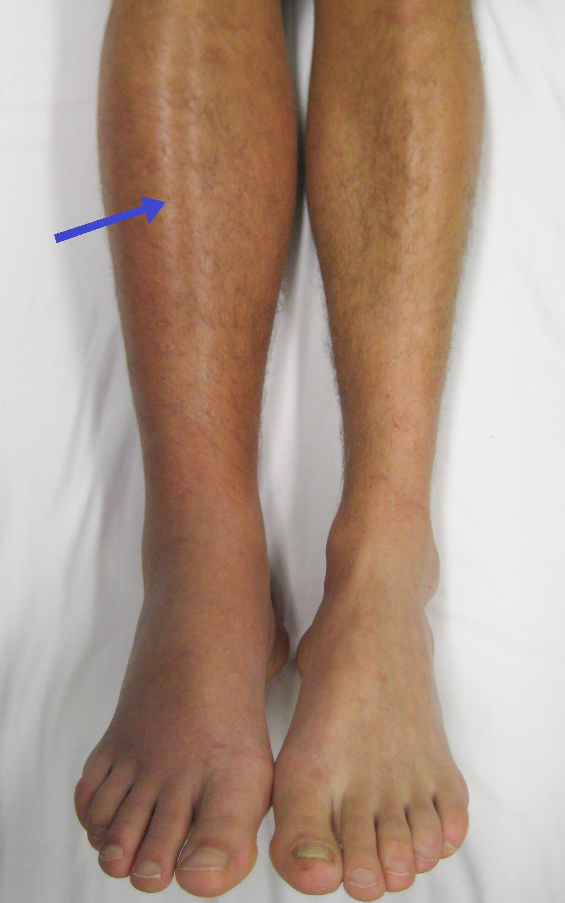
Pulmonary embolism (PE) is a blockage of an pulmonary artery, artery in the lungs by a substance that has moved from elsewhere in the body through the bloodstream (embolism). Symptoms of a PE may include dyspnea, shortness of breath, chest pain particularly upon breathing in, and coughing up blood. Symptoms of a deep vein thrombosis, blood clot in the leg may also be present, such as a erythema, red, warm, swollen, and painful leg. Signs of a PE include low blood oxygen saturation, oxygen levels, tachypnea, rapid breathing, tachycardia, rapid heart rate, and sometimes a mild fever. Severe cases can lead to Syncope (medicine), passing out, shock (circulatory), abnormally low blood pressure, obstructive shock, and cardiac arrest, sudden death. PE usually results from a blood clot in the leg that travels to the lung. The risk of blood clots is increased by advanced age, cancer, prolonged bed rest and immobilization, smoking, stroke, long-haul travel over 4 hours, certain genetics, g ... [...More Info...] [...Related Items...] OR: [Wikipedia] [Google] [Baidu] |
Arterial Blood Gas
An arterial blood gas (ABG) test, or arterial blood gas analysis (ABGA) measures the amounts of arterial gases, such as oxygen and carbon dioxide. An ABG test requires that a small volume of blood be drawn from the radial artery with a syringe and a thin needle, but sometimes the femoral artery in the groin or another site is used. The blood can also be drawn from an arterial catheter. An ABG test measures the blood gas tension values of the arterial partial pressure of oxygen (PaO2), and the arterial partial pressure of carbon dioxide (PaCO2), and the blood's pH. In addition, the arterial oxygen saturation (SaO2) can be determined. Such information is vital when caring for patients with critical illnesses or respiratory disease. Therefore, the ABG test is one of the most common tests performed on patients in intensive-care units. In other levels of care, pulse oximetry plus transcutaneous carbon-dioxide measurement is a less invasive, alternative method of obtaining similar ... [...More Info...] [...Related Items...] OR: [Wikipedia] [Google] [Baidu] |
Ventilation/perfusion Scan
A ventilation/perfusion lung scan, also called a V/Q lung scan, or ventilation/perfusion scintigraphy, is a type of medical imaging using scintigraphy and medical isotopes to evaluate the circulation of air and blood within a patient's lungs, in order to determine the ventilation/perfusion ratio. The ventilation part of the test looks at the ability of air to reach all parts of the lungs, while the perfusion part evaluates how well blood circulates within the lungs. As Q in physiology is the letter used to describe bloodflow the term V/Q scan emerged. Uses This test is most commonly done in order to check for the presence of a blood clot or abnormal blood flow inside the lungs (such as a pulmonary embolism (PE) although computed tomography with radiocontrast is now more commonly used for this purpose. The V/Q scan may be used in some circumstances where radiocontrast would be inappropriate, as in Allergy to contrast agent or kidney failure. A V/Q lung scan may be performed in th ... [...More Info...] [...Related Items...] OR: [Wikipedia] [Google] [Baidu] |
Chronic Thromboembolic Pulmonary Hypertension
Chronic thromboembolic pulmonary hypertension (CTEPH) is a long-term disease caused by a blockage in the blood vessels that deliver blood from the heart to the lungs ( the pulmonary arterial tree). These blockages cause increased resistance to flow in the pulmonary arterial tree which in turn leads to rise in pressure in these arteries ( pulmonary hypertension). The blockages either result from organised (or hardened) blood clots that usually originate from the deep veins of the lower limbs of the body (thromboembolism) and lodge in the pulmonary arterial tree after passing through the right side of the heart. The blockages may also result from scar tissue that forms at the site where the clot has damaged the endothelial lining of the pulmonary arteries, causing permanent fibrous obstruction (blood flow blockage). Most patients have a combination of microvascular (small vessel) and macrovascular (large vessel) obstruction. Some patients may present with normal or near-normal pulmo ... [...More Info...] [...Related Items...] OR: [Wikipedia] [Google] [Baidu] |
Balloon Pulmonary Angioplasty
Balloon pulmonary angioplasty (BPA) is an emerging minimally invasive procedure to treat chronic thromboembolic pulmonary hypertension (CTEPH) in people who are not suitable for pulmonary thromboendarterectomy (PTE) or still have residual pulmonary hypertension and areas of narrowing in the pulmonary arterial tree following previous PTE. The procedure uses balloons to open pulmonary arteries that have been narrowed or blocked by webs, bands and fibrous tissue and therefore restores blood flow to the lungs, reduces shortness of breath, and improves exercise tolerance. More data on its safety and effectiveness are still required. Medical uses BPA may be considered in people with symptomatic CTEPH who are not suitable for surgery. PTE is an established treatment for CTEPH but is only performed at a few specialist centres, requires surgical competence and intermittent total circulatory arrest under deep hypothermia. However, co-existing illnesses and inaccessible clots may contra ... [...More Info...] [...Related Items...] OR: [Wikipedia] [Google] [Baidu] |
Inferior Vena Cava
The inferior vena cava is a large vein that carries the deoxygenated blood from the lower and middle body into the right atrium of the heart. It is formed by the joining of the right and the left common iliac veins, usually at the level of the fifth lumbar vertebra. The inferior vena cava is the lower (" inferior") of the two venae cavae, the two large veins that carry deoxygenated blood from the body to the right atrium of the heart: the inferior vena cava carries blood from the lower half of the body whilst the superior vena cava carries blood from the upper half of the body. Together, the venae cavae (in addition to the coronary sinus, which carries blood from the muscle of the heart itself) form the venous counterparts of the aorta. It is a large retroperitoneal vein that lies posterior to the abdominal cavity and runs along the right side of the vertebral column. It enters the right auricle at the lower right, back side of the heart. The name derives from la, vena, "vei ... [...More Info...] [...Related Items...] OR: [Wikipedia] [Google] [Baidu] |
Right Atrium
The atrium ( la, ātrium, , entry hall) is one of two upper chambers in the heart that receives blood from the circulatory system. The blood in the atria is pumped into the heart ventricles through the atrioventricular valves. There are two atria in the human heart – the left atrium receives blood from the pulmonary circulation, and the right atrium receives blood from the venae cavae of the systemic circulation. During the cardiac cycle the atria receive blood while relaxed in diastole, then contract in systole to move blood to the ventricles. Each atrium is roughly cube-shaped except for an ear-shaped projection called an atrial appendage, sometimes known as an auricle. All animals with a closed circulatory system have at least one atrium. The atrium was formerly called the 'auricle'. That term is still used to describe this chamber in some other animals, such as the ''Mollusca''. They have thicker muscular walls than the atria do. Structure Humans have a four-chambered h ... [...More Info...] [...Related Items...] OR: [Wikipedia] [Google] [Baidu] |


