|
Proximal Humerus Fracture
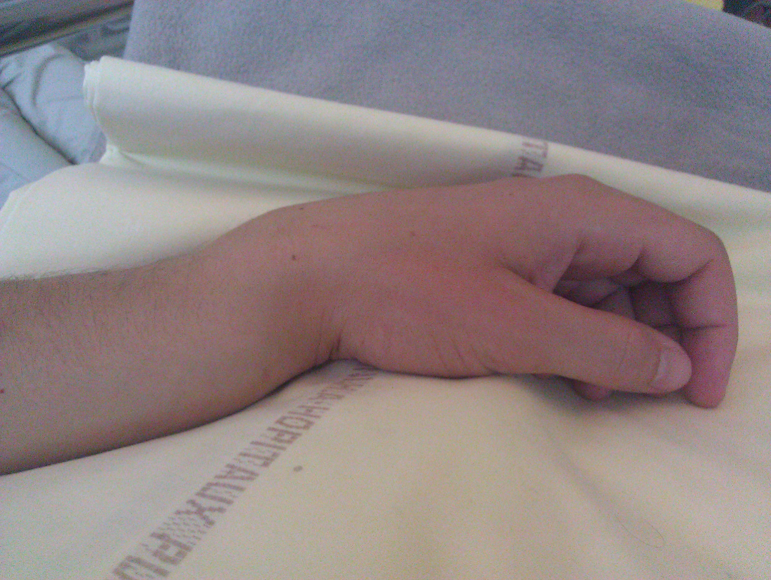
A proximal humerus fracture is a break of the upper part of the bone of the arm (humerus). Symptoms include pain, swelling, and a decreased ability to move the shoulder. Complications may include axillary nerve or axillary artery injury. The cause is generally a fall onto the arm or direct trauma to the arm. Risk factors include osteoporosis and diabetes. Diagnosis is generally based on X-rays or CT scan. It is a type of humerus fracture. A number of classification systems exist. Treatment is generally with an arm sling for a brief period of time followed by specific exercises. This appears appropriate in many cases even when the fragments are separated. Less commonly surgery is recommended. Proximal humerus fractures are common. Older people are most commonly affected. In this age group they are the third most common fractures after hip and Colles fractures. Women are more often affected than men. Signs and symptoms Typical signs and symptoms include pain, swelling, brui ... [...More Info...] [...Related Items...] OR: [Wikipedia] [Google] [Baidu] |
Greater Tuberosity
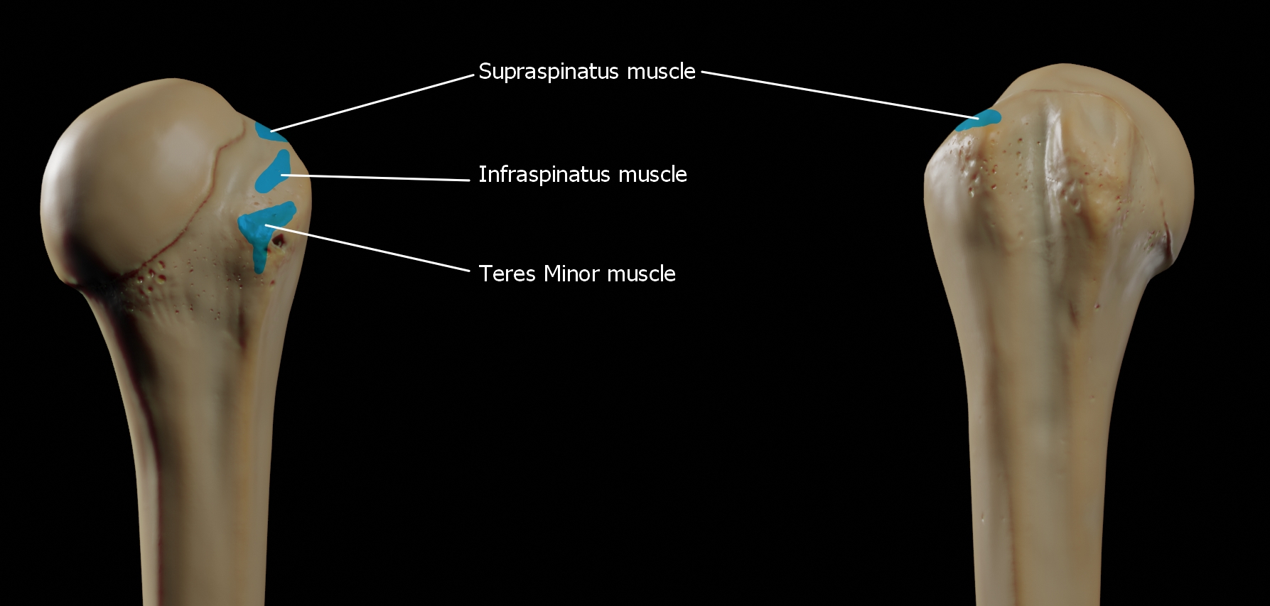
The greater tubercle of the humerus is the outward part the upper end of that bone, adjacent to the large rounded prominence of the humerus head. It provides attachment points for the supraspinatus, infraspinatus, and teres minor muscles, three of the four muscles of the rotator cuff, a muscle group that stabilizes the shoulder joint. In doing so the tubercle acts as a location for the transfer of forces from the rotator cuff muscles to the humerus. Structure The upper surface of the greater tubercle is rounded, and marked by three flat impressions: * the highest ("superior facet") gives insertion to the supraspinatus muscle. * the middle ("middle facet") gives insertion to the infraspinatus muscle. * the lowest ("inferior facet"), and the body of the bone for about 2.5 cm, gives insertion to the teres minor muscle. The lateral surface of the greater tubercle is convex, rough, and continuous with the lateral surface of the body of the humerus. It can be described as ... [...More Info...] [...Related Items...] OR: [Wikipedia] [Google] [Baidu] |
Colles Fracture
A Colles' fracture is a type of fracture of the distal forearm in which the broken end of the radius is bent backwards. Symptoms may include pain, swelling, deformity, and bruising. Complications may include damage to the median nerve. It typically occurs as a result of a fall on an outstretched hand. Risk factors include osteoporosis. The diagnosis may be confirmed via X-rays. The tip of the ulna may also be broken. Treatment may include casting or surgery. Surgical reduction and casting is possible in the majority of cases in people over the age of 50. Pain management can be achieved during the reduction with procedural sedation and analgesia or a hematoma block. A year or two may be required for healing to occur. About 15% of people have a Colles' fracture at some point in their life. They occur more commonly in young adults and older people than in children and middle-aged adults. Women are more frequently affected than men. The fracture is named after Abraham Colles ... [...More Info...] [...Related Items...] OR: [Wikipedia] [Google] [Baidu] |
Magnetic Resonance Imaging
Magnetic resonance imaging (MRI) is a medical imaging technique used in radiology to form pictures of the anatomy and the physiological processes inside the body. MRI scanners use strong magnetic fields, magnetic field gradients, and radio waves to generate images of the organs in the body. MRI does not involve X-rays or the use of ionizing radiation, which distinguishes it from computed tomography (CT) and positron emission tomography (PET) scans. MRI is a medical application of nuclear magnetic resonance (NMR) which can also be used for imaging in other NMR applications, such as NMR spectroscopy. MRI is widely used in hospitals and clinics for medical diagnosis, staging and follow-up of disease. Compared to CT, MRI provides better contrast in images of soft tissues, e.g. in the brain or abdomen. However, it may be perceived as less comfortable by patients, due to the usually longer and louder measurements with the subject in a long, confining tube, although "open ... [...More Info...] [...Related Items...] OR: [Wikipedia] [Google] [Baidu] |
X-ray
X-rays (or rarely, ''X-radiation'') are a form of high-energy electromagnetic radiation. In many languages, it is referred to as Röntgen radiation, after the German scientist Wilhelm Conrad Röntgen, who discovered it in 1895 and named it ''X-radiation'' to signify an unknown type of radiation.Novelline, Robert (1997). ''Squire's Fundamentals of Radiology''. Harvard University Press. 5th edition. . X-ray wavelengths are shorter than those of ultraviolet rays and longer than those of gamma rays. There is no universally accepted, strict definition of the bounds of the X-ray band. Roughly, X-rays have a wavelength ranging from 10 nanometers to 10 picometers, corresponding to frequencies in the range of 30 petahertz to 30 exahertz ( to ) and photon energies in the range of 100 eV to 100 keV, respectively. X-rays can penetrate many solid substances such as construction materials and living tissue, so X-ray radiography is widely used in medi ... [...More Info...] [...Related Items...] OR: [Wikipedia] [Google] [Baidu] |
Rotator Cuff
The rotator cuff is a group of muscles and their tendons that act to stabilize the human shoulder and allow for its extensive range of motion. Of the seven scapulohumeral muscles, four make up the rotator cuff. The four muscles are the supraspinatus muscle, the infraspinatus muscle, teres minor muscle, and the subscapularis muscle. Structure Muscles composing rotator cuff The supraspinatus muscle spreads out in a horizontal band to insert on the superior facet of the greater tubercle of the humerus. The greater tubercle projects as the most lateral structure of the humeral head. Medial to this, in turn, is the lesser tubercle of the humeral head. The subscapularis muscle origin is divided from the remainder of the rotator cuff origins as it is deep to the scapula. The four tendons of these muscles converge to form the rotator cuff tendon. These tendinous insertions along with the articular capsule, the coracohumeral ligament, and the glenohumeral ligament complex, b ... [...More Info...] [...Related Items...] OR: [Wikipedia] [Google] [Baidu] |
Deltoid Muscle
The deltoid muscle is the muscle forming the rounded contour of the human shoulder. It is also known as the 'common shoulder muscle', particularly in other animals such as the domestic cat. Anatomically, the deltoid muscle appears to be made up of three distinct sets of muscle fibers, namely the # anterior or clavicular part (pars clavicularis) # posterior or scapular part (pars scapularis) # intermediate or acromial part (pars acromialis) However, electromyography suggests that it consists of at least seven groups that can be independently coordinated by the nervous system. It was previously called the deltoideus (plural ''deltoidei'') and the name is still used by some anatomists. It is called so because it is in the shape of the Greek capital letter delta (Δ). Deltoid is also further shortened in slang as "delt". A study of 30 shoulders revealed an average mass of in humans, ranging from to . Structure Previous studies showed that the insertions of the tendons of the d ... [...More Info...] [...Related Items...] OR: [Wikipedia] [Google] [Baidu] |
Pectoralis Major
The pectoralis major () is a thick, fan-shaped or triangular convergent muscle, situated at the chest of the human body. It makes up the bulk of the chest muscles and lies under the breast. Beneath the pectoralis major is the pectoralis minor, a thin, triangular muscle. The pectoralis major's primary functions are flexion, adduction, and internal rotation of the humerus. The pectoral major may colloquially be referred to as "pecs", "pectoral muscle", or "chest muscle", because it is the largest and most superficial muscle in the chest area. Structure It arises from the anterior surface of the sternal half of the clavicle from breadth of the half of the anterior surface of the sternum, as low down as the attachment of the cartilage of the sixth or seventh rib; from the cartilages of all the true ribs, with the exception, frequently, of the first or seventh, and from the aponeurosis of the abdominal external oblique muscle. From this extensive origin the fibers converge toward ... [...More Info...] [...Related Items...] OR: [Wikipedia] [Google] [Baidu] |
Posterior Humeral Circumflex Artery
The posterior humeral circumflex artery (posterior circumflex artery, posterior circumflex humeral artery) arises from the third part of axillary artery at the lower border of the subscapularis, and runs posteriorly with the axillary nerve through the quadrangular space. It winds around the surgical neck of the humerus and is distributed to the deltoid muscle and shoulder-joint, anastomosing with the anterior humeral circumflex and deep artery of the arm. It supplies the teres major, teres minor, deltoid Deltoid (delta-shaped) can refer to: * The deltoid muscle, a muscle in the shoulder * Kite (geometry), also known as a deltoid, a type of quadrilateral * A deltoid curve, a three-cusped hypocycloid * A leaf shape * The deltoid tuberosity, a part of ..., and (long head only) triceps muscles. Additional images File:Gray810.png, Suprascapular and axillary nerves of right side, seen from behind. File:Slide4SSS.JPG, Posterior humeral circumflex artery See also * Anterior ... [...More Info...] [...Related Items...] OR: [Wikipedia] [Google] [Baidu] |
Anterior Humeral Circumflex Artery
The anterior humeral circumflex artery (anterior circumflex artery, anterior circumflex humeral artery) is an artery in the arm. It is one of two circumflexing arteries that branch from the axillary artery, the other being the posterior humeral circumflex artery. The anterior humeral circumflex artery is considerably smaller than the posterior and arises nearly opposite to it, from the lateral side of the axillary artery. The anterior humeral circumflex artery runs horizontally, beneath the coracobrachialis and short head of the biceps brachii muscle, in front of the neck of the humerus. On reaching the intertubercular sulcus, it gives off a branch which ascends in the sulcus to supply the head of the humerus and the shoulder-joint. The trunk of the vessel is then continued onward beneath the long head of the biceps brachii and the deltoideus muscle, and anastomoses with the posterior humeral circumflex artery. Additional Images File:Slide3SSS.JPG, Anterior humeral circumfl ... [...More Info...] [...Related Items...] OR: [Wikipedia] [Google] [Baidu] |
Ligament
A ligament is the fibrous connective tissue that connects bones to other bones. It is also known as ''articular ligament'', ''articular larua'', ''fibrous ligament'', or ''true ligament''. Other ligaments in the body include the: * Peritoneal ligament: a fold of peritoneum or other membranes. * Fetal remnant ligament: the remnants of a fetal tubular structure. * Periodontal ligament: a group of fibers that attach the cementum of teeth to the surrounding alveolar bone. Ligaments are similar to tendons and fasciae as they are all made of connective tissue. The differences among them are in the connections that they make: ligaments connect one bone to another bone, tendons connect muscle to bone, and fasciae connect muscles to other muscles. These are all found in the skeletal system of the human body. Ligaments cannot usually be regenerated naturally; however, there are periodontal ligament stem cells located near the periodontal ligament which are involved in the adult ... [...More Info...] [...Related Items...] OR: [Wikipedia] [Google] [Baidu] |
Scapula
The scapula (plural scapulae or scapulas), also known as the shoulder blade, is the bone that connects the humerus (upper arm bone) with the clavicle (collar bone). Like their connected bones, the scapulae are paired, with each scapula on either side of the body being roughly a mirror image of the other. The name derives from the Classical Latin word for trowel or small shovel, which it was thought to resemble. In compound terms, the prefix omo- is used for the shoulder blade in medical terminology. This prefix is derived from ὦμος (ōmos), the Ancient Greek word for shoulder, and is cognate with the Latin , which in Latin signifies either the shoulder or the upper arm bone. The scapula forms the back of the shoulder girdle. In humans, it is a flat bone, roughly triangular in shape, placed on a posterolateral aspect of the thoracic cage. Structure The scapula is a thick, flat bone lying on the thoracic wall that provides an attachment for three groups of muscles: i ... [...More Info...] [...Related Items...] OR: [Wikipedia] [Google] [Baidu] |
Shoulder Joint
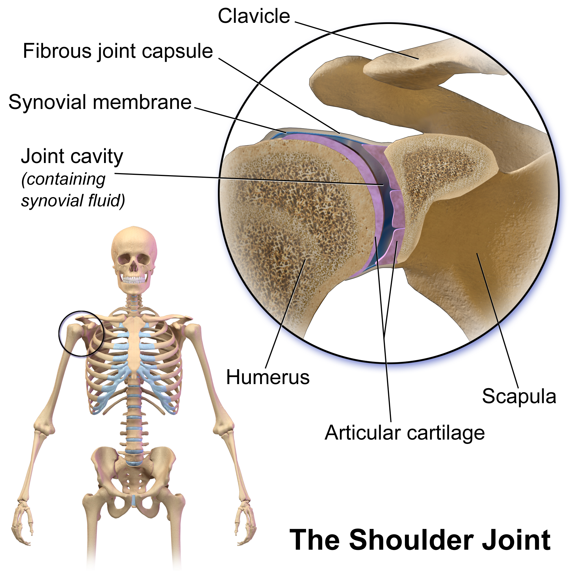
The shoulder joint (or glenohumeral joint from Greek ''glene'', eyeball, + -''oid'', 'form of', + Latin ''humerus'', shoulder) is structurally classified as a synovial ball-and-socket joint and functionally as a diarthrosis and multiaxial joint. It involves an articulation between the glenoid fossa of the scapula (shoulder blade) and the head of the humerus (upper arm bone). Due to the very loose joint capsule that gives a limited interface of the humerus and scapula, it is the most mobile joint of the human body. Structure The shoulder joint is a ball-and-socket joint between the scapula and the humerus. The socket of the glenoid fossa of the scapula is itself quite shallow, but it is made deeper by the addition of the glenoid labrum. The glenoid labrum is a ring of cartilaginous fibre attached to the circumference of the cavity. This ring is continuous with the tendon of the biceps brachii above. Spaces Significant joint spaces are: * The normal glenohumeral space is 4� ... [...More Info...] [...Related Items...] OR: [Wikipedia] [Google] [Baidu] |


