|
Pretectal Nuclei
In neuroanatomy, the pretectal area, or pretectum, is a midbrain structure composed of seven nuclei and comprises part of the subcortical visual system. Through reciprocal bilateral projections from the retina, it is involved primarily in mediating behavioral responses to acute changes in ambient light such as the pupillary light reflex, the optokinetic reflex, and temporary changes to the circadian rhythm. In addition to the pretectum's role in the visual system, the anterior pretectal nucleus has been found to mediate somatosensory and nociceptive information. Location and structure The pretectum is a bilateral group of highly interconnected nuclei located near the junction of the midbrain and forebrain. The pretectum is generally classified as a midbrain structure, although because of its proximity to the forebrain it is sometimes classified as part of the caudal diencephalon (forebrain). Within vertebrates, the pretectum is located directly anterior to the superior colliculus ... [...More Info...] [...Related Items...] OR: [Wikipedia] [Google] [Baidu] |
Midbrain
The midbrain or mesencephalon is the forward-most portion of the brainstem and is associated with vision, hearing, motor control, sleep and wakefulness, arousal ( alertness), and temperature regulation. The name comes from the Greek ''mesos'', "middle", and ''enkephalos'', "brain". Structure The principal regions of the midbrain are the tectum, the cerebral aqueduct, tegmentum, and the cerebral peduncles. Rostrally the midbrain adjoins the diencephalon ( thalamus, hypothalamus, etc.), while caudally it adjoins the hindbrain (pons, medulla and cerebellum). In the rostral direction, the midbrain noticeably splays laterally. Sectioning of the midbrain is usually performed axially, at one of two levels – that of the superior colliculi, or that of the inferior colliculi. One common technique for remembering the structures of the midbrain involves visualizing these cross-sections (especially at the level of the superior colliculi) as the upside-down fa ... [...More Info...] [...Related Items...] OR: [Wikipedia] [Google] [Baidu] |
Photosensitive Ganglion Cell
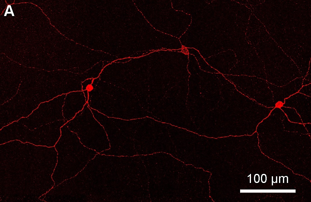
Intrinsically photosensitive retinal ganglion cells (ipRGCs), also called photosensitive retinal ganglion cells (pRGC), or melanopsin-containing retinal ganglion cells (mRGCs), are a type of neuron in the retina of the mammalian eye. The presence of (something like) ipRGCs was first suspected in 1927 when rodless, coneless mice still responded to a light stimulus through pupil constriction, This implied that rods and cones are not the only light-sensitive neurons in the retina. Yet research on these cells did not advance until the 1980s. Recent research has shown that these retinal ganglion cells, unlike other retinal ganglion cells, are intrinsically photosensitive due to the presence of melanopsin, a light-sensitive protein. Therefore they constitute a third class of photoreceptors, in addition to rod and cone cells. Overview Compared to the rods and cones, the ipRGCs respond more sluggishly and signal the presence of light over the long term. They represent a very small s ... [...More Info...] [...Related Items...] OR: [Wikipedia] [Google] [Baidu] |
Inferior Olivary Nucleus
The inferior olivary nucleus (ION), is a structure found in the medulla oblongata underneath the superior olivary nucleus.Gado, Thomas A. Woolsey; Joseph Hanaway; Mokhtar H. (2003). The brain atlas a visual guide to the human central nervous system (2nd ed.). Hoboken, NJ: Wiley. p. 206. . In vertebrates, the ION is known to coordinate signals from the spinal cord to the cerebellum to regulate motor coordination and learning.Schweighofer N, Lang EJ, Kawato M. Role of the olivo-cerebellar complex in motor learning and control. ''Frontiers in Neural Circuits''. 2013;7:94. . These connections have been shown to be tightly associated, as degeneration of either the cerebellum or the ION results in degeneration of the other. Neurons of the ION are glutamatergic and receive inhibitory input via GABA receptors. There are two distinct GABAα receptor populations that are spatially organized within each neuron present in the ION. The GABAα receptor make-up varies based on where the ... [...More Info...] [...Related Items...] OR: [Wikipedia] [Google] [Baidu] |
Eye Dilate
Eyes are organs of the visual system. They provide living organisms with vision, the ability to receive and process visual detail, as well as enabling several photo response functions that are independent of vision. Eyes detect light and convert it into electro-chemical impulses in neurons (neurones). In higher organisms, the eye is a complex optical system which collects light from the surrounding environment, regulates its intensity through a diaphragm, focuses it through an adjustable assembly of lenses to form an image, converts this image into a set of electrical signals, and transmits these signals to the brain through complex neural pathways that connect the eye via the optic nerve to the visual cortex and other areas of the brain. Eyes with resolving power have come in ten fundamentally different forms, and 96% of animal species possess a complex optical system. Image-resolving eyes are present in molluscs, chordates and arthropods. The most simple eyes, pit eyes, are ... [...More Info...] [...Related Items...] OR: [Wikipedia] [Google] [Baidu] |
Anatomy Of The Cerebellum
The anatomy of the cerebellum can be viewed at three levels. At the level of gross anatomy, the cerebellum consists of a tightly folded and crumpled layer of cortex, with white matter underneath, several deep nuclei embedded in the white matter, and a fluid-filled ventricle in the middle. At the intermediate level, the cerebellum and its auxiliary structures can be broken down into several hundred or thousand independently functioning modules or compartments known as microzones. At the microscopic level, each module consists of the same small set of neuronal elements, laid out with a highly stereotyped geometry. Gross anatomy The cerebellum is located at the base of the brain, with the large mass of the cerebral cortex above it and the portion of the brainstem called the pons in front of it. It is separated from the overlying cerebrum by a layer of tough dura mater; all of its connections with other parts of the brain travel through the pons. Anatomists classify the cerebellu ... [...More Info...] [...Related Items...] OR: [Wikipedia] [Google] [Baidu] |
Vestibular System
The vestibular system, in vertebrates, is a sensory system that creates the sense of balance and spatial orientation for the purpose of coordinating movement with balance. Together with the cochlea, a part of the auditory system, it constitutes the labyrinth of the inner ear in most mammals. As movements consist of rotations and translations, the vestibular system comprises two components: the semicircular canals, which indicate rotational movements; and the otoliths, which indicate linear accelerations. The vestibular system sends signals primarily to the neural structures that control eye movement; these provide the anatomical basis of the vestibulo-ocular reflex, which is required for clear vision. Signals are also sent to the muscles that keep an animal upright and in general control posture; these provide the anatomical means required to enable an animal to maintain its desired position in space. The brain uses information from the vestibular system in the head a ... [...More Info...] [...Related Items...] OR: [Wikipedia] [Google] [Baidu] |
Pulvinar Nuclei
The pulvinar nuclei or nuclei of the pulvinar (nuclei pulvinares) are the nuclei ( cell bodies of neurons) located in the thalamus (a part of the vertebrate brain). As a group they make up the collection called the pulvinar of the thalamus (pulvinar thalami), usually just called the pulvinar. The pulvinar is usually grouped as one of the ''lateral thalamic nuclei'' in rodents and carnivores, and stands as an independent complex in primates. Structure By convention, the pulvinar is divided into four nuclei: Their connectomic details are as follows: * The ''lateral'' and ''inferior'' pulvinar nuclei have widespread connections with early visual cortical areas. * The dorsal part of the ''lateral'' pulvinar nucleus predominantly has connections with posterior parietal cortex and the dorsal stream cortical areas. * The ''medial'' pulvinar nucleus has widespread connections with cingulate, posterior parietal, premotor and prefrontal cortical areas. * The pulvinar also has in ... [...More Info...] [...Related Items...] OR: [Wikipedia] [Google] [Baidu] |
Inferior Olive
The inferior olivary nucleus (ION), is a structure found in the medulla oblongata underneath the superior olivary nucleus.Gado, Thomas A. Woolsey; Joseph Hanaway; Mokhtar H. (2003). The brain atlas a visual guide to the human central nervous system (2nd ed.). Hoboken, NJ: Wiley. p. 206. . In vertebrates, the ION is known to coordinate signals from the spinal cord to the cerebellum to regulate motor coordination and learning.Schweighofer N, Lang EJ, Kawato M. Role of the olivo-cerebellar complex in motor learning and control. ''Frontiers in Neural Circuits''. 2013;7:94. . These connections have been shown to be tightly associated, as degeneration of either the cerebellum or the ION results in degeneration of the other. Neurons of the ION are glutamatergic and receive inhibitory input via GABA receptors. There are two distinct GABAα receptor populations that are spatially organized within each neuron present in the ION. The GABAα receptor make-up varies based on where the rec ... [...More Info...] [...Related Items...] OR: [Wikipedia] [Google] [Baidu] |
Pons
The pons (from Latin , "bridge") is part of the brainstem that in humans and other bipeds lies inferior to the midbrain, superior to the medulla oblongata and anterior to the cerebellum. The pons is also called the pons Varolii ("bridge of Varolius"), after the Italian anatomist and surgeon Costanzo Varolio (1543–75). This region of the brainstem includes neural pathways and tracts that conduct signals from the brain down to the cerebellum and medulla, and tracts that carry the sensory signals up into the thalamus.Saladin Kenneth S.(2007) Anatomy & physiology the unity of form and function. Dubuque, IA: McGraw-Hill Structure The pons is in the brainstem situated between the midbrain and the medulla oblongata, and in front of the cerebellum. A separating groove between the pons and the medulla is the inferior pontine sulcus. The superior pontine sulcus separates the pons from the midbrain. The pons can be broadly divided into two parts: the basilar part of the pons ( ... [...More Info...] [...Related Items...] OR: [Wikipedia] [Google] [Baidu] |
Reticular Formation
The reticular formation is a set of interconnected nuclei that are located throughout the brainstem. It is not anatomically well defined, because it includes neurons located in different parts of the brain. The neurons of the reticular formation make up a complex set of networks in the core of the brainstem that extend from the upper part of the midbrain to the lower part of the medulla oblongata. The reticular formation includes ascending pathways to the cortex in the ascending reticular activating system (ARAS) and descending pathways to the spinal cord via the reticulospinal tracts. Neurons of the reticular formation, particularly those of the ascending reticular activating system, play a crucial role in maintaining behavioral arousal and consciousness. The overall functions of the reticular formation are modulatory and premotor, involving somatic motor control, cardiovascular control, pain modulation, sleep and consciousness, and habituation. The modulatory functions are pri ... [...More Info...] [...Related Items...] OR: [Wikipedia] [Google] [Baidu] |
Nucleus Prepositus Hypoglossi
The nucleus prepositus (also nucleus prepositus hypoglossi), located in the caudal pons and upper medulla oblongata, contributes to several aspects of gaze control including the Horizontal gaze nystagmus test, horizontal gaze holding system. It functions as a neural integrator. References [...More Info...] [...Related Items...] OR: [Wikipedia] [Google] [Baidu] |


