|
Pp1a
ORF1ab (also ORF1a/b) refers collectively to two open reading frames (ORFs), ORF1a and ORF1b, that are conserved in the genomes of nidoviruses, a group of viruses that includes coronaviruses. The genes express large polyproteins that undergo proteolysis to form several nonstructural proteins with various functions in the viral life cycle, including proteases and the components of the replicase-transcriptase complex (RTC). Together the two ORFs are sometimes referred to as the replicase gene. They are related by a programmed ribosomal frameshift that allows the ribosome to continue translating past the stop codon at the end of ORF1a, in a -1 reading frame. The resulting polyproteins are known as pp1a and pp1ab. Expression ORF1a is the first open reading frame at the 5' end of the genome. Together ORF1ab occupies about two thirds of the genome, with the remaining third at the 3' end encoding the structural proteins and accessory proteins. It is translated from a 5' c ... [...More Info...] [...Related Items...] OR: [Wikipedia] [Google] [Baidu] |
SARS-CoV
Severe acute respiratory syndrome coronavirus 1 (SARS-CoV-1; or Severe acute respiratory syndrome coronavirus, SARS-CoV) is a strain of coronavirus that causes severe acute respiratory syndrome (SARS), the respiratory illness responsible for the 2002–2004 SARS outbreak. It is an enveloped, positive-sense, single-stranded RNA virus that infects the epithelial cells within the lungs. The virus enters the host cell by binding to angiotensin-converting enzyme 2. It infects humans, bats, and palm civets. On April 16, 2003, following the outbreak of SARS in Asia and secondary cases elsewhere in the world, the World Health Organization (WHO) issued a press release stating that the coronavirus identified by a number of laboratories was the official cause of SARS. The Centers for Disease Control and Prevention (CDC) in the United States and the National Microbiology Laboratory (NML) in Canada identified the SARS-CoV-1 genome in April 2003. Scientists at Erasmus University in Ro ... [...More Info...] [...Related Items...] OR: [Wikipedia] [Google] [Baidu] |
Reading Frame
In molecular biology, a reading frame is a way of dividing the sequence of nucleotides in a nucleic acid ( DNA or RNA) molecule into a set of consecutive, non-overlapping triplets. Where these triplets equate to amino acids or stop signals during translation, they are called codons. A single strand of a nucleic acid molecule has a phosphoryl end, called the 5′-end, and a hydroxyl or 3′-end. These define the 5′→3′ direction. There are three reading frames that can be read in this 5′→3′ direction, each beginning from a different nucleotide in a triplet. In a double stranded nucleic acid, an additional three reading frames may be read from the other, complementary strand in the 5′→3′ direction along this strand. As the two strands of a double-stranded nucleic acid molecule are antiparallel, the 5′→3′ direction on the second strand corresponds to the 3′→5′ direction along the first strand. In general, at the most, one reading frame in a given se ... [...More Info...] [...Related Items...] OR: [Wikipedia] [Google] [Baidu] |
Murine Coronavirus
Murine coronavirus (M-CoV) is a virus in the genus '' Betacoronavirus'' that infects mice. Belonging to the subgenus '' Embecovirus'', murine coronavirus strains are enterotropic or polytropic. Enterotropic strains include mouse hepatitis virus (MHV) strains D, Y, RI, and DVIM, whereas polytropic strains, such as JHM and A59, primarily cause hepatitis, enteritis, and encephalitis. Murine coronavirus is an important pathogen in the laboratory mouse and the laboratory rat. It is the most studied coronavirus in animals other than humans, and has been used as an animal disease model for many virological and clinical studies. Types Murine hepatitis virus Murine coronavirus was first discovered in 1949. The researchers isolated the virus from the brain, spinal cord, liver, lung, spleen, and kidney of a rat with symptoms of encephalitis and severe myelin injury, and gave it the strain name mouse hepatitis virus (MHV)-JHM. MHV is now the most studied coronavirus in animals other than ... [...More Info...] [...Related Items...] OR: [Wikipedia] [Google] [Baidu] |
RNA Secondary Structure
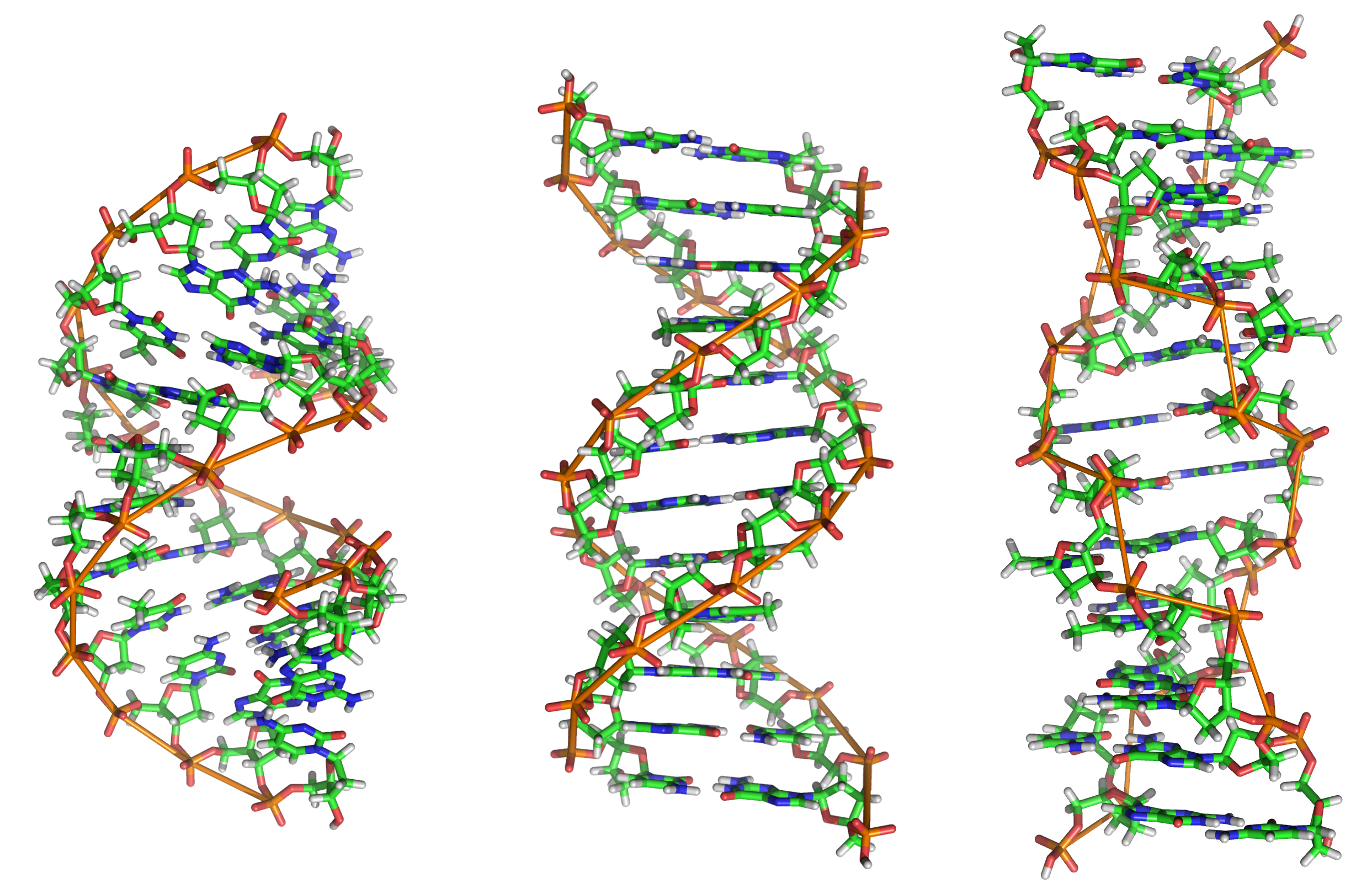
Nucleic acid secondary structure is the basepairing interactions within a single nucleic acid polymer or between two polymers. It can be represented as a list of bases which are paired in a nucleic acid molecule. The secondary structures of biological DNAs and RNAs tend to be different: biological DNA mostly exists as fully base paired double helices, while biological RNA is single stranded and often forms complex and intricate base-pairing interactions due to its increased ability to form hydrogen bonds stemming from the extra hydroxyl group in the ribose sugar. In a non-biological context, secondary structure is a vital consideration in the nucleic acid design of nucleic acid structures for DNA nanotechnology and DNA computing, since the pattern of basepairing ultimately determines the overall structure of the molecules. Fundamental concepts Base pairing In molecular biology, two nucleotides on opposite complementary DNA or RNA strands that are connected via hydrogen ... [...More Info...] [...Related Items...] OR: [Wikipedia] [Google] [Baidu] |
Pseudoknot
__NOTOC__ A pseudoknot is a nucleic acid secondary structure containing at least two stem-loop structures in which half of one stem is intercalated between the two halves of another stem. The pseudoknot was first recognized in the turnip yellow mosaic virus in 1982. Pseudoknots fold into knot-shaped three-dimensional conformations but are not true topological knots. Prediction and identification The structural configuration of pseudoknots does not lend itself well to bio-computational detection due to its context-sensitivity or "overlapping" nature. The base pairing in pseudoknots is not well nested; that is, base pairs occur that "overlap" one another in sequence position. This makes the presence of pseudoknots in RNA sequences more difficult to predict by the standard method of dynamic programming, which use a recursive scoring system to identify paired stems and consequently, most cannot detect non-nested base pairs. The newer method of stochastic context-free grammars ... [...More Info...] [...Related Items...] OR: [Wikipedia] [Google] [Baidu] |
Slippery Sequence
A slippery sequence is a small section of codon nucleotide sequences (usually UUUAAAC) that controls the rate and chance of ribosomal frameshifting. A slippery sequence causes a faster ribosomal transfer which in turn can cause the reading ribosome to "slip." This allows a tRNA to shift by 1 base (−1) after it has paired with its anticodon, changing the reading frame. A −1 frameshift triggered by such a sequence is a Programmed −1 Ribosomal Frameshift. It is followed by a spacer region, and an RNA secondary structure. Such sequences are common in virus polyproteins. The frameshift occurs due to wobble pairing. The Gibbs free energy of secondary structures downstream give a hint at how often frameshift happens. Tension on the mRNA molecule also plays a role. A list of slippery sequences found in animal viruses is available from Huang et al. Slippery sequences that cause a 2-base slip (−2 frameshift) have been constructed out of the HIV UUUUUUA sequence. See also *Nucle ... [...More Info...] [...Related Items...] OR: [Wikipedia] [Google] [Baidu] |
Kilobase
A base pair (bp) is a fundamental unit of double-stranded nucleic acids consisting of two nucleobases bound to each other by hydrogen bonds. They form the building blocks of the DNA double helix and contribute to the folded structure of both DNA and RNA. Dictated by specific hydrogen bonding patterns, "Watson–Crick" (or "Watson–Crick–Franklin") base pairs (guanine–cytosine and adenine– thymine) allow the DNA helix to maintain a regular helical structure that is subtly dependent on its nucleotide sequence. The complementary nature of this based-paired structure provides a redundant copy of the genetic information encoded within each strand of DNA. The regular structure and data redundancy provided by the DNA double helix make DNA well suited to the storage of genetic information, while base-pairing between DNA and incoming nucleotides provides the mechanism through which DNA polymerase replicates DNA and RNA polymerase transcribes DNA into RNA. Many DNA-binding pr ... [...More Info...] [...Related Items...] OR: [Wikipedia] [Google] [Baidu] |
Subgenomic RNA
Subgenomic mRNAs are essentially smaller sections of the original transcribed template strand. 3' to 5' DNA or RNA During transcription, the original template strand is usually read from the 3' to the 5' end from beginning to end. Subgenomic mRNAs are created when transcription begins at the 3' end of the template strand (or 5' of the to-be-newly synthesized template) and begins to copy towards the 5' end of the template strand before "jumping" to the end of the template and copying the last nucleotides of the 5' end of the template, (finishing the 3' tail for the newly created strand). As a result, the translated strand will have a similar 5' end to varying degrees with the original template (depending on which part of the template the transcription jumped over) and a similar 3' end to the template. 5' to 3' (positive sense) viral RNA Positive-sense (5' to 3') viral RNA which may be directly translated into the desired viral proteins, undergoes a similar process as describe ... [...More Info...] [...Related Items...] OR: [Wikipedia] [Google] [Baidu] |
Cap-dependent Translation
Eukaryotic translation is the biological process by which messenger RNA is translated into proteins in eukaryotes. It consists of four phases: gene translation, elongation, termination, and recapping. Initiation Translation initiation is the process by which the ribosome and its associated factors bind to an mRNA and are assembled at the start codon. This process is defined as either cap-dependent, in which the ribosome binds initially at the 5' cap and then travels to the stop codon, or as cap-independent, where the ribosome does not initially bind the 5' cap. Cap-dependent initiation Initiation of translation usually involves the interaction of certain key proteins, the initiation factors, with a special tag bound to the 5'-end of an mRNA molecule, the 5' cap, as well as with the 5' UTR. These proteins bind the small (40S) ribosomal subunit and hold the mRNA in place. eIF3 is associated with the 40S ribosomal subunit and plays a role in keeping the large (60S) ribosomal s ... [...More Info...] [...Related Items...] OR: [Wikipedia] [Google] [Baidu] |
5' Cap
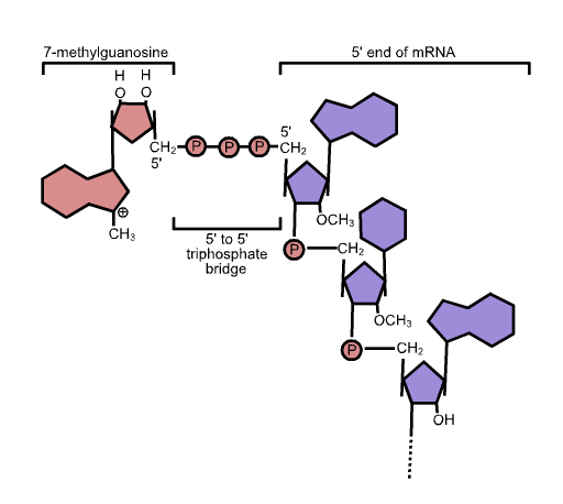
In molecular biology, the five-prime cap (5′ cap) is a specially altered nucleotide on the 5′ end of some primary transcripts such as precursor messenger RNA. This process, known as mRNA capping, is highly regulated and vital in the creation of stable and mature messenger RNA able to undergo translation during protein synthesis. Mitochondrial mRNA and chloroplastic mRNA are not capped. Structure In eukaryotes, the 5′ cap (cap-0), found on the 5′ end of an mRNA molecule, consists of a guanine nucleotide connected to mRNA via an unusual 5′ to 5′ triphosphate linkage. This guanosine is methylated on the 7 position directly after capping ''in vivo'' by a methyltransferase. It is referred to as a 7-methylguanylate cap, abbreviated m7G. In multicellular eukaryotes and some viruses, further modifications exist, including the methylation of the 2′ hydroxy-groups of the first 2 ribose sugars of the 5′ end of the mRNA. cap-1 has a methylated 2′-hydroxy group ... [...More Info...] [...Related Items...] OR: [Wikipedia] [Google] [Baidu] |
Viral Accessory Protein
A viral regulatory and accessory protein is a type of viral protein that can play an indirect role in the function of a virus. An example is Nef Nef or NEF may refer to: Businesses and organizations * National Energy Foundation, a British charity * National Enrichment Facility, an American uranium enrichment plant * New Economics Foundation, a British think-tank * Near East Foundation, .... References Further reading * Viral proteins {{virus-stub ... [...More Info...] [...Related Items...] OR: [Wikipedia] [Google] [Baidu] |
Viral Structural Protein
A viral structural protein is a viral protein that is a structural component of the mature virus. Examples include the SARS coronavirus 3a and 7a accessory proteins. Bacteriophage T4 structural proteins During assembly of the bacteriophage (phage) T4 virion, the structural proteins encoded by the phage genes interact with each other in a characteristic sequence. Maintaining an appropriate balance in the amounts of each of these structural proteins produced during viral infection appears to be critical for normal phage T4 morphogenesis. Phage T4 encoded proteins that determine virion structure include major structural components, minor structural components and non-structural proteins that catalyze specific steps in the morphogenesis sequence. Phage T4 morphogenesis is divided into three independent pathways: the head, the tail and the long tail fibres as detailed by Yap and Rossman.Yap ML, Rossmann MG. Structure and function of bacteriophage T4. Future Microbiol. 2014;9(12):13 ... [...More Info...] [...Related Items...] OR: [Wikipedia] [Google] [Baidu] |


