|
Post-LASIK Ectasia
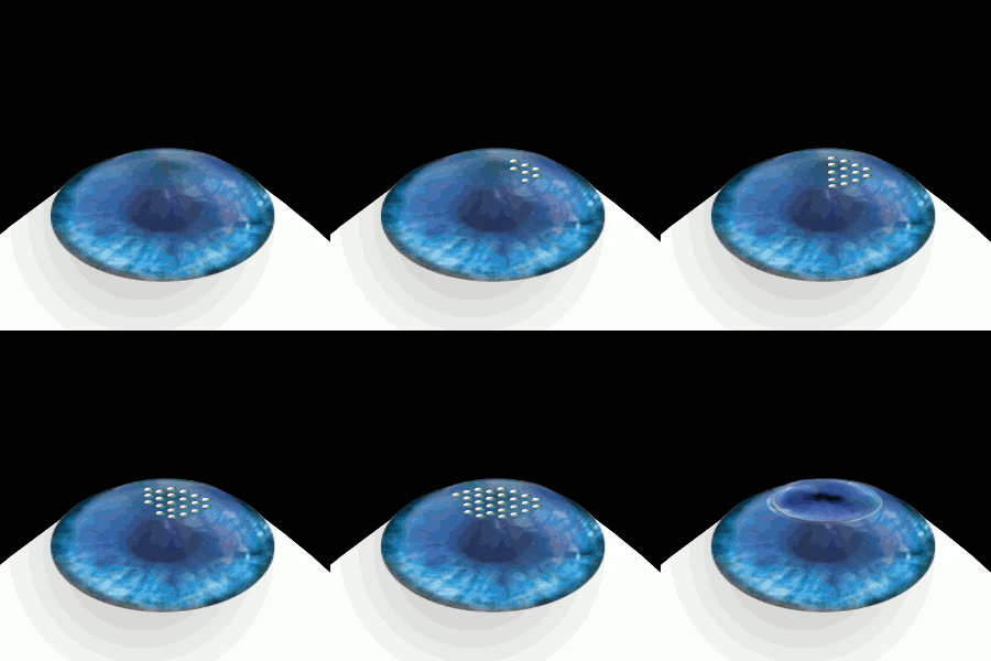
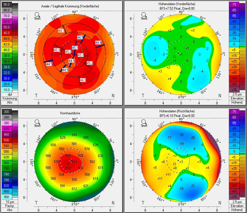
Post-LASIK ectasia is a condition similar to keratoconus where the cornea starts to bulge forwards at a variable time after LASIK, PRK, or SMILE corneal laser eye surgery. However, the physiological processes of post-LASIK ectasia seem to be different from keratoconus. The visible changes in the basal epithelial cell and anterior and posterior keratocytes linked with keratoconus were not observed in post-LASIK ectasia. __TOC__ Risk factors Before corneal refractive surgery such as LASIK, SMILE, and PRK, people must be examined for possible risk factors such as keratoconus. Abnormal corneal topography compromises of keratoconus, pellucid marginal degeneration, or forme fruste keratoconus with an I-S value of 1.4 or more is the most significant risk factor. Low age, low residual stromal bed (RSB) thickness, low preoperative corneal thickness, and high myopia are other important risk factors. Treatments Treatment options include contact lenses Contact lenses, or sim ... [...More Info...] [...Related Items...] OR: [Wikipedia] [Google] [Baidu] |
Keratoconus
Keratoconus (KC) is a disorder of the eye that results in progressive thinning of the cornea. This may result in blurry vision, double vision, nearsightedness, irregular astigmatism, and light sensitivity leading to poor quality-of-life. Usually both eyes are affected. In more severe cases a scarring or a circle may be seen within the cornea. While the cause is unknown, it is believed to occur due to a combination of genetic, environmental, and hormonal factors. Patients with a parent, sibling, or child who has keratoconus have 15 to 67 times higher risk in developing corneal ectasia compared to patients with no affected relatives. Proposed environmental factors include rubbing the eyes and allergies. The underlying mechanism involves changes of the cornea to a cone shape. Diagnosis is most often by topography. Topography measures the curvature of the cornea and creates a colored "map" of the cornea. Keratoconus causes very distinctive changes in the appearance of these ma ... [...More Info...] [...Related Items...] OR: [Wikipedia] [Google] [Baidu] |
LASIK
LASIK or Lasik (''laser-assisted in situ keratomileusis''), commonly referred to as laser eye surgery or laser vision correction, is a type of refractive surgery for the correction of myopia, hyperopia, and an actual cure for astigmatism, since it is in the cornea. LASIK surgery is performed by an ophthalmologist who uses a laser or microkeratome to reshape the eye's cornea in order to improve visual acuity. For most people, LASIK provides a long-lasting alternative to eyeglasses or contact lenses. LASIK is very similar to another surgical corrective procedure, photorefractive keratectomy (PRK), and LASEK. All represent advances over radial keratotomy in the surgical treatment of refractive errors of vision. For patients with moderate to high myopia or thin corneas which cannot be treated with LASIK and PRK, the phakic intraocular lens is an alternative. As of 2018, roughly 9.5 million Americans have had LASIK and, globally, between 1991 and 2016, more than 40 million proced ... [...More Info...] [...Related Items...] OR: [Wikipedia] [Google] [Baidu] |
Photorefractive Keratectomy
Photorefractive keratectomy (PRK) and laser-assisted sub-epithelial keratectomy (or laser epithelial keratomileusis) (LASEK) are laser eye surgery procedures intended to correct a person's vision, reducing dependency on glasses or contact lenses. LASEK and PRK permanently change the shape of the anterior central cornea using an excimer laser to ablate (remove by vaporization) a small amount of tissue from the corneal stroma at the front of the eye, just under the corneal epithelium. The outer layer of the cornea is removed prior to the ablation. A computer system tracks the patient's eye position 60 to 4,000 times per second, depending on the specifications of the laser that is used. The computer system redirects laser pulses for precise laser placement. Most modern lasers will automatically center on the patient's visual axis and will pause if the eye moves out of range and then resume ablating at that point after the patient's eye is re-centered. The outer layer of the cornea, ... [...More Info...] [...Related Items...] OR: [Wikipedia] [Google] [Baidu] |
Small Incision Lenticule Extraction
ReLEx Small incision lenticule extraction (SMILE), second generation of ReLEx Femtosecond lenticule extraction (FLEx), is a form of laser based refractive eye surgery developed by Carl Zeiss Meditec used to correct myopia, and cure astigmatism. Although similar to LASIK laser surgery, the intrastromal procedure is novel in that it uses a single femtosecond laser referenced to the corneal surface to cleave a thin lenticule from the corneal stroma for manual extraction and SMILE. It has been described as a painless procedure. For candidates to qualify for this treatment, they have their corneal stroma thickness checked to make sure that post operative thickness won't be too thin. The lenticule to be extracted is accurately cut to the correction prescription required by the patient using a photodisruption laser-tissue interaction. The method of extraction was via a LASIK-type flap in ReLEx FLEx, but in SMILE a flapless technique makes a small tunnel incision in the corneal periph ... [...More Info...] [...Related Items...] OR: [Wikipedia] [Google] [Baidu] |
Corneal Topography
Corneal topography, also known as photokeratoscopy or videokeratography, is a non-invasive medical imaging technique for mapping the anterior curvature of the cornea, the outer structure of the eye. Since the cornea is normally responsible for some 70% of the eye's refractive power, its topography is of critical importance in determining the quality of vision and corneal health. The three-dimensional map is therefore a valuable aid to the examining ophthalmologist or optometrist and can assist in the diagnosis and treatment of a number of conditions; in planning cataract surgery and intraocular lens implantation; in planning refractive surgery such as LASIK, and evaluating its results; or in assessing the fit of contact lenses. A development of keratoscopy, corneal topography extends the measurement range from the four points a few millimeters apart that is offered by keratometry to a grid of thousands of points covering the entire cornea. The procedure is carried out in seconds ... [...More Info...] [...Related Items...] OR: [Wikipedia] [Google] [Baidu] |
Pellucid Marginal Degeneration
Pellucid marginal degeneration (PMD) is a degenerative corneal condition, often confused with keratoconus. It typically presents with painless vision loss affecting both eyes. Rarely, it may cause acute vision loss with severe pain due to perforation of the cornea. It is typically characterized by a clear, bilateral thinning (ectasia) in the inferior and peripheral region of the cornea, although some cases affect only one eye. The cause of the disease remains unclear. Pellucid marginal degeneration is diagnosed by corneal topography. Corneal pachymetry may be useful in confirming the diagnosis. Treatment usually consists of vision correction with eyeglasses or contact lenses. Intacs implants, corneal collagen cross-linking, and corneal transplant surgery are additional options. Surgery is reserved for individuals who do not tolerate contact lenses. The term "pellucid marginal degeneration" was first coined in 1957 by the ophthalmologist Schalaeppi. The word "pellucid" means c ... [...More Info...] [...Related Items...] OR: [Wikipedia] [Google] [Baidu] |
Forme Fruste
In medicine, a ''forme fruste'' ( French, "crude, or unfinished, form"; ''pl.'', ''formes frustes'') is an atypical or attenuated manifestation of a disease or syndrome, with the implications of incompleteness, partial presence or aborted state. The context is usually one of a well defined clinical or pathological entity, which the case at hand almost — but not quite — fits. An opposite term in medicine, ''forme pleine'' — seldom used by English-speaking physicians — means the complete, or full-blown, form of a disease. Use According to gastroenterologist William Haubrich: A patient may exhibit sudden, intense, epigastric pain and a rigid abdomen. He is thought to have a perforated peptic ulcer. But at operation, only a penetrating ulcer is found, sealed off by adhesion to the omentum or anterior abdominal wall. Such a patient is said to have a forme fruste of acute free perforation as a complication of his peptic ulcer disease. History The Latin phrase ''frustra esse'' m ... [...More Info...] [...Related Items...] OR: [Wikipedia] [Google] [Baidu] |
Myopia
Near-sightedness, also known as myopia and short-sightedness, is an eye disease where light focuses in front of, instead of on, the retina. As a result, distant objects appear blurry while close objects appear normal. Other symptoms may include headaches and eye strain. Severe near-sightedness is associated with an increased risk of retinal detachment, cataracts, and glaucoma. The underlying mechanism involves the length of the eyeball growing too long or less commonly the lens being too strong. It is a type of refractive error. Diagnosis is by eye examination. Tentative evidence indicates that the risk of near-sightedness can be decreased by having young children spend more time outside. This decrease in risk may be related to natural light exposure. Near-sightedness can be corrected with eyeglasses, contact lenses, or a refractive surgery. Eyeglasses are the easiest and safest method of correction. Contact lenses can provide a wider field of vision, but are associated with ... [...More Info...] [...Related Items...] OR: [Wikipedia] [Google] [Baidu] |
Contact Lenses
Contact lenses, or simply contacts, are thin lenses placed directly on the surface of the eyes. Contact lenses are ocular prosthetic devices used by over 150 million people worldwide, and they can be worn to correct vision or for cosmetic or therapeutic reasons. In 2010, the worldwide market for contact lenses was estimated at $6.1 billion, while the US soft lens market was estimated at $2.1 billion.Nichols, Jason J., et a"ANNUAL REPORT: Contact Lenses 2010" January 2011. Multiple analysts estimated that the global market for contact lenses would reach $11.7 billion by 2015. , the average age of contact lens wearers globally was 31 years old, and two-thirds of wearers were female.Morgan, Philip B., et al"International Contact Lens Prescribing in 2010" ''Contact Lens Spectrum''. October 2011. People choose to wear contact lenses for many reasons. Aesthetics and cosmetics are main motivating factors for people who want to avoid wearing glasses or to change the appearance or c ... [...More Info...] [...Related Items...] OR: [Wikipedia] [Google] [Baidu] |
Intrastromal Corneal Ring Segments
An intrastromal corneal ring segment (ICRS) (also known as intrastromal corneal ring, corneal implant or corneal insert) is a small device implanted in the eye to correct vision. The procedure involves an ophthalmologist who makes a small incision in the cornea of the eye and inserts two crescent or semi-circular shaped ring segments between the layers of the corneal stroma, one on each side of the pupil. The embedding of the two rings in the cornea is intended to flatten the cornea and change the refraction of light passing through the cornea on its way into the eye. __TOC__ Design Intrastromal corneal ring segments have many different types and designs, including Intacs (US), Cornealring (Brazil), Keraring (Brazil), Ferrara ring (Brazil), Myoring (Austria) and Intraseg (UK). Medical uses Intrastromal corneal rings were originally used to treat mild myopia. For this purpose, they have largely been superseded by excimer lasers, which have better accuracy. They are now mostly used ... [...More Info...] [...Related Items...] OR: [Wikipedia] [Google] [Baidu] |
Corneal Collagen Cross-linking
Corneal cross-linking (CXL) with riboflavin (vitamin B2) and UV-A light is a surgical treatment for corneal ectasia such as keratoconus, PMD, and post-LASIK ectasia. It is used in an attempt to make the cornea stronger. According to a 2015 Cochrane review, there is insufficient evidence to determine if it is useful in keratoconus. In 2016, the US Food and Drug Administration approved riboflavin ophthalmic solution crosslinking based on three 12-month clinical trials. Medical uses A 2015 Cochrane review found that the evidence on corneal cross-linking was insufficient to determine if it is an effective procedure for the treatment of keratoconus. Adverse effects Among those with keratoconus who worsen CXL may be used. In this group, the most common side effects are haziness of the cornea, punctate keratitis, corneal striae, corneal epithelium defect, and eye pain. In those who use it after post-LASIK ectasia, the most common side effects are haziness of the cornea, corn ... [...More Info...] [...Related Items...] OR: [Wikipedia] [Google] [Baidu] |
Corneal Transplant
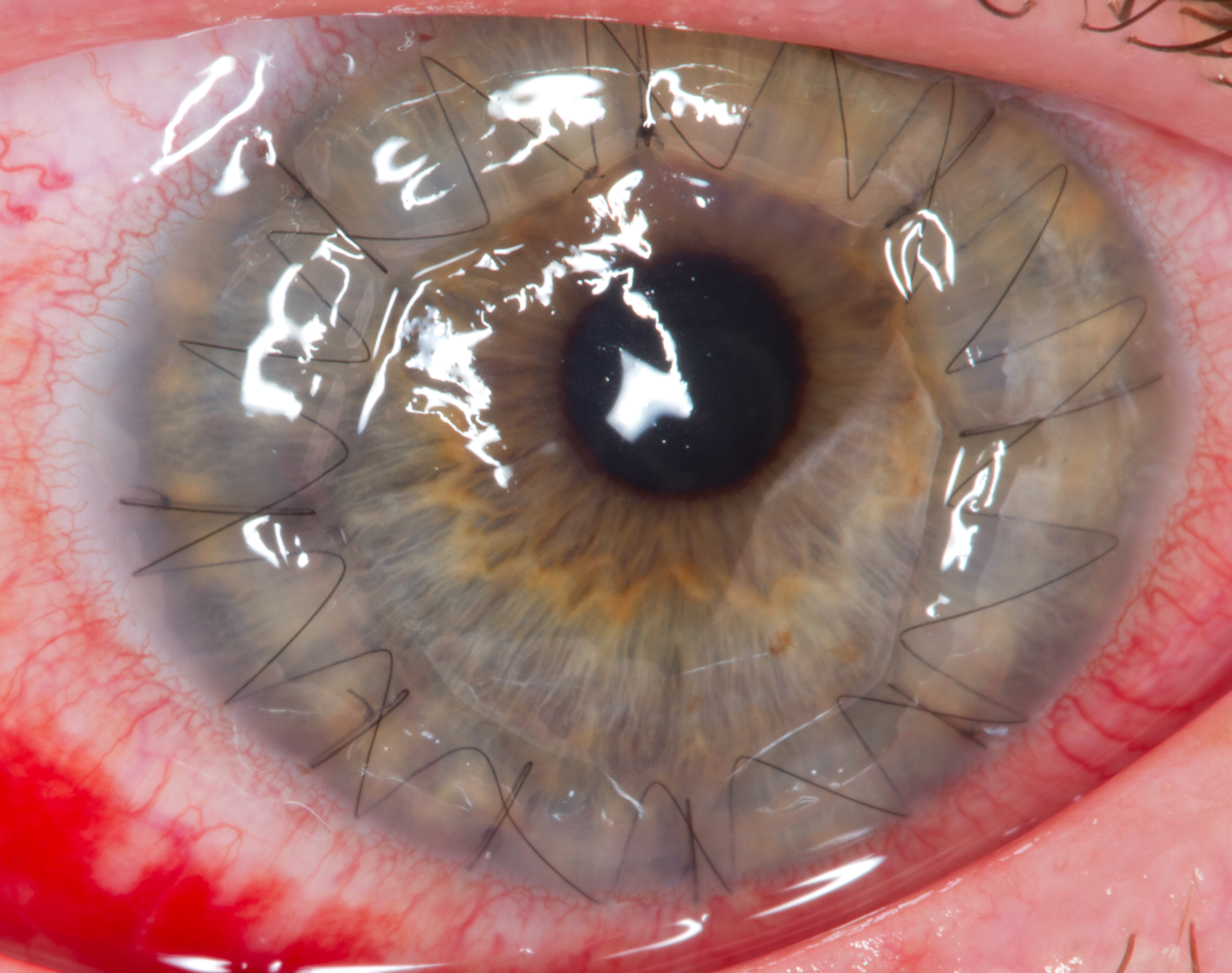
Corneal transplantation, also known as corneal grafting, is a surgical procedure where a damaged or diseased cornea is replaced by donated corneal tissue (the graft). When the entire cornea is replaced it is known as penetrating keratoplasty and when only part of the cornea is replaced it is known as lamellar keratoplasty. Keratoplasty simply means surgery to the cornea. The graft is taken from a recently deceased individual with no known diseases or other factors that may affect the chance of survival of the donated tissue or the health of the recipient. The cornea is the transparent front part of the eye that covers the iris, pupil and anterior chamber. The surgical procedure is performed by ophthalmologists, physicians who specialize in eyes, and is often done on an outpatient basis. Donors can be of any age, as is shown in the case of Janis Babson, who donated her eyes after dying at the age of 10. Corneal transplantation is performed when medicines, keratoconus conserva ... [...More Info...] [...Related Items...] OR: [Wikipedia] [Google] [Baidu] |


