|
Portacaval Anastomosis
A portocaval anastomosis or porto-systemic anastomosis is a specific type of anastomosis that occurs between the veins of the portal circulation and those of the systemic circulation. When there is a blockage of the portal system, portocaval anastomosis enables the blood to still reach the systemic venous circulation. The inferior end of the esophagus and the superior part of the rectum are potential sites of a harmful portocaval anastomosis. In portal hypertension, as in the case of cirrhosis of the liver, the anastomoses become congested and form venous dilatations. Such dilatation can lead to esophageal varices and anorectal varices. Caput medusae can also result.''Gray's Anatomy for Students'' Gray H, Drake R, Vogl W, Mitchell A, Tibbitts R, Richardson P. Philadelphia: Elsevier/Churchill Livingstone; 2010. p. 226 __TOC__ Presentation Clinical presentations of portal hypertension include: A dilated inferior mesenteric vein may or may not be related to portal hypertension. Ot ... [...More Info...] [...Related Items...] OR: [Wikipedia] [Google] [Baidu] |
Anastomosis
An anastomosis (, plural anastomoses) is a connection or opening between two things (especially cavities or passages) that are normally diverging or branching, such as between blood vessels, leaf#Veins, leaf veins, or streams. Such a connection may be normal (such as the foramen ovale (heart), foramen ovale in a fetus's heart) or abnormal (such as the atrial septal defect#Patent foramen ovale, patent foramen ovale in an adult's heart); it may be acquired (such as an arteriovenous fistula) or innate (such as the arteriovenous shunt of a metarteriole); and it may be natural (such as the aforementioned examples) or artificial (such as a surgical anastomosis). The reestablishment of an anastomosis that had become blocked is called a reanastomosis. Anastomoses that are abnormal, whether congenital disorder, congenital or acquired, are often called fistulas. The term is used in medicine, biology, mycology, geology, and geography. Etymology Anastomosis: medical or Modern Latin, from Gre ... [...More Info...] [...Related Items...] OR: [Wikipedia] [Google] [Baidu] |
Superior Rectal Vein
The inferior mesenteric vein begins in the rectum as the superior rectal vein (superior hemorrhoidal vein), which has its origin in the hemorrhoidal plexus, and through this plexus communicates with the middle and inferior hemorrhoidal veins. The superior rectal vein leaves the lesser pelvis and crosses the left common iliac vessels with the superior rectal artery, and is continued upward as the inferior mesenteric vein In human anatomy, the inferior mesenteric vein (IMV) is a blood vessel that drains blood from the large intestine. It usually terminates when reaching the splenic vein, which goes on to form the portal vein with the superior mesenteric vein (SMV) .... References Veins of the torso Rectum {{circulatory-stub ... [...More Info...] [...Related Items...] OR: [Wikipedia] [Google] [Baidu] |
Middle Colic Vein
The middle colic vein drains the transverse colon. It is a tributary of the superior mesenteric vein, and follows the path of its corresponding artery, the middle colic artery The middle colic artery is an artery of the abdomen; a branch of the superior mesenteric artery distributed to parts of the ascending and transverse colon. It usually divides into two terminal branches - a left one and a right one - which go on .... Veins of the torso {{Circulatory-stub ... [...More Info...] [...Related Items...] OR: [Wikipedia] [Google] [Baidu] |
Right Colic Vein
The right colic vein drains the ascending colon, and is a tributary of the superior mesenteric vein. It travels with its corresponding artery, the right colic artery The right colic artery is an artery of the abdomen, a branch of the superior mesenteric artery supplying the ascending colon. It divides into two terminal branches - an ascending branch and a descending branch - which form anastomoses with the .... Veins of the torso {{Circulatory-stub ... [...More Info...] [...Related Items...] OR: [Wikipedia] [Google] [Baidu] |
Gonadal Vein
In medicine, gonadal vein refers to the blood vessel that carries blood away from the gonad (testis, ovary) toward the heart. These are different arteries in women (ovarian vein) and men (testicular vein), but share the same embryological origin. The termination of the two gonadal veins in an individual is usually asymmetrical, with the left one draining into the left renal vein, and the right one draining into the inferior vena cava. Anatomy Fate The left gonadal vein usually empties into (inferior aspect of) the ipsilateral renal vein proximally to where the renal vein crossing over the aorta. The right gonadal vein typically empties directly into the (right anterolateral aspect of) inferior vena cava, joining it at an acute angle, some 2 cm inferior to the ipsilateral renal vein The renal veins are large-calibre veins that drain blood filtered by the kidneys into the inferior vena cava. There is one renal vein draining each kidney. Because the inferior vena cava is ... [...More Info...] [...Related Items...] OR: [Wikipedia] [Google] [Baidu] |
Suprarenal Vein
The suprarenal veins are two in number: * the ''right'' ends in the inferior vena cava. * the ''left'' ends in the left renal or left inferior phrenic vein. They receive blood from the adrenal glands and will sometimes form anastomoses An anastomosis (, plural anastomoses) is a connection or opening between two things (especially cavities or passages) that are normally diverging or branching, such as between blood vessels, leaf veins, or streams. Such a connection may be norm ... with the inferior phrenic veins. Additional images File:Gray480.png, Diagram showing completion of development of the parietal veins File:Gray1183.png, Suprarenal glands viewed from the front File:Gray1184.png, Suprarenal glands viewed from behind References External links Veins of the torso Adrenal gland {{circulatory-stub ... [...More Info...] [...Related Items...] OR: [Wikipedia] [Google] [Baidu] |
Renal Vein
The renal veins are large-calibre veins that drain blood filtered by the kidneys into the inferior vena cava. There is one renal vein draining each kidney. Because the inferior vena cava is on the right half of the body, the left renal vein is longer than the right one. Structure One renal vein drains each kidney. A renal vein is situated anterior to its corresponding accompanying renal artery. The renal veins empty into the inferior vena cava, entering it at nearly a 90° angle. Due to the right-ward displacement of the inferior vena cava from the midline, the left renal vein is some 3 times longer than the right one (~7.5 cm and ~2.5 cm, respectively). The renal vein divides into 4 divisions upon entering the kidney: * the anterior branch which receives blood from the anterior portion of the kidney and, * the posterior branch which receives blood from the posterior portion. Tributaries Because the tributaries of the inferior vena cava are not bilaterally symmetrical, the l ... [...More Info...] [...Related Items...] OR: [Wikipedia] [Google] [Baidu] |
Splenic Vein
The spleen is an organ (biology), organ found in almost all vertebrates. Similar in structure to a large lymph node, it acts primarily as a blood filter. The word spleen comes .σπλήν Henry George Liddell, Robert Scott, ''A Greek-English Lexicon'', on Perseus Digital Library The spleen plays very important roles in regard to red blood cells (erythrocytes) and the immune system. It removes old red blood cells and holds a reserve of blood, which can be valuable in case of Shock (circulatory), hemorrhagic shock, and also Human iron metabolism, recycles iron. As a part of the mononuclear phagocyte system, it metabolizes hemoglobin removed from senescent red blood cells. The globin portion of hemoglobin is degraded to its constitutive amino acids, and the h ... [...More Info...] [...Related Items...] OR: [Wikipedia] [Google] [Baidu] |
Retroperitoneal
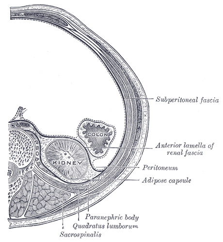
The retroperitoneal space (retroperitoneum) is the anatomical space (sometimes a potential space) behind (''retro'') the peritoneum. It has no specific delineating anatomical structures. Organs are retroperitoneal if they have peritoneum on their anterior side only. Structures that are not suspended by mesentery in the abdominal cavity and that lie between the parietal peritoneum and abdominal wall are classified as retroperitoneal. This is different from organs that are not retroperitoneal, which have peritoneum on their posterior side and are suspended by mesentery in the abdominal cavity. The retroperitoneum can be further subdivided into the following: *Perirenal (or perinephric) space *Anterior pararenal (or paranephric) space *Posterior pararenal (or paranephric) space Retroperitoneal structures Structures that lie behind the peritoneum are termed "retroperitoneal". Organs that were once suspended within the abdominal cavity by mesentery but migrated posterior to the ... [...More Info...] [...Related Items...] OR: [Wikipedia] [Google] [Baidu] |
Superior Epigastric Vein
In human anatomy, superior epigastric veins are two or more venae comitantes which accompany either superior epigastric artery before emptying into the internal thoracic vein. They participate in the drainage of the superior surface of the diaphragm. Structure Course The superior epigastric vein originates from the internal thoracic vein. The superior epigastric veins first run between the sternal margin and the costal margin of the diaphragm, then enter the rectus sheath. They run inferiorly, coursing superficially to the fibrous layer forming the posterior leaflet of the rectus sheath, and deep to the rectus abdominis muscle. The superior epigastric veins are venae comitantes of the superior epigastric artery, and mirror its course. Distribution The superior epigastric veins participate in the drainage of the superior surface of the diaphragm. Fate The superior epigastric veins drain into the internal thoracic vein. See also *Terms for anatomical location ... [...More Info...] [...Related Items...] OR: [Wikipedia] [Google] [Baidu] |
Paraumbilical Veins
In the course of the round ligament of the liver, small paraumbilical veins are found which establish an anastomosis between the veins of the anterior abdominal wall and the portal vein, hypogastric, and iliac veins. These veins include Burrow's veins, and the veins of Sappey – superior veins of Sappey and the inferior veins of Sappey. The best marked of these small veins is one which commences at the navel (umbilicus) and runs backward and upward in, or on the surface of, the round ligament (ligamentum teres) between the layers of the falciform ligament to end in the left portal vein. Pathophysiology In cases of portal hypertension, the paraumbilical veins may become enlarged in order to reduce hepatic portal vein pressure by shunting blood to the superficial epigastric vein. The superficial epigastric vein drains to the femoral vein which ultimately drains into the inferior vena cava directly through the external iliac and common iliac vein, thereby bypassing the liver. ... [...More Info...] [...Related Items...] OR: [Wikipedia] [Google] [Baidu] |


