|
Pores Of Kohn
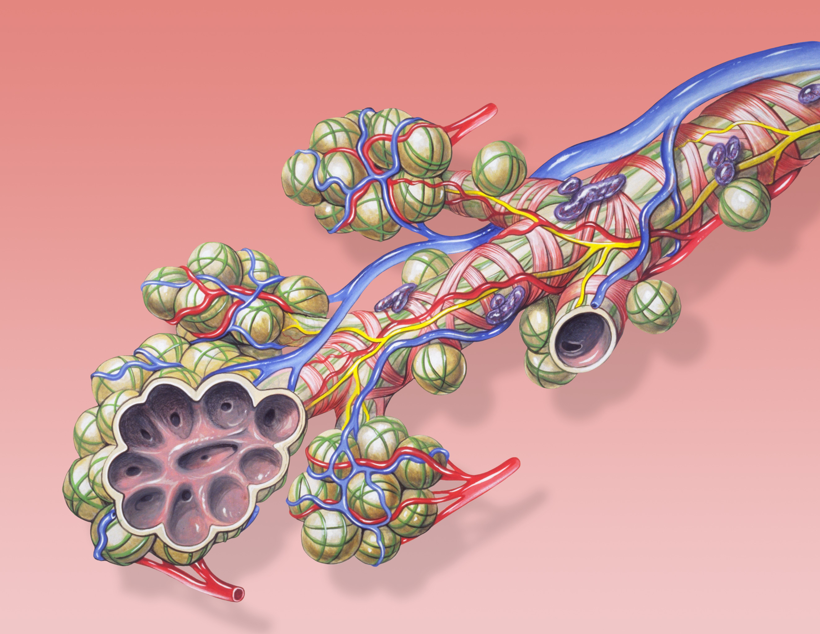
The pores of Kohn (also known as interalveolar connections or alveolar pores) are discrete holes in walls of adjacent alveoli. Cuboidal type II alveolar cells, which produce surfactant, usually form part of aperture. Etymology The pores of Kohn take their name from the German physician and pathologist Hans Nathan Kohn (1866–1935) who first described them in 1893. Development They are absent in human newborns. They develop at 3–4 years of age along with canals of Lambert during the process of thinning of alveolar septa. Function The pores allow the passage of other materials such as fluid and bacteria, which is an important mechanism of spread of infection in lobar pneumonia and spread of fibrin in the grey hepatisation phase of recovery from the same. They also equalize the pressure in adjacent alveoli and, combined with increased distribution of surfactant, thus play an important role in prevention of collapse of the lung. Unlike adults, in children these inter-alveolar con ... [...More Info...] [...Related Items...] OR: [Wikipedia] [Google] [Baidu] |
Alveolar Wall
A pulmonary alveolus (plural: alveoli, from Latin ''alveolus'', "little cavity"), also known as an air sac or air space, is one of millions of hollow, distensible cup-shaped cavities in the lungs where oxygen is exchanged for carbon dioxide. Alveoli make up the functional tissue of the mammalian lungs known as the lung parenchyma, which takes up 90 percent of the total lung volume. Alveoli are first located in the respiratory bronchioles that mark the beginning of the respiratory zone. They are located sparsely in these bronchioles, line the walls of the alveolar ducts, and are more numerous in the blind-ended alveolar sacs. The acini are the basic units of respiration, with gas exchange taking place in all the alveoli present. The alveolar membrane is the gas exchange surface, surrounded by a network of capillaries. Across the membrane oxygen is diffused into the capillaries and carbon dioxide released from the capillaries into the alveoli to be breathed out. Alveoli are pa ... [...More Info...] [...Related Items...] OR: [Wikipedia] [Google] [Baidu] |
Pulmonary Alveolus
A pulmonary alveolus (plural: alveoli, from Latin ''alveolus'', "little cavity"), also known as an air sac or air space, is one of millions of hollow, distensible cup-shaped cavities in the lungs where oxygen is exchanged for carbon dioxide. Alveoli make up the functional tissue of the mammalian lungs known as the lung parenchyma, which takes up 90 percent of the total lung volume. Alveoli are first located in the respiratory bronchioles that mark the beginning of the respiratory zone. They are located sparsely in these bronchioles, line the walls of the alveolar ducts, and are more numerous in the blind-ended alveolar sacs. The acini are the basic units of respiration, with gas exchange taking place in all the alveoli present. The alveolar membrane is the gas exchange surface, surrounded by a network of capillaries. Across the membrane oxygen is diffused into the capillaries and carbon dioxide released from the capillaries into the alveoli to be breathed out. Alveoli are pa ... [...More Info...] [...Related Items...] OR: [Wikipedia] [Google] [Baidu] |
Type II Alveolar Cell
A pulmonary alveolus (plural: alveoli, from Latin ''alveolus'', "little cavity"), also known as an air sac or air space, is one of millions of hollow, distensible cup-shaped cavities in the lungs where oxygen is exchanged for carbon dioxide. Alveoli make up the functional tissue of the mammalian lungs known as the lung parenchyma, which takes up 90 percent of the total lung volume. Alveoli are first located in the respiratory bronchioles that mark the beginning of the respiratory zone. They are located sparsely in these bronchioles, line the walls of the alveolar ducts, and are more numerous in the blind-ended alveolar sacs. The acini are the basic units of respiration, with gas exchange taking place in all the alveoli present. The alveolar membrane is the gas exchange surface, surrounded by a network of capillaries. Across the membrane oxygen is diffused into the capillaries and carbon dioxide released from the capillaries into the alveoli to be breathed out. Alveoli are part ... [...More Info...] [...Related Items...] OR: [Wikipedia] [Google] [Baidu] |
Pulmonary Surfactant
Pulmonary surfactant is a surface-active complex of phospholipids and proteins formed by type II alveolar cells. The proteins and lipids that make up the surfactant have both hydrophilic and hydrophobic In chemistry, hydrophobicity is the physical property of a molecule that is seemingly repelled from a mass of water (known as a hydrophobe). In contrast, hydrophiles are attracted to water. Hydrophobic molecules tend to be nonpolar and, t ... regions. By adsorption, adsorbing to the air-water Interface (chemistry), interface of pulmonary alveolus, alveoli, with hydrophilic head groups in the water and the hydrophobic tails facing towards the air, the main lipid component of surfactant, dipalmitoylphosphatidylcholine (DPPC), reduces surface tension. As a medication, Pulmonary surfactant (medication), pulmonary surfactant is on the WHO Model List of Essential Medicines, the most important medications needed in a basic health system. Function * To increase pulmonary com ... [...More Info...] [...Related Items...] OR: [Wikipedia] [Google] [Baidu] |
Canals Of Lambert
Collateral ventilation (CV) is a back-up system of alveolar ventilation that can bypass the normal route of airflow when airways are restricted or obstructed. The pathways involved include those between adjacent alveoli (pores of Kohn), between bronchioles and alveoli (canals of Lambert), and those between bronchioles (channels of Martin). Collateral ventilation also serves to modulate imbalances in ventilation and perfusion a feature of many diseases. The pathways are altered in lung diseases particularly asthma, and emphysema. A similar functional pattern of collateralisation is seen in the circulatory system of the heart. Interlobar collateral ventilation has also been noted and is a major unwanted factor in the consideration of lung volume reduction surgery and some lung volume reduction procedures. Pathways In normal respiratory conditions airflow is through the pathway of least resistance offered by the bronchial tree, to the alveoli and back to the bronchi and trachea.In ... [...More Info...] [...Related Items...] OR: [Wikipedia] [Google] [Baidu] |
Lobar Pneumonia
Lobar pneumonia is a form of pneumonia characterized by inflammatory exudate within the intra-alveolar space resulting in consolidation that affects a large and continuous area of the lobe of a lung. It is one of three anatomic classifications of pneumonia (the other being bronchopneumonia and atypical pneumonia). In children round pneumonia develops instead because the pores of Kohn which allow the lobar spread of infection are underdeveloped. Mechanism The invading organism starts multiplying, thereby releasing toxins that cause inflammation and edema of the lung parenchyma. This leads to the accumulation of cellular debris within the lungs. This leads to consolidation or solidification, which is a term that is used for macroscopic or radiologic appearance of the lungs affected by pneumonia. Bacterial pneumonia is mainly classified into lobar and diffuse depending on the degree of lung irritation or damage. Stages Lobar pneumonia usually has an acute progression. Classica ... [...More Info...] [...Related Items...] OR: [Wikipedia] [Google] [Baidu] |
Hepatization Of Lungs
Hepatization is conversion into a substance resembling the liver; a state of the lungs when gorged with effuse matter, so that they are no longer pervious to the air. Red hepatization is when there are red blood cells, neutrophils, and fibrin in the pulmonary alveolus/ alveoli; it precedes gray hepatization, where the red cells have been broken down leaving a fibrinosuppurative exudate. The main cause is lobar pneumonia Lobar pneumonia is a form of pneumonia characterized by inflammatory exudate within the intra-alveolar space resulting in consolidation that affects a large and continuous area of the lobe of a lung. It is one of three anatomic classifications o .... Transformation from Red hepatization to gray hepatization is an example for acute inflammation turning into a chronic inflammation. References Further readingLectures on the diseases of the lungs and heartby Thomas DaviesMedical Times 1841 [...More Info...] [...Related Items...] OR: [Wikipedia] [Google] [Baidu] |
Atelectasis
Atelectasis is the collapse or closure of a lung resulting in reduced or absent gas exchange. It is usually unilateral, affecting part or all of one lung. It is a condition where the alveoli are deflated down to little or no volume, as distinct from pulmonary consolidation, in which they are filled with liquid. It is often called a ''collapsed lung'', although that term may also refer to pneumothorax. It is a very common finding in chest X-rays and other radiological studies, and may be caused by normal exhalation or by various medical conditions. Although frequently described as a ''collapse of lung tissue'', atelectasis is not synonymous with a pneumothorax, which is a more specific condition that can cause atelectasis. Acute atelectasis may occur as a post-operative complication or as a result of surfactant deficiency. In premature babies, this leads to infant respiratory distress syndrome. The term uses combining forms of ''atel-'' + ''ectasis'', from el, ἀτελής, ... [...More Info...] [...Related Items...] OR: [Wikipedia] [Google] [Baidu] |
Round Pneumonia
Lobar pneumonia is a form of pneumonia characterized by inflammatory exudate within the intra-alveolar space resulting in consolidation that affects a large and continuous area of the Lung#Anatomy , lobe of a lung. It is one of three anatomic classifications of pneumonia (the other being bronchopneumonia and atypical pneumonia). In children round pneumonia develops instead because the pores of Kohn which allow the lobar spread of infection are underdeveloped. Mechanism The invading organism starts multiplying, thereby releasing toxins that cause inflammation and edema of the lung parenchyma. This leads to the accumulation of cellular debris within the lungs. This leads to consolidation or solidification, which is a term that is used for macroscopic or radiologic appearance of the lungs affected by pneumonia. Bacterial pneumonia is mainly classified into lobar and diffuse depending on the degree of lung irritation or damage. Stages Lobar pneumonia usually has an acute progre ... [...More Info...] [...Related Items...] OR: [Wikipedia] [Google] [Baidu] |


