|
Popliteus
The popliteus muscle in the leg is used for unlocking the knees when walking, by laterally rotating the femur on the tibia during the closed chain portion of the gait cycle (one with the foot in contact with the ground). In open chain movements (when the involved limb is not in contact with the ground), the popliteus muscle medially rotates the tibia on the femur. It is also used when sitting down and standing up. It is the only muscle in the posterior (back) compartment of the lower leg that acts just on the knee and not on the ankle. The gastrocnemius muscle acts on both joints. Structure The popliteus muscle originates from the lateral surface of the lateral condyle of the femur by a rounded tendon. Its fibers pass downward and medially. It inserts onto the posterior surface of tibia, above the soleal line. The popliteus tendon runs beneath the lateral collateral ligament and tendon of biceps femoris. The muscle also runs above the lateral meniscus but has no connection wi ... [...More Info...] [...Related Items...] OR: [Wikipedia] [Google] [Baidu] |
Popliteus Minor
The popliteus muscle in the leg is used for unlocking the knees when walking, by laterally rotating the femur on the tibia during the closed chain portion of the gait cycle (one with the foot in contact with the ground). In open chain movements (when the involved limb is not in contact with the ground), the popliteus muscle medially rotates the tibia on the femur. It is also used when sitting down and standing up. It is the only muscle in the posterior (back) compartment of the lower leg that acts just on the knee and not on the ankle. The gastrocnemius muscle acts on both joints. Structure The popliteus muscle originates from the lateral surface of the lateral condyle of the femur by a rounded tendon. Its fibers pass downward and medially. It inserts onto the posterior surface of tibia, above the soleal line. The popliteus tendon runs beneath the lateral collateral ligament and tendon of biceps femoris. The muscle also runs above the lateral meniscus but has no connection wit ... [...More Info...] [...Related Items...] OR: [Wikipedia] [Google] [Baidu] |
Posterolateral Knee
Posterolateral corner injuries (PLC injuries) of the knee are injuries to a complex area formed by the interaction of multiple structures. Injuries to the posterolateral corner can be debilitating to the person and require recognition and treatment to avoid long term consequences. Injuries to the PLC often occur in combination with other ligamentous injuries to the knee; most commonly the anterior cruciate ligament (ACL) and posterior cruciate ligament (PCL). As with any injury, an understanding of the anatomy and functional interactions of the posterolateral corner is important to diagnosing and treating the injury. Signs and symptoms Patients often complain of pain and instability at the joint. With concurrent Nerve injury, nerve injuries, patients may experience numbness, tingling and weakness of the Ankle dorsiflexion, ankle dorsiflexors and great toe extensors, or a footdrop. Complications Follow-up studies by Levy et al. and Stannard at al. both examined failure rates for ... [...More Info...] [...Related Items...] OR: [Wikipedia] [Google] [Baidu] |
Knees
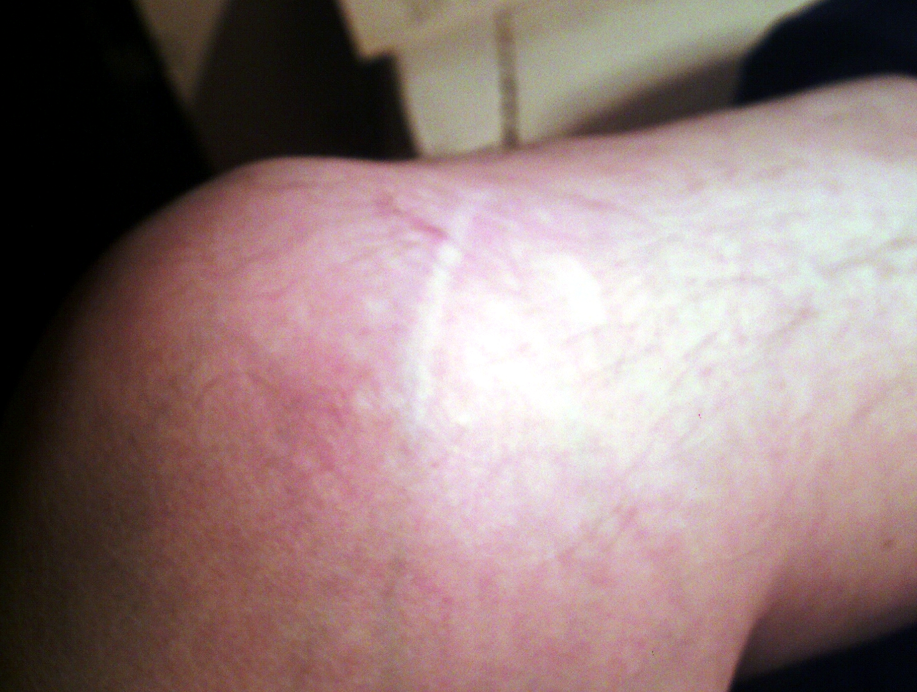
In humans and other primates, the knee joins the thigh with the leg and consists of two joints: one between the femur and tibia (tibiofemoral joint), and one between the femur and patella (patellofemoral joint). It is the largest joint in the human body. The knee is a modified hinge joint, which permits flexion and extension as well as slight internal and external rotation. The knee is vulnerable to injury and to the development of osteoarthritis. It is often termed a ''compound joint'' having tibiofemoral and patellofemoral components. (The fibular collateral ligament is often considered with tibiofemoral components.) Structure The knee is a modified hinge joint, a type of synovial joint, which is composed of three functional compartments: the patellofemoral articulation, consisting of the patella, or "kneecap", and the patellar groove on the front of the femur through which it slides; and the medial and lateral tibiofemoral articulations linking the femur, or thigh bon ... [...More Info...] [...Related Items...] OR: [Wikipedia] [Google] [Baidu] |
Tibia
The tibia (; ), also known as the shinbone or shankbone, is the larger, stronger, and anterior (frontal) of the two bones in the leg below the knee in vertebrates (the other being the fibula, behind and to the outside of the tibia); it connects the knee with the ankle. The tibia is found on the medial side of the leg next to the fibula and closer to the median plane. The tibia is connected to the fibula by the interosseous membrane of leg, forming a type of fibrous joint called a syndesmosis with very little movement. The tibia is named for the flute '' tibia''. It is the second largest bone in the human body, after the femur. The leg bones are the strongest long bones as they support the rest of the body. Structure In human anatomy, the tibia is the second largest bone next to the femur. As in other vertebrates the tibia is one of two bones in the lower leg, the other being the fibula, and is a component of the knee and ankle joints. The ossification or formation of the bo ... [...More Info...] [...Related Items...] OR: [Wikipedia] [Google] [Baidu] |
Sesamoid Bone
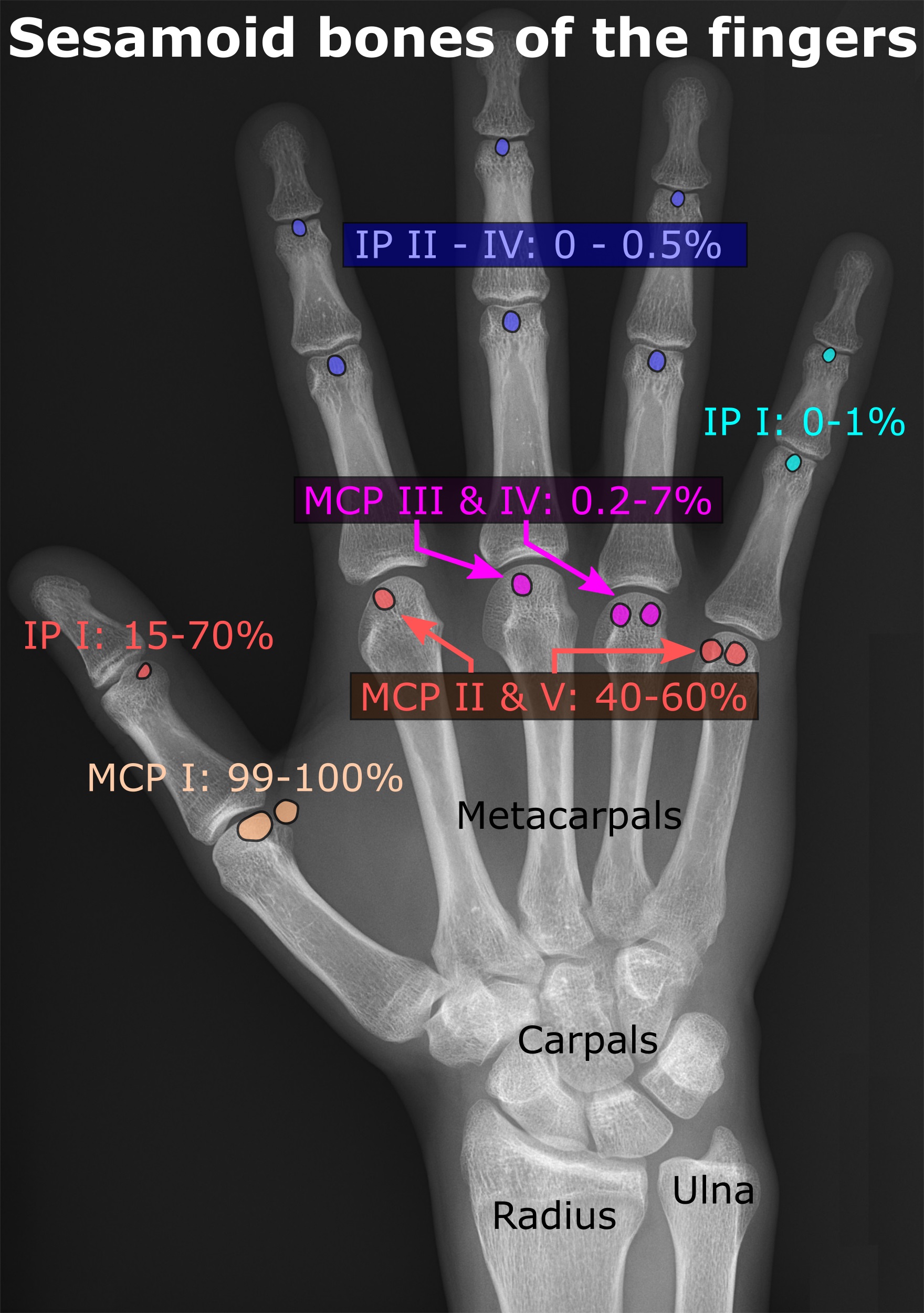
In anatomy, a sesamoid bone () is a bone embedded within a tendon or a muscle. Its name is derived from the Arabic word for 'sesame seed', indicating the small size of most sesamoids. Often, these bones form in response to strain, or can be present as a normal variant. The patella is the largest sesamoid bone in the body. Sesamoids act like pulleys, providing a smooth surface for tendons to slide over, increasing the tendon's ability to transmit muscular forces. Structure Sesamoid bones can be found on joints throughout the body, including: * In the knee—the patella (within the quadriceps tendon). This is the largest sesamoid bone. * In the hand—two sesamoid bones are commonly found in the distal portions of the first metacarpal bone (within the tendons of adductor pollicis and flexor pollicis brevis). There is also commonly a sesamoid bone in distal portions of the second metacarpal bone. * In the wrist—The pisiform of the wrist is a sesamoid bone (within t ... [...More Info...] [...Related Items...] OR: [Wikipedia] [Google] [Baidu] |
Femur
The femur (; ), or thigh bone, is the proximal bone of the hindlimb in tetrapod vertebrates. The head of the femur articulates with the acetabulum in the pelvic bone forming the hip joint, while the distal part of the femur articulates with the tibia (shinbone) and patella (kneecap), forming the knee joint. By most measures the two (left and right) femurs are the strongest bones of the body, and in humans, the largest and thickest. Structure The femur is the only bone in the upper leg. The two femurs converge medially toward the knees, where they articulate with the proximal ends of the tibiae. The angle of convergence of the femora is a major factor in determining the femoral-tibial angle. Human females have thicker pelvic bones, causing their femora to converge more than in males. In the condition ''genu valgum'' (knock knee) the femurs converge so much that the knees touch one another. The opposite extreme is ''genu varum'' (bow-leggedness). In the general pop ... [...More Info...] [...Related Items...] OR: [Wikipedia] [Google] [Baidu] |
Deep Posterior Compartment
The posterior compartment of the leg is one of the fascial compartments of leg, fascial compartments of the leg and is divided further into deep and superficial compartments. Structure Muscles Superficial posterior compartment Deep posterior compartment Blood supply Posterior tibial artery Innervation The posterior compartment of the leg is supplied by the tibial nerve. Function * It contains the plantar flexors: Additional images File:From below - Superficial posterior compartment of leg - animation.gif, Superficial posterior compartment. Animation. File:From below - Deep posterior compartment of leg - animation.gif, Deep posterior compartment. Animation. References External links Diagram at patientcareonline.com {{DEFAULTSORT:Posterior Compartment Of Leg Muscles of the lower limb ... [...More Info...] [...Related Items...] OR: [Wikipedia] [Google] [Baidu] |
Popliteal Artery
The popliteal artery is a deeply placed continuation of the femoral artery opening in the distal portion of the adductor magnus muscle. It courses through the popliteal fossa and ends at the lower border of the popliteus muscle, where it branches into the anterior and posterior tibial arteries. The deepest (most anterior) structure in the fossa, the popliteal artery runs close to the joint capsule of the knee as it spans the intercondylar fossa. Five genicular branches of the popliteal artery supply the capsule and ligaments of the knee joint. The genicular arteries are the superior lateral, superior medial, middle, inferior lateral, and inferior medial genicular arteries. They participate in the formation of the periarticular genicular anastomosis, a network of vessels surrounding the knee that provides collateral circulation capable of maintaining blood supply to the leg during full knee flexion, which may kink the popliteal artery. Structure The popliteal artery is the contin ... [...More Info...] [...Related Items...] OR: [Wikipedia] [Google] [Baidu] |
Lumbar Spinal Nerve 5
The lumbar nerves are the five pairs of spinal nerves emerging from the lumbar vertebrae. They are divided into posterior and anterior divisions. Structure The lumbar nerves are five spinal nerves which arise from either side of the spinal cord below the thoracic spinal cord and above the sacral spinal cord. They arise from the spinal cord between each pair of lumbar spinal vertebrae and travel through the intervertebral foramina. The nerves then split into an anterior branch, which travels forward, and a posterior branch, which travels backwards and supplies the area of the back. Posterior divisions The middle divisions of the posterior branches run close to the articular processes of the vertebrae and end in the multifidus muscle. The outer branches supply the erector spinae muscles. The nerves give off branches to the skin. These pierce the aponeurosis of the greater trochanter. Anterior divisions The anterior divisions of the lumbar nerves ( la, rami anteriores) ... [...More Info...] [...Related Items...] OR: [Wikipedia] [Google] [Baidu] |
Meniscus (anatomy)
A meniscus is a crescent-shaped fibrocartilaginous anatomical structure that, in contrast to an articular disc, only partly divides a joint cavity.Platzer (2004), p 208 In humans they are present in the knee, wrist, acromioclavicular, sternoclavicular, and temporomandibular joints; in other animals they may be present in other joints. Generally, the term "meniscus" is used to refer to the cartilage of the knee, either to the lateral or medial meniscus. Both are cartilaginous tissues that provide structural integrity to the knee when it undergoes tension and torsion. The menisci are also known as "semi-lunar" cartilages, referring to their half-moon, crescent shape. The term "meniscus" is from the Ancient Greek word (), meaning "crescent". Structure The menisci of the knee are two pads of fibrocartilaginous tissue which serve to disperse friction in the knee joint between the lower leg (tibia) and the thigh (femur). They are concave on the top and flat on the bott ... [...More Info...] [...Related Items...] OR: [Wikipedia] [Google] [Baidu] |
Inward Rotation
Motion, the process of movement, is described using specific anatomical terms. Motion includes movement of organs, joints, limbs, and specific sections of the body. The terminology used describes this motion according to its direction relative to the anatomical position of the body parts involved. Anatomists and others use a unified set of terms to describe most of the movements, although other, more specialized terms are necessary for describing unique movements such as those of the hands, feet, and eyes. In general, motion is classified according to the anatomical plane it occurs in. ''Flexion'' and ''extension'' are examples of ''angular'' motions, in which two axes of a joint are brought closer together or moved further apart. ''Rotational'' motion may occur at other joints, for example the shoulder, and are described as ''internal'' or ''external''. Other terms, such as ''elevation'' and ''depression'', describe movement above or below the horizontal plane. Many anat ... [...More Info...] [...Related Items...] OR: [Wikipedia] [Google] [Baidu] |
Thigh
In human anatomy, the thigh is the area between the hip ( pelvis) and the knee. Anatomically, it is part of the lower limb. The single bone in the thigh is called the femur. This bone is very thick and strong (due to the high proportion of bone tissue), and forms a ball and socket joint at the hip, and a modified hinge joint at the knee. Structure Bones The femur is the only bone in the thigh and serves as an attachment site for all muscles in the thigh. The head of the femur articulates with the acetabulum in the pelvic bone forming the hip joint, while the distal part of the femur articulates with the tibia and patella forming the knee. By most measures, the femur is the strongest bone in the body. The femur is also the longest bone in the body. The femur is categorised as a long bone and comprises a diaphysis, the shaft (or body) and two epiphysis or extremities that articulate with adjacent bones in the hip and knee. Muscular compartments In cross-section, ... [...More Info...] [...Related Items...] OR: [Wikipedia] [Google] [Baidu] |


