|
Pinealoblastoma
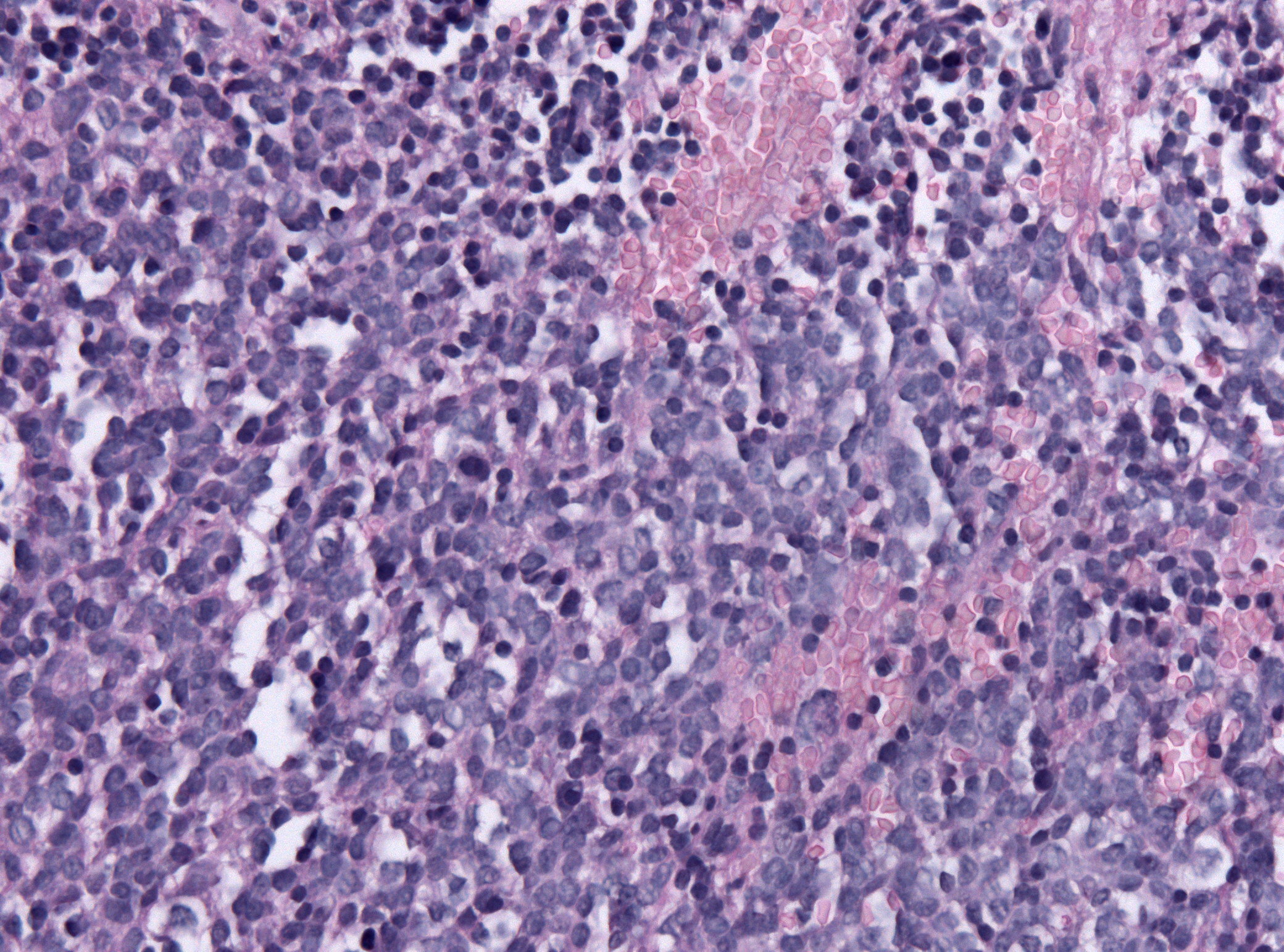
Pineoblastoma is a malignant tumor of the pineal gland. A pineoblastoma is a supratentorial midline primitive neuroectodermal tumor. Pineoblastoma can present at any age, but is most common in young children. They account for 0.001% of all primary CNS neoplasms. Epidemiology Pineoblastomas typically occur at very young ages. One study found the average age of presentation to be 4.3 years, with peaks at age 3 and 8. Another cites cases to more commonly occur in patients under 2 years of age. Rates of occurrence for males and females are similar, but may be slightly more common in females. One study found incidence of pineoblastoma to be increased in black patients compared to white patients by around 71%. This difference was most apparent in patients aged 5 to 9 years old. Pathophysiology The pineal gland is a small organ in the center of the brain that is responsible for controlling melatonin secretion. Several tumors can occur in the area of the pineal gland, with the most ... [...More Info...] [...Related Items...] OR: [Wikipedia] [Google] [Baidu] |
Pineoblastoma
Pineoblastoma is a malignant tumor of the pineal gland. A pineoblastoma is a supratentorial midline primitive neuroectodermal tumor. Pineoblastoma can present at any age, but is most common in young children. They account for 0.001% of all primary CNS neoplasms. Epidemiology Pineoblastomas typically occur at very young ages. One study found the average age of presentation to be 4.3 years, with peaks at age 3 and 8. Another cites cases to more commonly occur in patients under 2 years of age. Rates of occurrence for males and females are similar, but may be slightly more common in females. One study found incidence of pineoblastoma to be increased in black patients compared to white patients by around 71%. This difference was most apparent in patients aged 5 to 9 years old. Pathophysiology The pineal gland is a small organ in the center of the brain that is responsible for controlling melatonin secretion. Several tumors can occur in the area of the pineal gland, with the most a ... [...More Info...] [...Related Items...] OR: [Wikipedia] [Google] [Baidu] |
Magnetic Resonance Imaging
Magnetic resonance imaging (MRI) is a medical imaging technique used in radiology to form pictures of the anatomy and the physiological processes of the body. MRI scanners use strong magnetic fields, magnetic field gradients, and radio waves to generate images of the organs in the body. MRI does not involve X-rays or the use of ionizing radiation, which distinguishes it from CT and PET scans. MRI is a medical application of nuclear magnetic resonance (NMR) which can also be used for imaging in other NMR applications, such as NMR spectroscopy. MRI is widely used in hospitals and clinics for medical diagnosis, staging and follow-up of disease. Compared to CT, MRI provides better contrast in images of soft-tissues, e.g. in the brain or abdomen. However, it may be perceived as less comfortable by patients, due to the usually longer and louder measurements with the subject in a long, confining tube, though "Open" MRI designs mostly relieve this. Additionally, implants and oth ... [...More Info...] [...Related Items...] OR: [Wikipedia] [Google] [Baidu] |
Chemotherapy
Chemotherapy (often abbreviated to chemo and sometimes CTX or CTx) is a type of cancer treatment that uses one or more anti-cancer drugs (chemotherapeutic agents or alkylating agents) as part of a standardized chemotherapy regimen. Chemotherapy may be given with a curative intent (which almost always involves combinations of drugs) or it may aim to prolong life or to reduce symptoms ( palliative chemotherapy). Chemotherapy is one of the major categories of the medical discipline specifically devoted to pharmacotherapy for cancer, which is called ''medical oncology''. The term ''chemotherapy'' has come to connote non-specific usage of intracellular poisons to inhibit mitosis (cell division) or induce DNA damage, which is why inhibition of DNA repair can augment chemotherapy. The connotation of the word chemotherapy excludes more selective agents that block extracellular signals (signal transduction). The development of therapies with specific molecular or genetic targets, wh ... [...More Info...] [...Related Items...] OR: [Wikipedia] [Google] [Baidu] |
Radiation Therapy
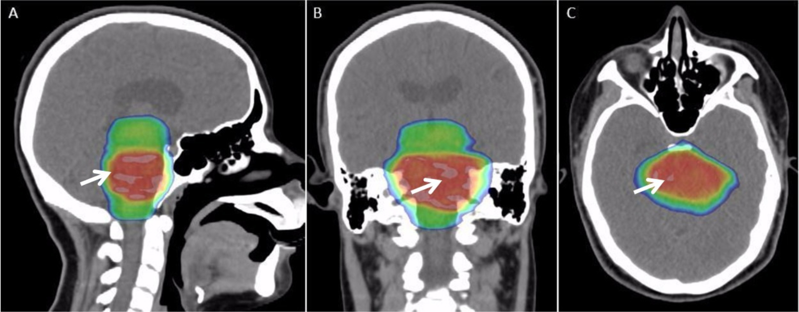
Radiation therapy or radiotherapy, often abbreviated RT, RTx, or XRT, is a therapy using ionizing radiation, generally provided as part of cancer treatment to control or kill malignant cells and normally delivered by a linear accelerator. Radiation therapy may be curative in a number of types of cancer if they are localized to one area of the body. It may also be used as part of adjuvant therapy, to prevent tumor recurrence after surgery to remove a primary malignant tumor (for example, early stages of breast cancer). Radiation therapy is synergistic with chemotherapy, and has been used before, during, and after chemotherapy in susceptible cancers. The subspecialty of oncology concerned with radiotherapy is called radiation oncology. A physician who practices in this subspecialty is a radiation oncologist. Radiation therapy is commonly applied to the cancerous tumor because of its ability to control cell growth. Ionizing radiation works by damaging the DNA of cancerous tissue ... [...More Info...] [...Related Items...] OR: [Wikipedia] [Google] [Baidu] |
Retina
The retina (from la, rete "net") is the innermost, light-sensitive layer of tissue of the eye of most vertebrates and some molluscs. The optics of the eye create a focused two-dimensional image of the visual world on the retina, which then processes that image within the retina and sends nerve impulses along the optic nerve to the visual cortex to create visual perception. The retina serves a function which is in many ways analogous to that of the film or image sensor in a camera. The neural retina consists of several layers of neurons interconnected by synapses and is supported by an outer layer of pigmented epithelial cells. The primary light-sensing cells in the retina are the photoreceptor cells, which are of two types: rods and cones. Rods function mainly in dim light and provide monochromatic vision. Cones function in well-lit conditions and are responsible for the perception of colour through the use of a range of opsins, as well as high-acuity vision used for task ... [...More Info...] [...Related Items...] OR: [Wikipedia] [Google] [Baidu] |
CRX (gene)
Cone-rod homeobox protein is a protein that in humans is encoded by the ''CRX'' gene. Function The protein encoded by this gene is a photoreceptor-specific transcription factor which plays a role in the differentiation of photoreceptor cells. This homeodomain protein is necessary for the maintenance of normal cone and rod function. Mutations in this gene are associated with photoreceptor degeneration, Leber's congenital amaurosis type III and the autosomal dominant cone-rod dystrophy 2. Several alternatively spliced transcript variants of this gene have been described, but the full-length nature of some variants has not been determined. Mammalian CRX encodes a 299 amino acid protein containing a DNA binding homeodomain (HD) near its N-terminus followed by glutamine rich (Gln), and basic amino acid regions, then a C-terminal transactivation domain (AD). While structural biochemistry has demonstrated that the CRX HD adopts a canonical homeodomain protein fold, the AD is predict ... [...More Info...] [...Related Items...] OR: [Wikipedia] [Google] [Baidu] |
Neurofilament
Neurofilaments (NF) are classed as type IV intermediate filaments found in the cytoplasm of neurons. They are protein polymers measuring 10 nm in diameter and many micrometers in length. Together with microtubules (~25 nm) and microfilaments (7 nm), they form the neuronal cytoskeleton. They are believed to function primarily to provide structural support for axons and to regulate axon diameter, which influences nerve conduction velocity. The proteins that form neurofilaments are members of the intermediate filament protein family, which is divided into six types based on their gene organization and protein structure. Types I and II are the keratins which are expressed in epithelia. Type III contains the proteins vimentin, desmin, peripherin and glial fibrillary acidic protein (GFAP). Type IV consists of the neurofilament proteins L, M, H and internexin. Type V consists of the Lamin, nuclear lamins, and type VI consists of the protein Nestin (protein), nesti ... [...More Info...] [...Related Items...] OR: [Wikipedia] [Google] [Baidu] |
Synaptophysin
Synaptophysin, also known as the major synaptic vesicle protein p38, is a protein that in humans is encoded by the ''SYP'' gene. Genomics The gene is located on the short arm of X chromosome (Xp11.23-p11.22). It is 12,406 bases in length and lies on the minus strand. The encoded protein has 313 amino acids with a predicted molecular weight of 33.845 kDa. Molecular biology The protein is a synaptic vesicle glycoprotein with four transmembrane domains weighing 38kDa. It is present in neuroendocrine cells and in virtually all neurons in the brain and spinal cord that participate in synaptic transmission. It acts as a marker for neuroendocrine tumors, and its ubiquity at the synapse has led to the use of synaptophysin immunostaining for quantification of synapses. The exact function of the protein is unknown: it interacts with the essential synaptic vesicle protein synaptobrevin, but when the synaptophysin gene is experimentally inactivated in animals, they still develop and ... [...More Info...] [...Related Items...] OR: [Wikipedia] [Google] [Baidu] |
Photoreceptor Cell
A photoreceptor cell is a specialized type of neuroepithelial cell found in the retina that is capable of visual phototransduction. The great biological importance of photoreceptors is that they convert light (visible electromagnetic radiation) into signals that can stimulate biological processes. To be more specific, photoreceptor proteins in the cell absorb photons, triggering a change in the cell's membrane potential. There are currently three known types of photoreceptor cells in mammalian eyes: rods, cones, and intrinsically photosensitive retinal ganglion cells. The two classic photoreceptor cells are rods and cones, each contributing information used by the visual system to form an image of the environment, sight. Rods primarily mediate scotopic vision (dim conditions) whereas cones primarily mediate to photopic vision (bright conditions), but the processes in each that supports phototransduction is similar. A third class of mammalian photoreceptor cell was discovered ... [...More Info...] [...Related Items...] OR: [Wikipedia] [Google] [Baidu] |
Glia
Glia, also called glial cells (gliocytes) or neuroglia, are non-neuronal cells in the central nervous system (brain and spinal cord) and the peripheral nervous system that do not produce electrical impulses. They maintain homeostasis, form myelin in the peripheral nervous system, and provide support and protection for neurons. In the central nervous system, glial cells include oligodendrocytes, astrocytes, ependymal cells, and microglia, and in the peripheral nervous system they include Schwann cells and satellite cells. Function They have four main functions: *to surround neurons and hold them in place *to supply nutrients and oxygen to neurons *to insulate one neuron from another *to destroy pathogens and remove dead neurons. They also play a role in neurotransmission and synaptic connections, and in physiological processes such as breathing. While glia were thought to outnumber neurons by a ratio of 10:1, recent studies using newer methods and reappraisal of historical quan ... [...More Info...] [...Related Items...] OR: [Wikipedia] [Google] [Baidu] |
Neuronal Lineage Marker
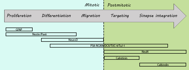
A neuronal lineage marker is an endogenous tag that is expressed in different cells along neurogenesis and differentiated cells such as neurons. It allows detection and identification of cells by using different techniques. A neuronal lineage marker can be either DNA, mRNA or RNA expressed in a cell of interest. It can also be a protein tag, as a partial protein, a protein or an epitope that discriminates between different cell types or different states of a common cell. An ideal marker is specific to a given cell type in normal conditions and/or during injury. Cell markers are very valuable tools for examining the function of cells in normal conditions as well as during disease. The discovery of various proteins specific to certain cells led to the production of cell-type-specific antibodies that have been used to identify cells. The techniques used for its detection can be immunohistochemistry, immunocytochemistry, methods that utilize transcriptional modulators and site-specif ... [...More Info...] [...Related Items...] OR: [Wikipedia] [Google] [Baidu] |
Immunohistochemistry
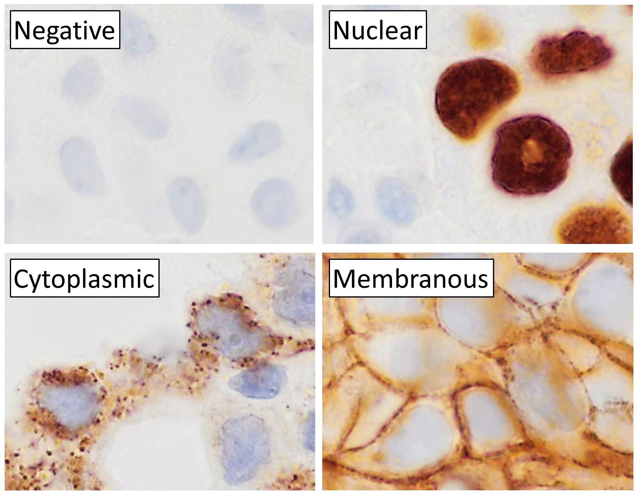
Immunohistochemistry (IHC) is the most common application of immunostaining. It involves the process of selectively identifying antigens (proteins) in cells of a tissue section by exploiting the principle of antibodies binding specifically to antigens in biological tissues. IHC takes its name from the roots "immuno", in reference to antibodies used in the procedure, and "histo", meaning tissue (compare to immunocytochemistry). Albert Coons conceptualized and first implemented the procedure in 1941. Visualising an antibody-antigen interaction can be accomplished in a number of ways, mainly either of the following: * ''Chromogenic immunohistochemistry'' (CIH), wherein an antibody is conjugated to an enzyme, such as peroxidase (the combination being termed immunoperoxidase), that can catalyse a colour-producing reaction. * '' Immunofluorescence'', where the antibody is tagged to a fluorophore, such as fluorescein or rhodamine. Immunohistochemical staining is widely used in the dia ... [...More Info...] [...Related Items...] OR: [Wikipedia] [Google] [Baidu] |


