|
Perineal Tissue
The perineum in humans is the space between the anus and scrotum in the male, or between the anus and the vulva in the female. The perineum is the region of the body between the pubic symphysis (pubic arch) and the coccyx (tail bone), including the perineal body and surrounding structures. There is some variability in how the boundaries are defined. The perineal raphe is visible and pronounced to varying degrees. The perineum is an erogenous zone. The word perineum entered English from late Latin via Greek περίναιος ~ περίνεος ''perinaios, perineos'', itself from περίνεος, περίνεοι 'male genitals' and earlier περίς ''perís'' 'penis' through influence from πηρίς ''pērís'' 'scrotum'. The term was originally understood as a purely male body-part with the perineal raphe seen as a continuation of the scrotal septum since masculinization causes the development of a large anogenital distance in men, in comparison to the corresponding la ... [...More Info...] [...Related Items...] OR: [Wikipedia] [Google] [Baidu] |
Human Musculoskeletal System
The human musculoskeletal system (also known as the human locomotor system, and previously the activity system) is an organ system that gives humans the ability to move using their muscular and skeletal systems. The musculoskeletal system provides form, support, stability, and movement to the body. It is made up of the bones of the skeleton, muscles, cartilage, tendons, ligaments, joints, and other connective tissue that supports and binds tissues and organs together. The musculoskeletal system's primary functions include supporting the body, allowing motion, and protecting vital organs. The skeletal portion of the system serves as the main storage system for calcium and phosphorus and contains critical components of the hematopoietic system. This system describes how bones are connected to other bones and muscle fibers via connective tissue such as tendons and ligaments. The bones provide stability to the body. Muscles keep bones in place and also play a role in the movement ... [...More Info...] [...Related Items...] OR: [Wikipedia] [Google] [Baidu] |
Scrotal Septum
The septum of the scrotum is a vertical layer of fibrous tissue that divides the two compartments of the scrotum. It consists of flexible connective tissue. Its structure extends to the skin surface of the scrotum as the scrotal raphe. It is an incomplete wall of connective tissue and nonstriated muscle (dartos fascia) dividing the scrotum into two sacs, each containing a testis. Histological septa are seen throughout most tissues of the body, particularly where they are needed to stiffen soft cellular tissue, and they also provide planes of ingress for small blood vessels. Because the dense collagen fibres of a septum usually extend out into the softer adjacent tissues. A septum is a cross-wall. Thus it divides a structure into smaller parts. The scrotal septum is used in reconstructive surgery to restore tissue and or reproductive organs injured or severed by trauma.Male Sexual Dysfunction: Pathophysiology and Treatment, edited by Fouad R. Kandeel, Edition: 1st, 2007. ... [...More Info...] [...Related Items...] OR: [Wikipedia] [Google] [Baidu] |
Anal Triangle
The anal triangle is the posterior part of the perineum. It contains the anal canal. Structure The anal triangle can be defined either by its vertices or its sides. * ''Vertices'' ** one vertex at the coccyx bone ** the two ischial tuberosities of the pelvic bone * ''Sides'' ** perineal membrane (posterior border of perineal membrane forms anterior border of anal triangle) ** the two sacrotuberous ligaments Contents Some components of the anal triangle include:Daftary, Shirish; Chakravarti, Sudip (2011). Manual of Obstetrics, 3rd Edition. Elsevier. pp. 1-16. . * Ischioanal fossa * Anococcygeal body * Sacrotuberous ligament * Sacrospinous ligament * Pudendal nerve * Internal pudendal artery and Internal pudendal vein * Anal canal * Muscles ** Sphincter ani externus muscle ** Gluteus maximus muscle ** Obturator internus muscle ** Levator ani muscle ** Coccygeus muscle Additional images Image:Gray320.png, Articulations of pelvis. Posterior view. Image:Gray542.png, The superfi ... [...More Info...] [...Related Items...] OR: [Wikipedia] [Google] [Baidu] |
Human Penis
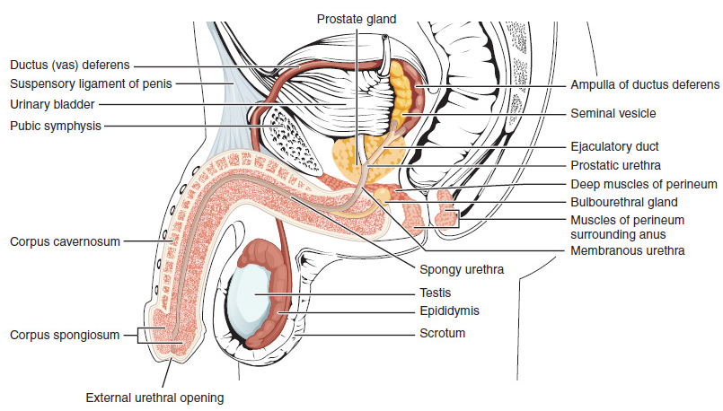
The human penis is an external male intromittent organ that additionally serves as the urinary duct. The main parts are the root (radix); the body (corpus); and the epithelium of the penis including the shaft skin and the foreskin (prepuce) covering the glans penis. The body of the penis is made up of three columns of tissue: two corpora cavernosa on the dorsal side and corpus spongiosum between them on the ventral side. The human male urethra passes through the prostate gland, where it is joined by the ejaculatory duct, and then through the penis. The urethra traverses the corpus spongiosum, and its opening, the meatus (), lies on the tip of the glans penis. It is a passage both for urination and ejaculation of semen (''see'' male reproductive system.) Most of the penis develops from the same embryonic tissue as the clitoris in females. The skin around the penis and the urethra share the same embryonic origin as the labia minora in females. An erection is the stiffening e ... [...More Info...] [...Related Items...] OR: [Wikipedia] [Google] [Baidu] |
Urogenital Triangle
The urogenital triangle is the anterior part of the perineum. In female mammals, it contains the vagina and associated parts of the internal genitalia. Structure The urogenital triangle is the area bound by a triangle with one vertex at the pubic symphysis and the two other vertices at the iliac tuberosities of the pelvic bone. Components As might be expected, the contents of the urogenital triangle differ greatly between the male and the female. Some of the components include:Daftary, Shirish; Chakravarti, Sudip (2011). Manual of Obstetrics, 3rd Edition. Elsevier. pp. 1-16. . * Posterior scrotal nerves / Posterior labial nerves * Urethra * Vagina * Bulbourethral gland / Bartholin's gland * Muscles ** Superficial transverse perineal muscle ** Ischiocavernosus muscle ** Bulbospongiosus muscle * Crus penis / Clitoral crura * Bulb of penis / vestibular bulb * Urogenital diaphragm * Muscular perineal body * Superficial and Deep perineal pouch The deep perineal pouch (also deep ... [...More Info...] [...Related Items...] OR: [Wikipedia] [Google] [Baidu] |
Tuberosity Of The Ischium
The ischial tuberosity (or tuberosity of the ischium, tuber ischiadicum), also known colloquially as the sit bones or sitz bones, or as a pair the sitting bones, is a large swelling posteriorly on the superior ramus of the ischium. It marks the lateral boundary of the pelvic outlet. When sitting, the weight is frequently placed upon the ischial tuberosity. The gluteus maximus provides cover in the upright posture, but leaves it free in the seated position.Platzer (2004), p 236 The distance between a cyclist's ischial tuberosities is one of the factors in the choice of a bicycle saddle. Divisions The tuberosity is divided into two portions: a lower, rough, somewhat triangular part, and an upper, smooth, quadrilateral portion. * The ''lower portion'' is subdivided by a prominent longitudinal ridge, passing from base to apex, into two parts: ** The outer gives attachment to the adductor magnus ** The inner to the sacrotuberous ligament * The ''upper portion'' is subdivided into ... [...More Info...] [...Related Items...] OR: [Wikipedia] [Google] [Baidu] |
Anatomical Line
{{short description, None Anatomical "lines", or "reference lines," are theoretical lines drawn through anatomical structures and are used to describe anatomical location. The following reference lines are identified in ''Terminologia Anatomica'': * Anterior median line * Lateral sternal line: A vertical line corresponding to the lateral margin of the sternum. * Parasternal line: A vertical line equidistant from the sternal and mid-clavicular lines. * Mid-clavicular line: A vertical line passing through the midpoint of the clavicle. * Mammillary line * Anterior axillary line: A vertical line on the anterior torso marked by the anterior axillary fold. * Midaxillary line: A vertical line passing through the apex of the axilla. * Posterior axillary line: A vertical line passing through the posterior axillary fold. * Scapular line: A vertical line passing through the inferior angle of the scapula. * Paravertebral line: A vertical line corresponding to the tips of the transverse proces ... [...More Info...] [...Related Items...] OR: [Wikipedia] [Google] [Baidu] |
Outlet Of The Pelvis
The lower circumference of the lesser pelvis is very irregular; the space enclosed by it is named the inferior aperture or pelvic outlet. It is an important component of pelvimetry. Boundaries It has the following boundaries: * anteriorly: the pubic arch * laterally: the ischial tuberosities * posterolaterally: the inferior margin of the sacrotuberous ligament * posteriorly: the anterior border of the middle of the coccyx. Notches These eminences are separated by three notches: * one in front, the pubic arch, formed by the convergence of the inferior rami of the ischium and pubis on either side. * The other notches, one on either side, are formed by the sacrum and coccyx behind, the ischium in front, and the ilium above; they are called the sciatic notches; in the natural state they are converted into foramina by the sacrotuberous and sacrospinous ligaments. In situ When the ligaments are in situ, the inferior aperture of the pelvis is lozenge-shaped, bounded as follows: * i ... [...More Info...] [...Related Items...] OR: [Wikipedia] [Google] [Baidu] |
Henry Gray
Henry Gray (1827 – 13 June 1861) was a British anatomist and surgery, surgeon most notable for publishing the book ''Gray's Anatomy''. He was elected a Fellow of the Royal Society (FRS) at the age of 25. Biography Gray was born in Belgravia, London, in 1827 and lived most of his life in London. In 1842, he entered as a student at St George's Hospital, St. George's Hospital, London (then situated in Belgravia, now moved to Tooting), and he is described by those who knew him as a most painstaking and methodical worker, and one who learned his anatomy by the slow but invaluable method of making dissections for himself. While still a student, Gray secured the triennial prize of Royal College of Surgeons of England, Royal College of Surgeons in 1848 for an essay entitled ''The Origin, Connexions and Distribution of nerves to the human eye and its appendages, illustrated by comparative dissections of the eye in other vertebrate animals.'' In 1852, at the early age of 25 ... [...More Info...] [...Related Items...] OR: [Wikipedia] [Google] [Baidu] |
Vagina
In mammals, the vagina is the elastic, muscular part of the female genital tract. In humans, it extends from the vestibule to the cervix. The outer vaginal opening is normally partly covered by a thin layer of mucosal tissue called the hymen. At the deep end, the cervix (neck of the uterus) bulges into the vagina. The vagina allows for sexual intercourse and birth. It also channels menstrual flow, which occurs in humans and closely related primates as part of the menstrual cycle. Although research on the vagina is especially lacking for different animals, its location, structure and size are documented as varying among species. Female mammals usually have two external openings in the vulva; these are the urethral opening for the urinary tract and the vaginal opening for the genital tract. This is different from male mammals, who usually have a single urethral opening for both urination and reproduction. The vaginal opening is much larger than the nearby urethral opening, an ... [...More Info...] [...Related Items...] OR: [Wikipedia] [Google] [Baidu] |
Human Anus
In humans, the anus (from Latin '' anus'' meaning "ring", "circle") is the external opening of the rectum, located inside the intergluteal cleft and separated from the genitals by the perineum. Two sphincters control the exit of feces from the body during an act of defecation, which is the primary function of the anus. These are the internal anal sphincter and the external anal sphincter, which are circular muscles that normally maintain constriction of the orifice and which relaxes as required by normal physiological functioning. The inner sphincter is involuntary and the outer is voluntary. It is located behind the perineum which is located behind the vagina or scrotum. In part owing to its exposure to feces, a number of medical conditions may affect the anus such as hemorrhoids. The anus is the site of potential infections and other conditions, including cancer (see Anal cancer). With anal sex, the anus can play a role in sexuality. Attitudes toward anal sex vary, an ... [...More Info...] [...Related Items...] OR: [Wikipedia] [Google] [Baidu] |
Surface Anatomy
Surface anatomy (also called superficial anatomy and visual anatomy) is the study of the external features of the body of an animal.Seeley (2003) chap.1 p.2 In birds this is termed ''topography''. Surface anatomy deals with anatomical features that can be studied by sight, without dissection. As such, it is a branch of gross anatomy, along with endoscopic and radiological anatomy.Standring (2008) ''Introduction'', ''Anatomical nomenclature'', p.2 Surface anatomy is a descriptive science. In particular, in the case of human surface anatomy, these are the form and proportions of the human body and the surface landmarks which correspond to deeper structures hidden from view, both in static pose and in motion. In addition, the science of surface anatomy includes the theories and systems of body proportions and related artistic canons. The study of surface anatomy is the basis for depicting the human body in classical art. Some pseudo-sciences such as physiognomy, phrenology and pa ... [...More Info...] [...Related Items...] OR: [Wikipedia] [Google] [Baidu] |


