|
Pathophysiology Of Heart Failure
The main pathophysiology of heart failure is a reduction in the efficiency of the heart muscle, through damage or overloading. As such, it can be caused by a wide number of conditions, including myocardial infarction (in which the heart muscle is starved of oxygen and dies), hypertension (which increases the force of contraction needed to pump blood) and cardiac amyloidosis (in which misfolded proteins are deposited in the heart muscle, causing it to stiffen). Over time these increases in workload will produce changes to the heart itself: The heart of a person with heart failure may have a reduced force of contraction due to overloading of the ventricle. In a healthy heart, increased filling of the ventricle results in increased contraction force (by the Frank–Starling law of the heart) and thus a rise in cardiac output. In heart failure, this mechanism fails, as the ventricle is loaded with blood to the point where heart muscle contraction becomes less efficient. This is due ... [...More Info...] [...Related Items...] OR: [Wikipedia] [Google] [Baidu] |
Pathophysiology
Pathophysiology ( physiopathology) – a convergence of pathology with physiology – is the study of the disordered physiological processes that cause, result from, or are otherwise associated with a disease or injury. Pathology is the medical discipline that describes conditions typically ''observed'' during a disease state, whereas physiology is the biological discipline that describes processes or mechanisms ''operating'' within an organism. Pathology describes the abnormal or undesired condition, whereas pathophysiology seeks to explain the functional changes that are occurring within an individual due to a disease or pathologic state. History Etymology The term ''pathophysiology'' comes from the Ancient Greek πάθος (''pathos'') and φυσιολογία (''phusiologia''). Nineteenth century Reductionism In Germany in the 1830s, Johannes Müller led the establishment of physiology research autonomous from medical research. In 1843, the Berlin Physical Socie ... [...More Info...] [...Related Items...] OR: [Wikipedia] [Google] [Baidu] |
Heart Arrhythmia
Arrhythmias, also known as cardiac arrhythmias, heart arrhythmias, or dysrhythmias, are irregularities in the heartbeat, including when it is too fast or too slow. A resting heart rate that is too fast – above 100 beats per minute in adults – is called tachycardia, and a resting heart rate that is too slow – below 60 beats per minute – is called bradycardia. Some types of arrhythmias have no symptoms. Symptoms, when present, may include palpitations or feeling a pause between heartbeats. In more serious cases, there may be lightheadedness, passing out, shortness of breath or chest pain. While most cases of arrhythmia are not serious, some predispose a person to complications such as stroke or heart failure. Others may result in sudden death. Arrhythmias are often categorized into four groups: extra beats, supraventricular tachycardias, ventricular arrhythmias and bradyarrhythmias. Extra beats include premature atrial contractions, premature ventricular contractions ... [...More Info...] [...Related Items...] OR: [Wikipedia] [Google] [Baidu] |
Interstitial Fluid
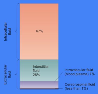
In cell biology, extracellular fluid (ECF) denotes all body fluid outside the cells of any multicellular organism. Total body water in healthy adults is about 60% (range 45 to 75%) of total body weight; women and the obese typically have a lower percentage than lean men. Extracellular fluid makes up about one-third of body fluid, the remaining two-thirds is intracellular fluid within cells. The main component of the extracellular fluid is the interstitial fluid that surrounds cells. Extracellular fluid is the internal environment of all multicellular animals, and in those animals with a blood circulatory system, a proportion of this fluid is blood plasma. Plasma and interstitial fluid are the two components that make up at least 97% of the ECF. Lymph makes up a small percentage of the interstitial fluid. The remaining small portion of the ECF includes the transcellular fluid (about 2.5%). The ECF can also be seen as having two components – plasma and lymph as a delivery system ... [...More Info...] [...Related Items...] OR: [Wikipedia] [Google] [Baidu] |
Catecholamines
A catecholamine (; abbreviated CA) is a monoamine neurotransmitter, an organic compound that has a catechol (benzene with two hydroxyl side groups next to each other) and a side-chain amine. Catechol can be either a free molecule or a substituent of a larger molecule, where it represents a 1,2-dihydroxybenzene group. Catecholamines are derived from the amino acid tyrosine, which is derived from dietary sources as well as synthesis from phenylalanine. Catecholamines are water-soluble and are 50% bound to plasma proteins in circulation. Included among catecholamines are epinephrine (adrenaline), norepinephrine (noradrenaline), and dopamine. Release of the hormones epinephrine and norepinephrine from the adrenal medulla of the adrenal glands is part of the fight-or-flight response. Tyrosine is created from phenylalanine by hydroxylation by the enzyme phenylalanine hydroxylase. Tyrosine is also ingested directly from dietary protein. Catecholamine-secreting cells use several re ... [...More Info...] [...Related Items...] OR: [Wikipedia] [Google] [Baidu] |
Adrenal Glands
The adrenal glands (also known as suprarenal glands) are endocrine glands that produce a variety of hormones including adrenaline and the steroids aldosterone and cortisol. They are found above the kidneys. Each gland has an outer cortex which produces steroid hormones and an inner medulla. The adrenal cortex itself is divided into three main zones: the zona glomerulosa, the zona fasciculata and the zona reticularis. The adrenal cortex produces three main types of steroid hormones: mineralocorticoids, glucocorticoids, and androgens. Mineralocorticoids (such as aldosterone) produced in the zona glomerulosa help in the regulation of blood pressure and electrolyte balance. The glucocorticoids cortisol and cortisone are synthesized in the zona fasciculata; their functions include the regulation of metabolism and immune system suppression. The innermost layer of the cortex, the zona reticularis, produces androgens that are converted to fully functional sex hormones in the gonads ... [...More Info...] [...Related Items...] OR: [Wikipedia] [Google] [Baidu] |
Aldosterone
Aldosterone is the main mineralocorticoid steroid hormone produced by the zona glomerulosa of the adrenal cortex in the adrenal gland. It is essential for sodium conservation in the kidney, salivary glands, sweat glands, and colon. It plays a central role in the homeostatic regulation of blood pressure, plasma sodium (Na+), and potassium (K+) levels. It does so primarily by acting on the mineralocorticoid receptors in the distal tubules and collecting ducts of the nephron. It influences the reabsorption of sodium and excretion of potassium (from and into the tubular fluids, respectively) of the kidney, thereby indirectly influencing water retention or loss, blood pressure, and blood volume.Marieb Human Anatomy & Physiology 9th edition, chapter:16, page:629, question number:14 When dysregulated, aldosterone is pathogenic and contributes to the development and progression of cardiovascular and kidney disease. Aldosterone has exactly the opposite function of the atrial natriure ... [...More Info...] [...Related Items...] OR: [Wikipedia] [Google] [Baidu] |
Angiotensin
Angiotensin is a peptide hormone that causes vasoconstriction and an increase in blood pressure. It is part of the renin–angiotensin system, which regulates blood pressure. Angiotensin also stimulates the release of aldosterone from the adrenal cortex to promote sodium retention by the kidneys. An oligopeptide, angiotensin is a hormone and a dipsogen. It is derived from the precursor molecule angiotensinogen, a serum globulin produced in the liver. Angiotensin was isolated in the late 1930s (first named 'angiotonin' or 'hypertensin') and subsequently characterized and synthesized by groups at the Cleveland Clinic and Ciba laboratories. Precursor and types Angiotensinogen Angiotensinogen is an α-2-globulin synthesized in the liver and is a precursor for angiotensin, but has also been indicated as having many other roles not related to angiotensin peptides. It is a member of the serpin family of proteins, leading to another name: Serpin A8, although it is not known to ... [...More Info...] [...Related Items...] OR: [Wikipedia] [Google] [Baidu] |
Renin
Renin (etymology and pronunciation), also known as an angiotensinogenase, is an aspartic protease protein and enzyme secreted by the kidneys that participates in the body's renin–angiotensin–aldosterone system (RAAS)—also known as the renin–angiotensin–aldosterone axis—that increases the volume of extracellular fluid (blood plasma, lymph and interstitial fluid) and causes arterial vasoconstriction. Thus, it increases the body's mean arterial blood pressure. Renin is not commonly referred to as a hormone, albeit it having a receptor, the (pro)renin receptor, also known as the renin receptor and prorenin receptor (see also below), as well as enzymatic activity with which it hydrolyzes angiotensinogen to angiotensin I. Biochemistry and physiology Structure The primary structure of renin precursor consists of 406 amino acids with a pre- and a pro-segment carrying 20 and 46 amino acids, respectively. Mature renin contains 340 amino acids and has a mass of 37 kDa. ... [...More Info...] [...Related Items...] OR: [Wikipedia] [Google] [Baidu] |
Vasopressin
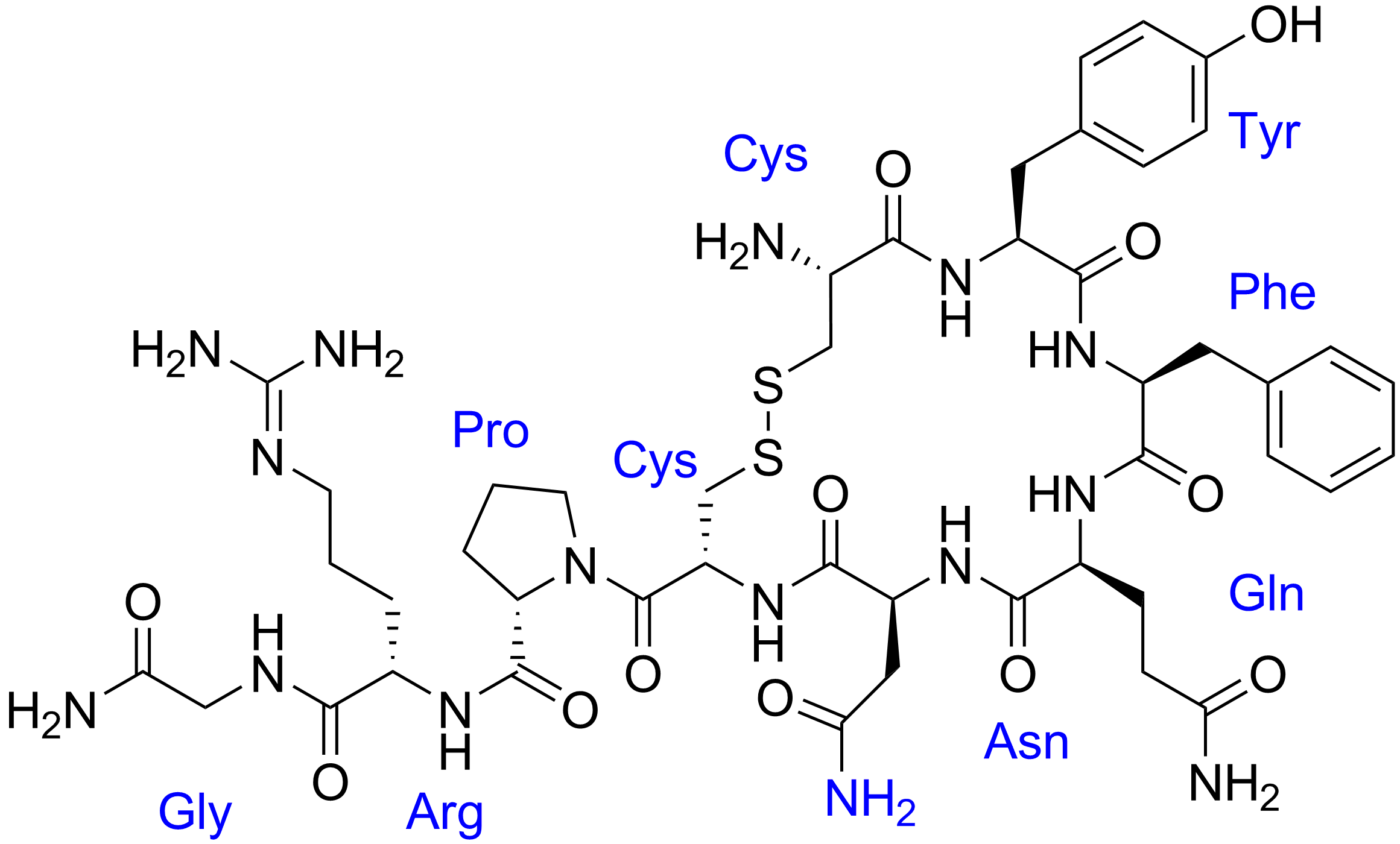
Human vasopressin, also called antidiuretic hormone (ADH), arginine vasopressin (AVP) or argipressin, is a hormone synthesized from the AVP gene as a peptide prohormone in neurons in the hypothalamus, and is converted to AVP. It then travels down the axon terminating in the posterior pituitary, and is released from vesicles into the circulation in response to extracellular fluid hypertonicity (hyperosmolality). AVP has two primary functions. First, it increases the amount of solute-free water reabsorbed back into the circulation from the filtrate in the kidney tubules of the nephrons. Second, AVP constricts arterioles, which increases peripheral vascular resistance and raises arterial blood pressure. A third function is possible. Some AVP may be released directly into the brain from the hypothalamus, and may play an important role in social behavior, sexual motivation and pair bonding, and maternal responses to stress. Vasopressin induces differentiation of stem cells in ... [...More Info...] [...Related Items...] OR: [Wikipedia] [Google] [Baidu] |
Vasoconstriction
Vasoconstriction is the narrowing of the blood vessels resulting from contraction of the muscular wall of the vessels, in particular the large arteries and small arterioles. The process is the opposite of vasodilation, the widening of blood vessels. The process is particularly important in controlling hemorrhage and reducing acute blood loss. When blood vessels constrict, the flow of blood is restricted or decreased, thus retaining body heat or increasing vascular resistance. This makes the skin turn paler because less blood reaches the surface, reducing the radiation of heat. On a larger level, vasoconstriction is one mechanism by which the body regulates and maintains mean arterial pressure. Medications causing vasoconstriction, also known as vasoconstrictors, are one type of medicine used to raise blood pressure. Generalized vasoconstriction usually results in an increase in systemic blood pressure, but it may also occur in specific tissues, causing a localized reduction in bl ... [...More Info...] [...Related Items...] OR: [Wikipedia] [Google] [Baidu] |
Nucleus Tractus Solitarii
In the human brainstem, the solitary nucleus, also called nucleus of the solitary tract, nucleus solitarius, and nucleus tractus solitarii, (SN or NTS) is a series of purely sensory nuclei (clusters of nerve cell bodies) forming a vertical column of grey matter embedded in the medulla oblongata. Through the center of the SN runs the solitary tract, a white bundle of nerve fibers, including fibers from the facial, glossopharyngeal and vagus nerves, that innervate the SN. The SN projects to, among other regions, the reticular formation, parasympathetic preganglionic neurons, hypothalamus and thalamus, forming circuits that contribute to autonomic regulation. Cells along the length of the SN are arranged roughly in accordance with function; for instance, cells involved in taste are located in the rostral part, while those receiving information from cardio-respiratory and gastrointestinal processes are found in the caudal part. Inputs * Taste information from the facial nerve via the ... [...More Info...] [...Related Items...] OR: [Wikipedia] [Google] [Baidu] |
Aortic Arch
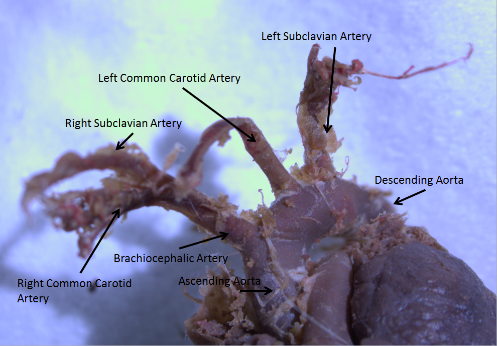
The aortic arch, arch of the aorta, or transverse aortic arch () is the part of the aorta between the ascending and descending aorta. The arch travels backward, so that it ultimately runs to the left of the trachea. Structure The aorta begins at the level of the upper border of the second/third sternocostal articulation of the right side, behind the ventricular outflow tract and pulmonary trunk. The right atrial appendage overlaps it. The first few centimeters of the ascending aorta and pulmonary trunk lies in the same pericardial sheath. and runs at first upward, arches over the pulmonary trunk, right pulmonary artery, and right main bronchus to lie behind the right second coastal cartilage. The right lung and sternum lies anterior to the aorta at this point. The aorta then passes posteriorly and to the left, anterior to the trachea, and arches over left main bronchus and left pulmonary artery, and reaches to the left side of the T4 vertebral body. Apart from T4 vertebral body ... [...More Info...] [...Related Items...] OR: [Wikipedia] [Google] [Baidu] |
.png)


