|
Pastern
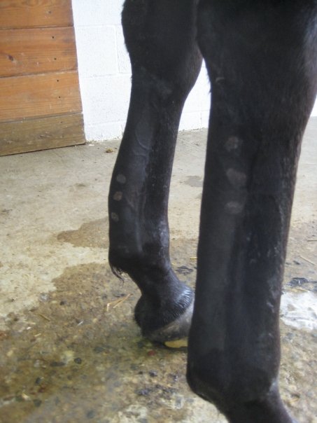
The pastern is a part of the leg of a horse between the fetlock and the top of the hoof. It incorporates the Equine_forelimb_anatomy#Metacarpal_bones, long pastern bone (proximal phalanx) and the Equine_forelimb_anatomy#Middle_phalanx, short pastern bone (middle phalanx), which are held together by two sets of paired ligaments to form the pastern joint (proximal interphalangeal joint). Anatomically homologous to the two largest bones found in the human finger, the pastern was famously mis-defined by Samuel Johnson in A Dictionary of the English Language, his dictionary as "the knee of a horse". When a lady asked Johnson how this had happened, he gave the much-quoted reply: "Ignorance, madam, pure ignorance." Anatomy and importance The pastern consists of two bones, the uppermost called the "large pastern bone" or proximal phalanx, which begins just under the fetlock joint, and the lower called the "small pastern bone" or middle phalanx, located between the large pastern bone an ... [...More Info...] [...Related Items...] OR: [Wikipedia] [Google] [Baidu] |
Ringbone
{{No footnotes, date=February 2020 Ringbone is exostosis (bone growth) in the pastern or coffin joint of a horse. In severe cases, the growth can encircle the bones, giving ringbone its name. It has been suggested by some authors that such a colloquial term, whilst commonly used, might be misleading and that it would be better to refer to this condition as osteoarthritis of the inter-phalanx bones, phalangeal joints in ungulates (Rogers and Waldron, 1995: 34–35). Ringbone can be classified by its location, with "high ringbone" occurring on the lower part of the large pastern bone or the upper part of the small pastern bone. "Low ringbone" occurs on the lower part of the small pastern bone or the upper part of the pedal bone, coffin bone. High ringbone is easier seen than low ringbone, as low ringbone occurs in the hoof of the horse. However, low ringbone may be seen if it becomes serious, as it creates a bony bump on the Limbs of the horse#Legs, coronet of the horse. Causes * ... [...More Info...] [...Related Items...] OR: [Wikipedia] [Google] [Baidu] |
Equine Forelimb Anatomy
The limbs of the horse are structures made of dozens of bones, joints, muscles, tendons, and ligaments that support the weight of the equine body. They include three apparatuses: the suspensory apparatus, which carries much of the weight, prevents overextension of the joint and absorbs shock, the stay apparatus, which locks major joints in the limbs, allowing horses to remain standing while relaxed or asleep, and the reciprocal apparatus, which causes the hock to follow the motions of the stifle. The limbs play a major part in the movement of the horse, with the legs performing the functions of absorbing impact, bearing weight, and providing thrust. In general, the majority of the weight is borne by the front legs, while the rear legs provide propulsion. The hooves are also important structures, providing support, traction and shock absorption, and containing structures that provide blood flow through the lower leg. As the horse developed as a cursorial animal, with a primary ... [...More Info...] [...Related Items...] OR: [Wikipedia] [Google] [Baidu] |
Navicular Disease
Navicular syndrome, often called navicular disease, is a syndrome of lameness problems in horses. It most commonly describes an inflammation or degeneration of the navicular bone and its surrounding tissues, usually on the front feet. It can lead to significant and even disabling lameness. Description of the navicular area Knowledge of equine forelimb anatomy is especially useful for understanding navicular syndrome. The navicular bone lies behind the coffin bone and under the small pastern bone (middle phalanx). The deep digital flexor (DDF) tendon runs down the back of the cannon and soft tissue in that area and under the navicular bone before attaching to the underside of the coffin bone. The DDF tendon flexes the coffin joint, and the navicular bone acts as a fulcrum that the DDF tendon runs over. The navicular bone is stabilized by the impar ligament distally at its attachment to the coffin bone (distal phalanx), and by the collateral ligaments of the navicular bone extend ... [...More Info...] [...Related Items...] OR: [Wikipedia] [Google] [Baidu] |
Bowed Tendon
Tendinitis/tendonitis is inflammation of a tendon, often involving torn collagen fibers. A bowed tendon is a horseman's term for a tendon after a horse has sustained an injury that causes swelling in one or more tendons creating a "bowed" appearance. Description of tendinitis in horses Tendinitis usually involves disruption of the tendon fibers. It is most commonly seen in the superficial digital flexor tendon (SDFT) in a front leg—the tendon that runs down the back of the leg, closest to the surface. Tendinitis creating a "bow" is uncommon in the deep digital flexor tendon (DDFT) of a front leg, but is not uncommon in the pastern and foot regions. Tendinitis of the SDFT or DDFT in the hind leg is less common. When the SDFT is damaged significantly, there is a thickening of the tendon, giving it a bowed appearance when the leg is viewed from the side. Bows usually occur in the middle of the tendon region, although they may also be seen in the upper third, right below the kne ... [...More Info...] [...Related Items...] OR: [Wikipedia] [Google] [Baidu] |
A Dictionary Of The English Language
''A Dictionary of the English Language'', sometimes published as ''Johnson's Dictionary'', was published on 15 April 1755 and written by Samuel Johnson. It is among the most influential dictionary, dictionaries in the history of the English language. There was dissatisfaction with the dictionaries of the period, so in June 1746 a group of London booksellers contracted Johnson to write a dictionary for the sum of 1,500 Guinea (British coin), guineas (£1,575), equivalent to about £ in . Johnson took seven years to complete the work, although he had claimed he could finish it in three. He did so single-handedly, with only clerical assistance to copy the illustrative quotations that he had marked in books. Johnson produced several revised editions during his life. Until the completion of the ''Oxford English Dictionary'' 173 years later, Johnson's was viewed as the pre-eminent English dictionary. According to Walter Jackson Bate, the Dictionary "easily ranks as one of the g ... [...More Info...] [...Related Items...] OR: [Wikipedia] [Google] [Baidu] |
Fetlock
Fetlock is the common name in horses, large animals, and sometimes dogs for the metacarpophalangeal and metatarsophalangeal joints (MCPJ and MTPJ). Although it somewhat resembles the human ankle in appearance, the joint is homologous to the ball of the foot. In anatomical terms, the hoof corresponds to the toe, rather than the whole human foot. Etymology and related terminology The word literally means "foot-lock" and refers to the small tuft of hair situated on the rear of the fetlock joint. "Feather" refers to the particularly long, luxuriant hair growth over the lower leg and fetlock that is characteristic of certain breeds. Formation A fetlock (a MCPJ or a MTPJ) is formed by the junction of the third metacarpal (in the forelimb) or metatarsal (in the hindlimb) bones, either of which are commonly called the cannon bones, proximally and the proximal phalanx distally, commonly called the pastern bone. Paired proximal sesamoid bones form the joint with the ... [...More Info...] [...Related Items...] OR: [Wikipedia] [Google] [Baidu] |
Sidebone
Sidebone is a common condition of horses, characterized by the ossification of the collateral cartilages of the coffin bone. These are found on either side of the foot protruding above the level of the coronary band. The lateral cartilages support the hoof wall and provide an important role in the support and cushioning provided to the heel. The front feet are most commonly affected. Causes Repeated concussion of the foot is probably the cause in many cases. Such concussion could be produced when a horse is always worked on a hard surface. There also appears to be a hereditary component to sidebone but this may be because bad conformation is hereditary and bad conformation appears to predispose to sidebone. Bad conformation would include * those with narrow, upright feet * those with unbalanced feet, especially if they have toe-in or toe-out conformation * draft horses, or horses with a heavy build, are more likely to develop sidebone than light horses or ponies Symptoms Sidebon ... [...More Info...] [...Related Items...] OR: [Wikipedia] [Google] [Baidu] |
Sesamoiditis
Sesamoiditis is inflammation of the sesamoid bones. Humans Sesamoiditis occurs on the bottom of the foot, just behind the big toe. There are normally two sesamoid bones on each foot; sometimes sesamoids can be bipartite, which means they each comprise two separate pieces. The sesamoids are roughly the size of jelly beans. The sesamoid bones act as a fulcrum for the flexor tendons, the tendons which bend the big toe downward. Symptoms include inflammation and pain. Sometimes a sesamoid bone is fractured. This can be difficult to pick up on X-ray, so a bone scan or MRI is a better alternative. Among those who are susceptible to the malady are dancers, catchers and pitchers in baseball, soccer players, and American football players. Horses In the horse it occurs at the horse's fetlock. The sesamoid bones lie behind the bones of the fetlock, at the back of the joint, and help to keep the tendons and ligaments that run between them correctly functioning. Usually periostitis ( ... [...More Info...] [...Related Items...] OR: [Wikipedia] [Google] [Baidu] |
Horse Anatomy
Equine anatomy encompasses the gross and microscopic anatomy of horses, ponies and other equids, including donkeys, mules and zebras. While all anatomical features of equids are described in the same terms as for other animals by the International Committee on Veterinary Gross Anatomical Nomenclature in the book '' Nomina Anatomica Veterinaria'', there are many horse-specific colloquial terms used by equestrians. External anatomy * Back: the area where the saddle sits, beginning at the end of the withers, extending to the last thoracic vertebrae (colloquially includes the loin or "coupling", though technically incorrect usage) * Barrel: the body of the horse, enclosing the rib cage and the major internal organs * Buttock: the part of the hindquarters behind the thighs and below the root of the tail * Cannon or cannon bone: the area between the knee or hock and the fetlock joint, sometimes called the "shin" of the horse, though technically it is the third metacarpal * Chestn ... [...More Info...] [...Related Items...] OR: [Wikipedia] [Google] [Baidu] |
Coffin Bone
The coffin bone, also known as the pedal bone (U.S.), is the Phalanx bones, distal phalanx, the bottommost bone in the Limbs of the horse#Legs, front and rear legs of horses, cattle, pigs and other ruminants. It is encased by the horse hoof, hoof capsule. In horses and other odd-toed ungulates it is the third phalanx, or "P3"; in even-toed ungulates such as cattle, it is the third and fourth (P3 and P4). The coffin bone meets the short pastern bone or second phalanx at the coffin joint. The coffin bone is connected to the inner wall of the horse hoof by a structure called the laminar layer. The insensitive laminae coming in from the hoof wall connects to the sensitive laminae layer, containing the blood supply and nerves, which is attached to the coffin bone. The lamina is a critical structure for hoof health, therefore any injury to the hoof or its support system can in turn affect the coffin bone. Despite the protection provided by the Horse hoof, hoof, the coffin bone can be ... [...More Info...] [...Related Items...] OR: [Wikipedia] [Google] [Baidu] |


