|
Paracentral Lobule
In neuroanatomy, the paracentral lobule is on the medial surface of the cerebral hemisphere and is the continuation of the precentral and postcentral gyri. The paracentral lobule controls motor and sensory innervations of the contralateral lower extremity. It is also responsible for control of defecation and urination. It includes portions of the frontal and parietal lobes: * The anterior portion of the paracentral lobule is part of the frontal lobe and contains a little portion of Brodmann's area 6 (SMA): this is because the paracentral sulcus (branch of the limbic sulcus) does not correspond to the precentral sulcus on the medial plane. * The posterior portion is considered part of the parietal lobe and deals with somatosensory of the distal limbs. While the boundary between the lobes, the central sulcus, is easy to locate on the lateral surface of the cerebral hemispheres, this boundary is often discerned in a cytoarchetectonic manner in cases where the central sulcus i ... [...More Info...] [...Related Items...] OR: [Wikipedia] [Google] [Baidu] |
Neuroanatomy
Neuroanatomy is the study of the structure and organization of the nervous system. In contrast to animals with radial symmetry, whose nervous system consists of a distributed network of cells, animals with bilateral symmetry have segregated, defined nervous systems. Their neuroanatomy is therefore better understood. In vertebrates, the nervous system is segregated into the internal structure of the brain and spinal cord (together called the central nervous system, or CNS) and the routes of the nerves that connect to the rest of the body (known as the peripheral nervous system, or PNS). The delineation of distinct structures and regions of the nervous system has been critical in investigating how it works. For example, much of what neuroscientists have learned comes from observing how damage or "lesions" to specific brain areas affects behavior or other neural functions. For information about the composition of non-human animal nervous systems, see nervous system. For information a ... [...More Info...] [...Related Items...] OR: [Wikipedia] [Google] [Baidu] |
Precentral Sulcus
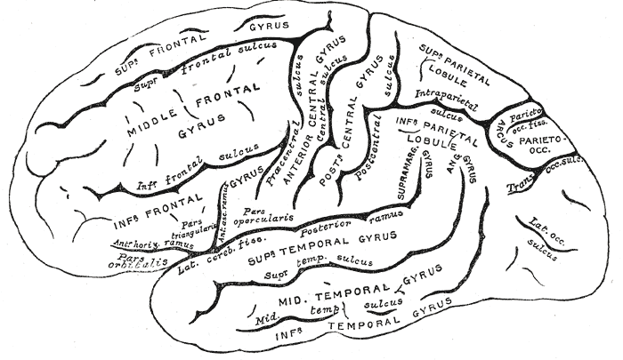
The precentral sulcus is a part of the human brain that lies parallel to, and in front of, the central sulcus. (A ''sulcus'' is one of the prominent grooves on the surface of the human brain.) The precentral sulcus divides the inferior, middle and superior frontal gyri from the precentral gyrus. In most brains, the precentral sulcus is divided into two parts: the inferior precentral sulcus and the superior precentral sulcus. However, the precentral sulcus may sometimes be divided into three parts or form one continuous sulcus. Additional images File:Precentral sulcus animation small.gif, Position of precentral sulcus (shown in red). File:FrontalCaptsLateral.png, Lateral surface of right frontal lobe The frontal lobe is the largest of the four major lobes of the brain in mammals, and is located at the front of each cerebral hemisphere (in front of the parietal lobe and the temporal lobe). It is parted from the parietal lobe by a groove be .... Precentral sulcus is lab ... [...More Info...] [...Related Items...] OR: [Wikipedia] [Google] [Baidu] |
Gyri
In neuroanatomy, a gyrus (pl. gyri) is a ridge on the cerebral cortex. It is generally surrounded by one or more sulci (depressions or furrows; sg. ''sulcus''). Gyri and sulci create the folded appearance of the brain in humans and other mammals. Structure The gyri are part of a system of folds and ridges that create a larger surface area for the human brain and other mammalian brains. Because the brain is confined to the skull, brain size is limited. Ridges and depressions create folds allowing a larger cortical surface area, and greater cognitive function, to exist in the confines of a smaller cranium. Development The human brain undergoes gyrification during fetal and neonatal development. In embryonic development, all mammalian brains begin as smooth structures derived from the neural tube. A cerebral cortex without surface convolutions is lissencephalic, meaning 'smooth-brained'. As development continues, gyri and sulci begin to take shape on the fetal brain, w ... [...More Info...] [...Related Items...] OR: [Wikipedia] [Google] [Baidu] |
Central Sulcus
In neuroanatomy, the central sulcus (also central fissure, fissure of Rolando, or Rolandic fissure, after Luigi Rolando) is a sulcus, or groove, in the cerebral cortex in the brains of vertebrates. It is sometimes confused with the longitudinal fissure. The central sulcus is a prominent landmark of the brain, separating the parietal lobe from the frontal lobe and the primary motor cortex from the primary somatosensory cortex. Evolution of the central sulcus The evolution of the central sulcus is theorized to have occurred in mammals when the complete dissociation of the original somatosensory cortex from its mirror duplicate developed in placental mammals such as primates, though the development did not stop there as time progressed the distinction between the two cortices grew. Evolution in primates The central sulcus is more prominent in apes as a result of fine-tuning of the motor system in apes. Hominins (bipedal apes) continued this trend through increased use of thei ... [...More Info...] [...Related Items...] OR: [Wikipedia] [Google] [Baidu] |
Paracentral Sulcus
The paracentral sulcus is a sulcus of the brain. It forms the paracentral lobule's anterior border. It is part of the cingulate sulcus. Gallery File:Gray727 cingulate sulcus.svg, Medial surface of left cerebral hemisphere. (Cingulate sulcus The cingulate sulcus is a sulcus (brain fold) on the cingulate cortex in the medial wall of the cerebral cortex. The frontal and parietal lobes are separated from the cingulate gyrus by the cingulate sulcus. It terminates as the marginal sulcus o ... shaded in red.) References External links * https://web.archive.org/web/20090505144011/http://anatomy.med.umich.edu/atlas/n1a2p12.html Cerebrum Sulci (neuroanatomy) {{neuroanatomy-stub ... [...More Info...] [...Related Items...] OR: [Wikipedia] [Google] [Baidu] |
Marginal Sulcus
In neuroanatomy, the marginal sulcus (margin of the cingulate sulcus) is a sulcus (crevice) that may be considered the termination of the cingulate sulcus. It separates the paracentral lobule anteriorly and the precuneus posteriorly. Additional images File:Marginal sulcus animation small.gif, Position of marginal sulcus (shown in red). File:Cingulate sulcus of Vervet Monkey.png, Transverse sections of brains of vervet monkey The vervet monkey (''Chlorocebus pygerythrus''), or simply vervet, is an Old World monkey of the family Cercopithecidae native to Africa. The term "vervet" is also used to refer to all the members of the genus ''Chlorocebus''. The five distinct .... It showing difference of the relative position of the left and right ascending ramus of the cingulate sulcus. References External links * https://web.archive.org/web/20070416083824/http://www.med.harvard.edu/AANLIB/cases/case3/mr1/033.html Parietal lobe Sulci (neuroanatomy) {{neuroanatom ... [...More Info...] [...Related Items...] OR: [Wikipedia] [Google] [Baidu] |
Cingulate Sulcus
The cingulate sulcus is a sulcus (brain fold) on the cingulate cortex in the medial wall of the cerebral cortex. The frontal and parietal lobes are separated from the cingulate gyrus by the cingulate sulcus. It terminates as the marginal sulcus of the cingulate sulcus. It sends a ramus to separate the paracentral lobule from the frontal gyri, the paracentral sulcus. Additional images File:Cingulate sulcus animation small.gif, Position of cingulate sulcus (shown in red). File:LobesCaptsMedial1.png, Medial surface of right cerebral hemisphere. Cingulate sulcus (labeled as sulcus cinguli) and brain lobes. File:Slide2ZEN.JPG, Medial surface of cerebral hemisphere.Medial view.Deep dissection. File:Slide3ZEN.JPG, Medial surface of cerebral hemisphere.Medial view.Deep dissection. File:Slide4ZE.JPG, Medial surface of cerebral hemisphere.Medial view.Deep dissection. External links * NIF Search - Cingulate Sulcusvia the Neuroscience Information Framework The Neuroscience Informatio ... [...More Info...] [...Related Items...] OR: [Wikipedia] [Google] [Baidu] |
Anterior Cerebral Artery
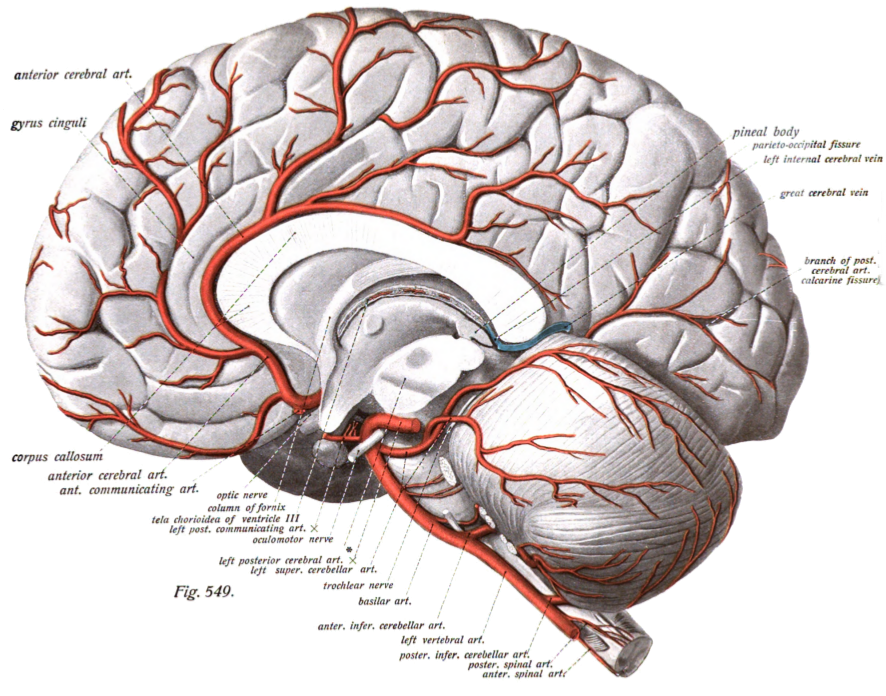
The anterior cerebral artery (ACA) is one of a pair of cerebral arteries that supplies oxygenated blood to most midline portions of the frontal lobes and superior medial parietal lobes of the brain. The two anterior cerebral arteries arise from the internal carotid artery and are part of the circle of Willis. The left and right anterior cerebral arteries are connected by the anterior communicating artery. Anterior cerebral artery syndrome refers to symptoms that follow a stroke occurring in the area normally supplied by one of the arteries. It is characterized by weakness and sensory loss in the lower leg and foot opposite to the lesion and behavioral changes. Structure The anterior cerebral artery is divided into 5 segments. Its smaller branches: the callosal (supracallosal) arteries are considered to be the A4 and A5 segments. *A1 originates from the internal carotid artery and extends to the ''anterior communicating artery'' (AComm). The ''anteromedial central'' (medial le ... [...More Info...] [...Related Items...] OR: [Wikipedia] [Google] [Baidu] |
Cytoarchitectonics
Cytoarchitecture (Greek '' κύτος''= "cell" + '' ἀρχιτεκτονική''= "architecture"), also known as cytoarchitectonics, is the study of the cellular composition of the central nervous system's tissues under the microscope. Cytoarchitectonics is one of the ways to parse the brain, by obtaining sections of the brain using a microtome and staining them with chemical agents which reveal where different neurons are located. The study of the parcellation of ''nerve fibers'' (primarily axons) into layers forms the subject of myeloarchitectonics ( History of the cerebral cytoarchitecture Defining cerebral cytoarchitecture began with the advent of —the science of slicing a ...[...More Info...] [...Related Items...] OR: [Wikipedia] [Google] [Baidu] |
Central Sulcus
In neuroanatomy, the central sulcus (also central fissure, fissure of Rolando, or Rolandic fissure, after Luigi Rolando) is a sulcus, or groove, in the cerebral cortex in the brains of vertebrates. It is sometimes confused with the longitudinal fissure. The central sulcus is a prominent landmark of the brain, separating the parietal lobe from the frontal lobe and the primary motor cortex from the primary somatosensory cortex. Evolution of the central sulcus The evolution of the central sulcus is theorized to have occurred in mammals when the complete dissociation of the original somatosensory cortex from its mirror duplicate developed in placental mammals such as primates, though the development did not stop there as time progressed the distinction between the two cortices grew. Evolution in primates The central sulcus is more prominent in apes as a result of fine-tuning of the motor system in apes. Hominins (bipedal apes) continued this trend through increased use of thei ... [...More Info...] [...Related Items...] OR: [Wikipedia] [Google] [Baidu] |
Somatosensory
In physiology, the somatosensory system is the network of neural structures in the brain and body that produce the perception of touch (haptic perception), as well as temperature (thermoception), body position (proprioception), and pain. It is a subset of the sensory nervous system, which also represents visual, auditory, olfactory, and gustatory stimuli. Somatosensation begins when mechano- and thermosensitive structures in the skin or internal organs sense physical stimuli such as pressure on the skin (see mechanotransduction, nociception). Activation of these structures, or receptors, leads to activation of peripheral sensory neurons that convey signals to the spinal cord as patterns of action potentials. Sensory information is then processed locally in the spinal cord to drive reflexes, and is also conveyed to the brain for conscious perception of touch and proprioception. Note, somatosensory information from the face and head enters the brain through peripheral se ... [...More Info...] [...Related Items...] OR: [Wikipedia] [Google] [Baidu] |
Paracentral Sulcus
The paracentral sulcus is a sulcus of the brain. It forms the paracentral lobule's anterior border. It is part of the cingulate sulcus. Gallery File:Gray727 cingulate sulcus.svg, Medial surface of left cerebral hemisphere. (Cingulate sulcus The cingulate sulcus is a sulcus (brain fold) on the cingulate cortex in the medial wall of the cerebral cortex. The frontal and parietal lobes are separated from the cingulate gyrus by the cingulate sulcus. It terminates as the marginal sulcus o ... shaded in red.) References External links * https://web.archive.org/web/20090505144011/http://anatomy.med.umich.edu/atlas/n1a2p12.html Cerebrum Sulci (neuroanatomy) {{neuroanatomy-stub ... [...More Info...] [...Related Items...] OR: [Wikipedia] [Google] [Baidu] |


