|
PAD1
Peptidyl arginine deiminase, type I, also known as PADI1, is a protein which in humans is encoded by the ''PADI1'' gene. This gene encodes a member of the peptidyl arginine deiminase family of enzymes, which catalyze the post-translational deimination of proteins by converting arginine residues into citrullines in the presence of calcium ions. The family members have distinct substrate specificities and tissue-specific expression patterns. The type I enzyme is involved in the late stages of epidermal differentiation, where it deiminates filaggrin and keratin K1, which maintains hydration of the stratum corneum, and hence the cutaneous barrier function. This enzyme may also play a role in hair follicle formation. This gene exists in a cluster with four other paralogous Sequence homology is the biological homology between DNA, RNA, or protein sequences, defined in terms of shared ancestry in the evolutionary history of life. Two segments of DNA can have shared ancestry becau ... [...More Info...] [...Related Items...] OR: [Wikipedia] [Google] [Baidu] |
Deimination
Citrullination or deimination is the conversion of the amino acid arginine in a protein into the amino acid citrulline. Citrulline is not one of the 20 standard amino acids encoded by DNA in the genetic code. Instead, it is the result of a post-translational modification. Citrullination is distinct from the formation of the free amino acid citrulline as part of the urea cycle or as a byproduct of enzymes of the nitric oxide synthase family. Enzymes called arginine deiminases (ADIs) catalyze the deimination of free arginine, while protein arginine deiminases or peptidylarginine deiminases (PADs) replace the primary ketimine group (>C=NH) by a ketone group (>C=O). Arginine is positively charged at a neutral pH, whereas citrulline has no net charge. This increases the hydrophobicity of the protein, which can lead to changes in protein folding, affecting the structure and function. The immune system can attack citrullinated proteins, leading to autoimmune diseases such as rheumatoid ... [...More Info...] [...Related Items...] OR: [Wikipedia] [Google] [Baidu] |
Protein
Proteins are large biomolecules and macromolecules that comprise one or more long chains of amino acid residues. Proteins perform a vast array of functions within organisms, including catalysing metabolic reactions, DNA replication, responding to stimuli, providing structure to cells and organisms, and transporting molecules from one location to another. Proteins differ from one another primarily in their sequence of amino acids, which is dictated by the nucleotide sequence of their genes, and which usually results in protein folding into a specific 3D structure that determines its activity. A linear chain of amino acid residues is called a polypeptide. A protein contains at least one long polypeptide. Short polypeptides, containing less than 20–30 residues, are rarely considered to be proteins and are commonly called peptides. The individual amino acid residues are bonded together by peptide bonds and adjacent amino acid residues. The sequence of amino acid residue ... [...More Info...] [...Related Items...] OR: [Wikipedia] [Google] [Baidu] |
Gene
In biology, the word gene (from , ; "...Wilhelm Johannsen coined the word gene to describe the Mendelian units of heredity..." meaning ''generation'' or ''birth'' or ''gender'') can have several different meanings. The Mendelian gene is a basic unit of heredity and the molecular gene is a sequence of nucleotides in DNA that is transcribed to produce a functional RNA. There are two types of molecular genes: protein-coding genes and noncoding genes. During gene expression, the DNA is first copied into RNA. The RNA can be directly functional or be the intermediate template for a protein that performs a function. The transmission of genes to an organism's offspring is the basis of the inheritance of phenotypic traits. These genes make up different DNA sequences called genotypes. Genotypes along with environmental and developmental factors determine what the phenotypes will be. Most biological traits are under the influence of polygenes (many different genes) as well as gen ... [...More Info...] [...Related Items...] OR: [Wikipedia] [Google] [Baidu] |
Peptidylarginine Deiminases
In enzymology, a protein-arginine deiminase () is an enzyme that catalyzes a form of post translational modification called arginine de-imination or citrullination: :protein L-arginine + H2O \rightleftharpoons protein L-citrulline + NH3 Thus, the two substrates of this enzyme are protein L-arginine (arginine residue inside a protein) and H2O, whereas its two products are protein L-citrulline and NH3: : This enzyme belongs to the family of hydrolases, those acting on carbon-nitrogen bonds other than peptide bonds, specifically in linear amidines. The systematic name of this enzyme class is protein-L-arginine iminohydrolase. This enzyme is also called peptidylarginine deiminase. Structural studies As of late 2007, seven structures have been solved for this class of enzymes, with PDB accession codes , , , , , , and . Mammalian proteins Mammals have 5 protein-arginine deiminases, with symbols *PADI1, PADI2, PADI3, PADI4, PADI6 except for rodent Rodents (from Lat ... [...More Info...] [...Related Items...] OR: [Wikipedia] [Google] [Baidu] |
Arginine
Arginine is the amino acid with the formula (H2N)(HN)CN(H)(CH2)3CH(NH2)CO2H. The molecule features a guanidino group appended to a standard amino acid framework. At physiological pH, the carboxylic acid is deprotonated (−CO2−) and both the amino and guanidino groups are protonated, resulting in a cation. Only the -arginine (symbol Arg or R) enantiomer is found naturally. Arg residues are common components of proteins. It is encoded by the codons CGU, CGC, CGA, CGG, AGA, and AGG. The guanidine group in arginine is the precursor for the biosynthesis of nitric oxide. Like all amino acids, it is a white, water-soluble solid. History Arginine was first isolated in 1886 from yellow lupin seedlings by the German chemist Ernst Schulze and his assistant Ernst Steiger. He named it from the Greek ''árgyros'' (ἄργυρος) meaning "silver" due to the silver-white appearance of arginine nitrate crystals. In 1897, Schulze and Ernst Winterstein (1865–1949) determined the structure ... [...More Info...] [...Related Items...] OR: [Wikipedia] [Google] [Baidu] |
Citrulline
The organic compound citrulline is an α-amino acid. Its name is derived from ''citrullus'', the Latin word for watermelon. Although named and described by gastroenterologists since the late 19th century, it was first isolated from watermelon in 1914 by Japanese researchers Yotaro Koga and Ryo OdakeEarly references spell Ryo Odake's name as ''Ryo Othake''. and further codified by Mitsunori Wada of Tokyo Imperial University in 1930. It has the formula H2NC(O)NH(CH2)3CH(NH2)CO2H. It is a key intermediate in the urea cycle, the pathway by which mammals excrete ammonia by converting it into urea. Citrulline is also produced as a byproduct of the enzymatic production of nitric oxide from the amino acid arginine, catalyzed by nitric oxide synthase. Biosynthesis Citrulline can be derived from: * from arginine via nitric oxide synthase, as a byproduct of the production of nitric oxide for signaling purposes * from ornithine through the breakdown of proline or glutamine/glutamate * from ... [...More Info...] [...Related Items...] OR: [Wikipedia] [Google] [Baidu] |
Epidermis (skin)
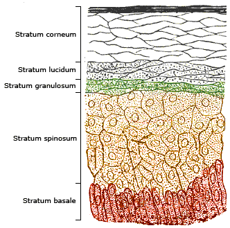
The epidermis is the outermost of the three layers that comprise the skin, the inner layers being the dermis and hypodermis. The epidermis layer provides a barrier to infection from environmental pathogens and regulates the amount of water released from the body into the atmosphere through transepidermal water loss. The epidermis is composed of multiple layers of flattened cells that overlie a base layer (stratum basale) composed of columnar cells arranged perpendicularly. The layers of cells develop from stem cells in the basal layer. The human epidermis is a familiar example of epithelium, particularly a stratified squamous epithelium. The word epidermis is derived through Latin , itself and . Something related to or part of the epidermis is termed epidermal. Structure Cellular components The epidermis primarily consists of keratinocytes ( proliferating basal and differentiated suprabasal), which comprise 90% of its cells, but also contains melanocytes, Langerhans c ... [...More Info...] [...Related Items...] OR: [Wikipedia] [Google] [Baidu] |
Filaggrin
Filaggrin (filament aggregating protein) is a filament-associated protein that binds to keratin fibers in epithelial cells. Ten to twelve filaggrin units are post-translationally hydrolyzed from a large profilaggrin precursor protein during terminal differentiation of epidermal cells. In humans, profilaggrin is encoded by the ''FLG'' gene, which is part of the S100 fused-type protein (SFTP) family within the epidermal differentiation complex on chromosome 1q21. Profilaggrin Filaggrin monomers are tandemly clustered into a large, 350kDa protein precursor known as profilaggrin. In the epidermis, these structures are present in the keratohyalin granules in cells of the stratum granulosum. Profilaggrin undergoes proteolytic processing to yield individual filaggrin monomers at the transition between the stratum granulosum and the stratum corneum, which may be facilitated by calcium-dependent enzymes. Structure Filaggrin is characterized by a particularly high isoelectric point due ... [...More Info...] [...Related Items...] OR: [Wikipedia] [Google] [Baidu] |
Keratin 1
Keratin 1 is a Type II intermediate filament (IFs) of the intracytoplasmatic cytoskeleton. Is co-expressed with and binds to Keratin 10, a Type I keratin, to form a coiled coil heterotypic keratin chain. Keratin 1 and Keratin 10 are specifically expressed in the spinous and granular layers of the epidermis. In contrast, basal layer keratinocytes express little to no Keratin 1. Mutations in ''KRT1'', the gene encoding Keratin 1, have been associated with variants of the disease bullous congenital ichthyosiform erythroderma in which the palms and soles of the feet are affected. Mutations in ''KRT10'' have also been associated with bullous congenital ichthyosiform erythroderma; however, in patients with KRT10 mutations the palms and soles are spared. This difference is likely due to Keratin 9, rather than Keratin 10, being the major binding partner of Keratin 1 in acral (palm and sole) keratinocytes. Type II cytokeratins are clustered in a region of chromosome 12q12-q13. Interactions ... [...More Info...] [...Related Items...] OR: [Wikipedia] [Google] [Baidu] |
Stratum Corneum
The stratum corneum (Latin for 'horny layer') is the outermost layer of the epidermis. The human stratum corneum comprises several levels of flattened corneocytes that are divided into two layers: the ''stratum disjunctum'' and ''stratum compactum''. The skin's protective acid mantle and lipid barrier sit on top of the stratum disjunctum. The stratum disjunctum is the uppermost and loosest layer of skin. The stratum compactum is the comparatively deeper, more compacted and more cohesive part of the stratum corneum. The corneocytes of the stratum disjunctum are larger, more rigid and more hydrophobic than that of the stratum compactum. The stratum corneum is the dead tissue that performs protective and adaptive physiological functions including mechanical shear, impact resistance, water flux and hydration regulation, microbial proliferation and invasion regulation, initiation of inflammation through cytokine activation and dendritic cell activity, and selective permeability to exc ... [...More Info...] [...Related Items...] OR: [Wikipedia] [Google] [Baidu] |
Hair Follicle
The hair follicle is an organ found in mammalian skin. It resides in the dermal layer of the skin and is made up of 20 different cell types, each with distinct functions. The hair follicle regulates hair growth via a complex interaction between hormones, neuropeptides, and immune cells. This complex interaction induces the hair follicle to produce different types of hair as seen on different parts of the body. For example, terminal hairs grow on the scalp and lanugo hairs are seen covering the bodies of fetuses in the uterus and in some newborn babies. The process of hair growth occurs in distinct sequential stages. The first stage is called ''anagen'' and is the active growth phase, ''telogen'' is the resting stage, ''catagen'' is the regression of the hair follicle phase, ''exogen'' is the active shedding of hair phase and lastly ''kenogen'' is the phase between the empty hair follicle and the growth of new hair. The function of hair in humans has long been a subject of interest ... [...More Info...] [...Related Items...] OR: [Wikipedia] [Google] [Baidu] |


