|
Ossifying Fibroma
Osteofibrous dysplasia is a rare, benign non-neoplastic condition with no known cause. It is considered a fibrovascular defect. Campanacci described this condition in two leg bones, the tibia and fibula, and coined the term. This condition should be differentiated from Nonossifying fibroma and fibrous dysplasia of bone. Presentation The tibia is the most commonly involved bone, accounting for 85% of cases. It is usually painless, although there may be localized pain or fracture, and presents as a localized firm swelling of the tibia in children less than two decades old (median age for males 10, females 13). Several authors have related this non-neoplastic lesion to adamantinoma - a tumor involving subcutaneous long bones - stating the common cause to be fibrovascular defect. However, the latter is distinguished from an osteofibrous dysplasia by the presence of soft tissue extension, intramedullary extension, periosteal reaction and presence of hyperchromic epithelial cells under ... [...More Info...] [...Related Items...] OR: [Wikipedia] [Google] [Baidu] |
Nonossifying Fibroma
A non-ossifying fibroma (NOF) is a benign bone tumor of the osteoclastic giant cell-rich tumor type. It generally occurs in the metaphysis of long bones in children and adolescents. Typically, there are no symptoms unless there is a fracture. It can occur as part of a syndrome such as when multiple non-ossifying fibromas occur in neurofibromatosis, or Jaffe-Campanacci syndrome in combination with cafe-au-lait spots, mental retardation, hypogonadism, eye and cardiovascular abnormalities. Diagnosis is by X-ray or MRI, usually when investigating a person for something else. Medical imaging typically shows a well defined radiolucent lesion, with a distinct multilocular appearance, sometimes looking like bubbles. It is usually around 1-2cm in size, but be as large as 7cm. They consist of foci consist of collagen rich connective tissue, fibroblasts, histiocytes and osteoclasts. Usually no treatment is required. Surgical curettage and bone grafting may be required if it is large. It ... [...More Info...] [...Related Items...] OR: [Wikipedia] [Google] [Baidu] |
Neoplasm
A neoplasm () is a type of abnormal and excessive growth of tissue. The process that occurs to form or produce a neoplasm is called neoplasia. The growth of a neoplasm is uncoordinated with that of the normal surrounding tissue, and persists in growing abnormally, even if the original trigger is removed. This abnormal growth usually forms a mass, when it may be called a tumor. ICD-10 classifies neoplasms into four main groups: benign neoplasms, in situ neoplasms, malignant neoplasms, and neoplasms of uncertain or unknown behavior. Malignant neoplasms are also simply known as cancers and are the focus of oncology. Prior to the abnormal growth of tissue, as neoplasia, cells often undergo an abnormal pattern of growth, such as metaplasia or dysplasia. However, metaplasia or dysplasia does not always progress to neoplasia and can occur in other conditions as well. The word is from Ancient Greek 'new' and 'formation, creation'. Types A neoplasm can be benign, potentially ma ... [...More Info...] [...Related Items...] OR: [Wikipedia] [Google] [Baidu] |
Tibia
The tibia (; ), also known as the shinbone or shankbone, is the larger, stronger, and anterior (frontal) of the two bones in the leg below the knee in vertebrates (the other being the fibula, behind and to the outside of the tibia); it connects the knee with the ankle. The tibia is found on the medial side of the leg next to the fibula and closer to the median plane. The tibia is connected to the fibula by the interosseous membrane of leg, forming a type of fibrous joint called a syndesmosis with very little movement. The tibia is named for the flute ''tibia''. It is the second largest bone in the human body, after the femur. The leg bones are the strongest long bones as they support the rest of the body. Structure In human anatomy, the tibia is the second largest bone next to the femur. As in other vertebrates the tibia is one of two bones in the lower leg, the other being the fibula, and is a component of the knee and ankle joints. The ossification or formation of the bone ... [...More Info...] [...Related Items...] OR: [Wikipedia] [Google] [Baidu] |
Fibula
The fibula or calf bone is a leg bone on the lateral side of the tibia, to which it is connected above and below. It is the smaller of the two bones and, in proportion to its length, the most slender of all the long bones. Its upper extremity is small, placed toward the back of the head of the tibia, below the knee joint and excluded from the formation of this joint. Its lower extremity inclines a little forward, so as to be on a plane anterior to that of the upper end; it projects below the tibia and forms the lateral part of the ankle joint. Structure The bone has the following components: * Lateral malleolus * Interosseous membrane connecting the fibula to the tibia, forming a syndesmosis joint * The superior tibiofibular articulation is an arthrodial joint between the lateral condyle of the tibia and the head of the fibula. * The inferior tibiofibular articulation (tibiofibular syndesmosis) is formed by the rough, convex surface of the medial side of the lower end of the f ... [...More Info...] [...Related Items...] OR: [Wikipedia] [Google] [Baidu] |
Fibrous Dysplasia Of Bone
Fibrous dysplasia is a disorder where normal bone and marrow is replaced with fibrous tissue, resulting in formation of bone that is weak and prone to expansion. As a result, most complications result from fracture, deformity, functional impairment, and pain. Disease occurs along a broad clinical spectrum ranging from asymptomatic, incidental lesions, to severe disabling disease. Disease can affect one bone ( monostotic), multiple ( polyostotic), or all bones (panostotic) and may occur in isolation or in combination with café au lait skin macules and hyperfunctioning endocrinopathies, termed McCune–Albright syndrome. More rarely, fibrous dysplasia may be associated with intramuscular myxomas, termed Mazabraud's syndrome. Fibrous dysplasia is very rare, and there is no known cure. Fibrous dysplasia is not a form of cancer. Presentation Fibrous dysplasia is a mosaic disease that can involve any part or combination of the craniofacial, axillary, and/or appendicular skeleton. The ... [...More Info...] [...Related Items...] OR: [Wikipedia] [Google] [Baidu] |
Adamantinoma
Adamantinoma (from the Greek word ''adamantinos'', meaning "very hard") is a rare bone cancer, making up less than 1% of all bone cancers. It almost always occurs in the bones of the lower leg and involves both epithelial and osteofibrous tissue. The condition was first described by Fischer in 1913. Presentation Patients typically present with swelling with or without pain. The slow-growing tumor predominantly arises in long bones in a subcortical location (95% in the tibia or fibula). Most commonly, patients are in their second or third decade, but adamantinoma can occur over a wide age range. Benign osteofibrous dysplasia may be a precursor of adamantinoma or a regressive phase of adamantinoma. Histologically, islands of epithelial cells are found in a fibrous stroma. The tumor is typically well-demarcated, osteolytic and eccentric, with cystic zones resembling soap bubbles. Diagnosis Diagnosis is on plain radiography, or CT scan Treatment Treatment consists of wide rese ... [...More Info...] [...Related Items...] OR: [Wikipedia] [Google] [Baidu] |
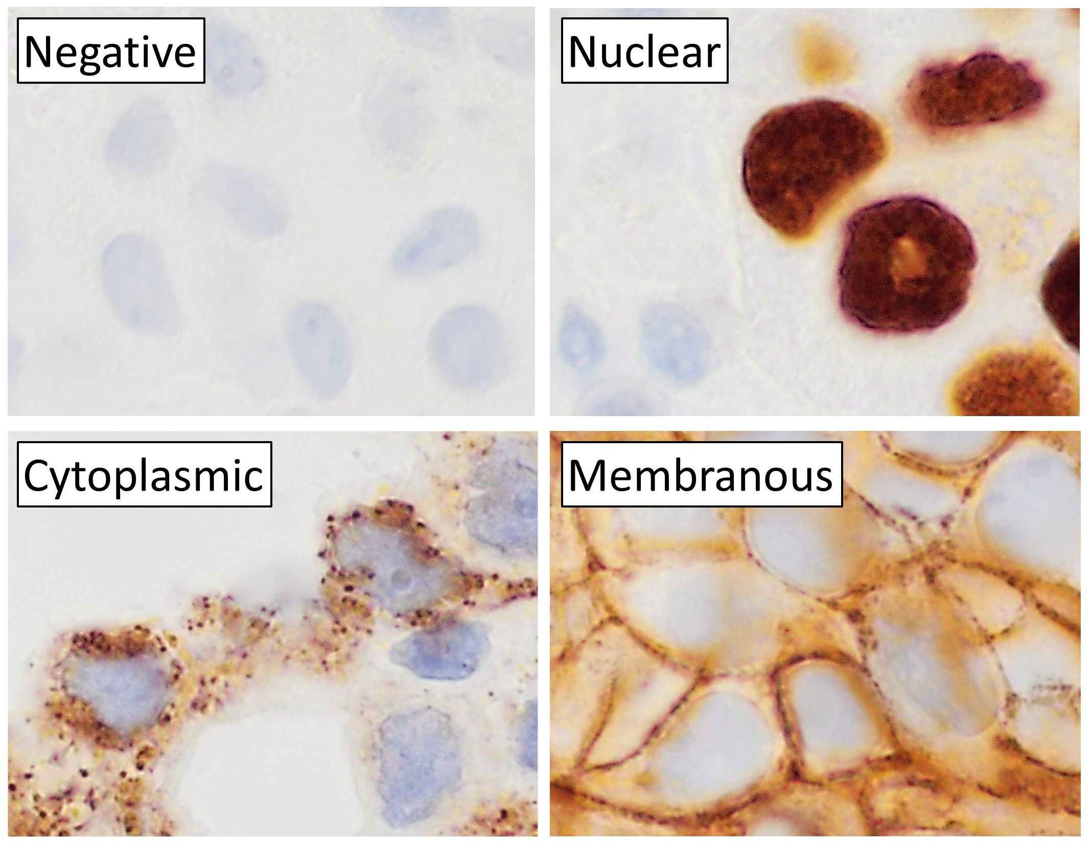
Immunohistochemistry
Immunohistochemistry (IHC) is the most common application of immunostaining. It involves the process of selectively identifying antigens (proteins) in cells of a tissue section by exploiting the principle of antibodies binding specifically to antigens in biological tissues. IHC takes its name from the roots "immuno", in reference to antibodies used in the procedure, and "histo", meaning tissue (compare to immunocytochemistry). Albert Coons conceptualized and first implemented the procedure in 1941. Visualising an antibody-antigen interaction can be accomplished in a number of ways, mainly either of the following: * ''Chromogenic immunohistochemistry'' (CIH), wherein an antibody is conjugated to an enzyme, such as peroxidase (the combination being termed immunoperoxidase), that can catalyse a colour-producing reaction. * '' Immunofluorescence'', where the antibody is tagged to a fluorophore, such as fluorescein or rhodamine. Immunohistochemical staining is widely used in the dia ... [...More Info...] [...Related Items...] OR: [Wikipedia] [Google] [Baidu] |
Osteonectin
Osteonectin (ON) also known as secreted protein acidic and rich in cysteine (SPARC) or basement-membrane protein 40 (BM-40) is a protein that in humans is encoded by the ''SPARC'' gene. Osteonectin is a glycoprotein in the bone that binds calcium. It is secreted by osteoblasts during bone formation, initiating mineralization and promoting mineral crystal formation. Osteonectin also shows affinity for collagen in addition to bone mineral calcium. A correlation between osteonectin over-expression and ampullary cancers and chronic pancreatitis has been found. Gene The human SPARC gene is 26.5 kb long, and contains 10 exons and 9 introns and is located on chromosome 5q31-q33. Structure Osteonectin is a 40 kDa acidic and cysteine-rich glycoprotein consisting of a single polypeptide chain that can be broken into 4 domains: 1) a Ca2+ binding domain near the glutamic acid-rich region at the amino terminus (domain I), 2) a cysteine-rich domain (II), 3) a hydrophilic region ( ... [...More Info...] [...Related Items...] OR: [Wikipedia] [Google] [Baidu] |
Neurofibromin 1
Neurofibromin 1 (''NF1'') is a gene in humans that is located on chromosome 17. ''NF1'' codes for neurofibromin, a GTPase-activating protein that negatively regulates RAS/MAPK pathway activity by accelerating the hydrolysis of Ras-bound GTP. ''NF1'' has a high mutation rate and mutations in ''NF1'' can alter cellular growth control, and neural development, resulting in neurofibromatosis type 1 (NF1, also known as von Recklinghausen syndrome). Symptoms of NF1 include disfiguring cutaneous neurofibromas (CNF), café au lait pigment spots, plexiform neurofibromas (PN), skeletal defects, optic nerve gliomas, life-threatening malignant peripheral nerve sheath tumors (MPNST), pheochromocytoma, attention deficits, learning deficits and other cognitive disabilities. Gene ''NF1'' was cloned in 1990 and its gene product neurofibromin was identified in 1992. Neurofibromin, a GTPase-activating protein, primarily regulates the protein Ras. ''NF1'' is located on the long arm of chromosom ... [...More Info...] [...Related Items...] OR: [Wikipedia] [Google] [Baidu] |
S-100 Protein
The S100 proteins are a family of low molecular-weight proteins found in vertebrates characterized by two calcium-binding sites that have helix-loop-helix ("EF-hand-type") conformation. At least 21 different S100 proteins are known. They are encoded by a family of genes whose symbols use the ''S100'' prefix, for example, ''S100A1'', ''S100A2'', ''S100A3''. They are also considered as damage-associated molecular pattern molecules (DAMPs), and knockdown of aryl hydrocarbon receptor downregulates the expression of S100 proteins in THP-1 cells. Structure Most S100 proteins consist of two identical polypeptides (homodimeric), which are held together by noncovalent bonds. They are structurally similar to calmodulin. They differ from calmodulin, though, on the other features. For instance, their expression pattern is cell-specific, i.e. they are expressed in particular cell types. Their expression depends on environmental factors. In contrast, calmodulin is a ubiquitous and univer ... [...More Info...] [...Related Items...] OR: [Wikipedia] [Google] [Baidu] |
Osteopathies
Osteopathic medicine is a branch of the medical profession in the United States that promotes the practice of allopathic medicine with a set of philosophy and principles set by its earlier form, osteopathy. Osteopathic physicians (DOs) are licensed to practice medicine and surgery in all 50 US states. Only graduates of American osteopathic medical colleges may practice the full scope of medicine and surgery generally considered to be medicine by the general public; US DO graduates have historically applied for medical licensure in 87 countries outside of the United States, 85 of which provided them with the full scope of medical and surgical practice. The field is distinct from osteopathic practices offered in nations outside of the U.S., whose practitioners are generally not considered part of core medical staff nor of medicine itself. The other major branch of medicine in the United States is referred to by practitioners of osteopathic medicine as allopathic medicine. By the ... [...More Info...] [...Related Items...] OR: [Wikipedia] [Google] [Baidu] |


