|
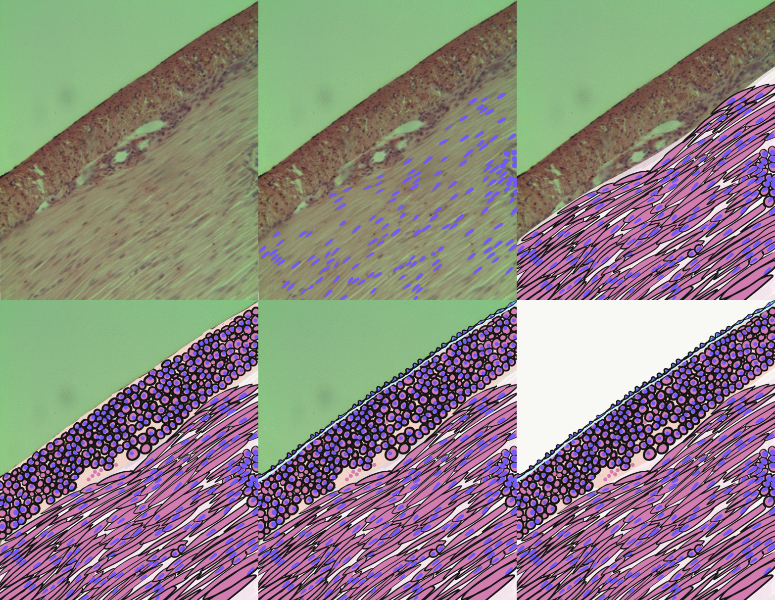
Orbitalis Muscle
The orbitalis muscle is a vestigial or rudimentary nonstriated muscle (smooth muscle) that crosses from the infraorbital groove and sphenomaxillary fissure and is intimately united with the periosteum of the orbit. It was described by Heinrich Müller (physiologist), Heinrich Müller and is often called Müller's muscle. It lies at the back of the orbit and spans the infraorbital fissure.Gray's Anatomy - 40th Ed/MINOR MUSCLES OF THE EYELIDS It is a thin layer of smooth muscle that bridges the inferior orbital fissure. It is supplied by sympathetic nerves, and its function is unknown. Function The muscle forms an important part of the lateral orbital wall in some animals and can act to change the wall's volume in lower mammals, while in humans it is not known to have any significant function, but its contraction may possibly produce a slight forward protrusion of the eyeball. Several sources have suggested a role in the autonomic regulation of the vascular system due to the patte ... [...More Info...] [...Related Items...] OR: [Wikipedia] [Google] [Baidu] |
Superior Tarsal Muscle
The superior tarsal muscle is a smooth muscle adjoining the levator palpebrae superioris muscle that helps to raise the upper eyelid. Structure The superior tarsal muscle originates on the underside of levator palpebrae superioris and inserts on the superior tarsal plate of the eyelid. Nerve supply The superior tarsal muscle receives its innervation from the sympathetic nervous system. Postganglionic sympathetic fibers originate in the superior cervical ganglion, and travel via the internal carotid plexus, where small branches communicate with the oculomotor nerve as it passes through the cavernous sinus. The sympathetic fibres continue to the superior division of the oculomotor nerve, where they enter the superior tarsal muscle on its inferior aspect. Function Its role is not fully clear, but may be an accessory muscle to raise the upper eyelid. Clinical significance Damage to some elements of the sympathetic nervous system can inhibit this muscle, causing a drooping ey ... [...More Info...] [...Related Items...] OR: [Wikipedia] [Google] [Baidu] |
Ciliary Muscle
The ciliary muscle is an intrinsic muscle of the eye formed as a ring of smooth muscleSchachar, Ronald A. (2012). "Anatomy and Physiology." (Chapter 4) . in the eye's middle layer, uvea ( vascular layer). It controls accommodation for viewing objects at varying distances and regulates the flow of aqueous humor into Schlemm's canal. It also changes the shape of the lens within the eye but not the size of the pupil which is carried out by the sphincter pupillae muscle and dilator pupillae. Structure Development The ciliary muscle develops from mesenchyme within the choroid and is considered a cranial neural crest derivative.Dudek RW, Fix JD (2004). "Eye" (chapter 9). ''Embryology - Board Review Series'' (3rd edition, illustrated). Lippincott Williams & Wilkins. p. 92. , . Books.Google.com. Retrieved on 2010-01-17 from https://books.google.com/books?id=MmoJQWsJteoC. Nerve supply The ciliary muscle receives parasympathetic fibers from the short ciliary nerves that arise fro ... [...More Info...] [...Related Items...] OR: [Wikipedia] [Google] [Baidu] |
Smooth Muscle
Smooth muscle is an involuntary non-striated muscle, so-called because it has no sarcomeres and therefore no striations (''bands'' or ''stripes''). It is divided into two subgroups, single-unit and multiunit smooth muscle. Within single-unit muscle, the whole bundle or sheet of smooth muscle cells contracts as a syncytium. Smooth muscle is found in the walls of hollow organs, including the stomach, intestines, bladder and uterus; in the walls of passageways, such as blood, and lymph vessels, and in the tracts of the respiratory, urinary, and reproductive systems. In the eyes, the ciliary muscles, a type of smooth muscle, dilate and contract the iris and alter the shape of the lens. In the skin, smooth muscle cells such as those of the arrector pili cause hair to stand erect in response to cold temperature or fear. Structure Gross anatomy Smooth muscle is grouped into two types: single-unit smooth muscle, also known as visceral smooth muscle, and multiunit smooth muscle. ... [...More Info...] [...Related Items...] OR: [Wikipedia] [Google] [Baidu] |
Infraorbital Groove
The infraorbital groove (or sulcus) is located in the middle of the posterior part of the orbital surface of the maxilla. Its function is to act as the passage of the infraorbital artery, the infraorbital vein, and the infraorbital nerve. Structure The infraorbital groove begins at the middle of the posterior border of the maxilla (with which it is continuous). This is near the upper edge of the infratemporal surface of the maxilla. It passes forward, and ends in a canal which subdivides into two branches. The infraorbital groove has an average length of 16.7 mm, with a small amount of variation between people. It is similar in men and women. Function The infraorbital groove creates space that allows for passage of the infraorbital artery, the infraorbital vein, and the infraorbital nerve. Clinical significance The infraorbital groove is an important surgical landmark for local anaesthesia of the infraorbital nerve. See also * Infraorbital foramen In human anatomy ... [...More Info...] [...Related Items...] OR: [Wikipedia] [Google] [Baidu] |
Sphenomaxillary Fissure
The inferior orbital fissure is formed by the sphenoid bone and the maxilla. It is located posteriorly along the boundary of the floor and lateral wall of the orbit. It transmits a number of structures, including: * the zygomatic branch of the maxillary nerve * the ascending branches from the pterygopalatine ganglion * the infraorbital vessels, which travel down the infraorbital groove into the infraorbital canal and exit through the infraorbital foramen * the inferior division of the ophthalmic vein Images File:Gray189.png, Left infratemporal fossa. File:Gray191.png, Horizontal section of nasal and orbital cavities. File:Gray787.png, Dissection showing origins of right ocular muscles, and nerves entering by the superior orbital fissure. File:Slide2rome.JPG, Inferior orbital fissure. See also *Foramina of skull *Superior orbital fissure The superior orbital fissure is a foramen or cleft of the skull between the lesser and greater wings of the sphenoid bone. It gives ... [...More Info...] [...Related Items...] OR: [Wikipedia] [Google] [Baidu] |
Periosteum
The periosteum is a membrane that covers the outer surface of all bones, except at the articular surfaces (i.e. the parts within a joint space) of long bones. Endosteum lines the inner surface of the medullary cavity of all long bones. Structure The periosteum consists of an outer fibrous layer, and an inner cambium layer (or osteogenic layer). The fibrous layer is of dense irregular connective tissue, containing fibroblasts, while the cambium layer is highly cellular containing progenitor cells that develop into osteoblasts. These osteoblasts are responsible for increasing the width of a long bone and the overall size of the other bone types. After a bone fracture, the progenitor cells develop into osteoblasts and chondroblasts, which are essential to the healing process. The outer fibrous layer and the inner cambium layer is differentiated under electron micrography. As opposed to osseous tissue, the periosteum has nociceptors, sensory neurons that make it very sensitive to ... [...More Info...] [...Related Items...] OR: [Wikipedia] [Google] [Baidu] |
Heinrich Müller (physiologist)
Heinrich Müller (17 December 1820 – 10 May 1864) was a German anatomist and professor at the University of Würzburg. He is best known for his work in comparative anatomy and his studies involving the eye. He was a native of Castell, Lower Franconia. He was a student at several universities, being influenced by Ignaz Dollinger (1770–1841) in Munich, Friedrich Arnold (1803–1890) in Freiburg, Jakob Henle (1809–1895) in Heidelberg and Carl von Rokitansky (1804–1878) in Vienna. In 1847 he received his habilitation at Würzburg, where from 1858 he served as a full professor of topographical and comparative anatomy. As an instructor, he also taught classes in systematic anatomy, histology and microscopy. In 1851 Müller noticed the red color in rod cells now known as rhodopsin or visual purple, which is a pigment that is present in the rods of the retina. However, Franz Christian Boll (1849–1879) is credited as the discoverer of rhodopsin because he was able to ... [...More Info...] [...Related Items...] OR: [Wikipedia] [Google] [Baidu] |
Horner's Syndrome
Horner's syndrome, also known as oculosympathetic paresis, is a combination of symptoms that arises when a group of nerves known as the sympathetic trunk is damaged. The signs and symptoms occur on the same side (ipsilateral) as it is a lesion of the sympathetic trunk. It is characterized by miosis (a constricted pupil), partial ptosis (a weak, droopy eyelid), apparent anhidrosis (decreased sweating), with apparent enophthalmos (inset eyeball). The nerves of the sympathetic trunk arise from the spinal cord in the chest, and from there ascend to the neck and face. The nerves are part of the sympathetic nervous system, a division of the autonomic (or involuntary) nervous system. Once the syndrome has been recognized, medical imaging and response to particular eye drops may be required to identify the location of the problem and the underlying cause. Signs and symptoms Signs that are found in people with Horner's syndrome on the affected side of the face include the following: * ... [...More Info...] [...Related Items...] OR: [Wikipedia] [Google] [Baidu] |
Enophthalmos
Enophthalmos is a posterior displacement of the eyeball within the orbit. It is due to either enlargement of the bony orbit and/or reduction of the orbital content, this in relation to each other. It should not be confused with its opposite, exophthalmos, which is the anterior displacement of the eye. It may be a congenital anomaly, or be acquired as a result of trauma (such as in a blowout fracture of the orbit), Horner's syndrome (apparent enophthalmos due to ptosis), Marfan syndrome, Duane's syndrome, silent sinus syndrome or phthisis bulbi Phthisis bulbi is a shrunken, non-functional eye. It may result from severe eye disease, inflammation or injury, or it may represent a complication of eye surgery. Treatment options include insertion of a prosthesis, which may be preceded by e .... References Further reading * External links Disorders of eyelid, lacrimal system and orbit {{med-sign-stub ... [...More Info...] [...Related Items...] OR: [Wikipedia] [Google] [Baidu] |
Horner's Syndrome
Horner's syndrome, also known as oculosympathetic paresis, is a combination of symptoms that arises when a group of nerves known as the sympathetic trunk is damaged. The signs and symptoms occur on the same side (ipsilateral) as it is a lesion of the sympathetic trunk. It is characterized by miosis (a constricted pupil), partial ptosis (a weak, droopy eyelid), apparent anhidrosis (decreased sweating), with apparent enophthalmos (inset eyeball). The nerves of the sympathetic trunk arise from the spinal cord in the chest, and from there ascend to the neck and face. The nerves are part of the sympathetic nervous system, a division of the autonomic (or involuntary) nervous system. Once the syndrome has been recognized, medical imaging and response to particular eye drops may be required to identify the location of the problem and the underlying cause. Signs and symptoms Signs that are found in people with Horner's syndrome on the affected side of the face include the following: * ... [...More Info...] [...Related Items...] OR: [Wikipedia] [Google] [Baidu] |
Ptosis (eyelid)
Ptosis, also known as blepharoptosis, is a drooping or falling of the upper eyelid. The drooping may be worse after being awake longer when the individual's muscles are tired. This condition is sometimes called "lazy eye", but that term normally refers to the condition amblyopia. If severe enough and left untreated, the drooping eyelid can cause other conditions, such as amblyopia or astigmatism. This is why it is especially important for this disorder to be treated in children at a young age, before it can interfere with vision development. The term is from Greek 'fall, falling'. Signs and symptoms Signs and symptoms typically seen in this condition include: * The eyelid(s) may appear to droop. * Droopy eyelids can give the face a false appearance of being fatigued, disinterested, or even sinister. * The eyelid may not protect the eye as effectively, allowing it to dry out. * Sagging upper eyelids can partially block the person's field of view. * Obstructed vision may cause ... [...More Info...] [...Related Items...] OR: [Wikipedia] [Google] [Baidu] |
Superior Tarsal Muscle
The superior tarsal muscle is a smooth muscle adjoining the levator palpebrae superioris muscle that helps to raise the upper eyelid. Structure The superior tarsal muscle originates on the underside of levator palpebrae superioris and inserts on the superior tarsal plate of the eyelid. Nerve supply The superior tarsal muscle receives its innervation from the sympathetic nervous system. Postganglionic sympathetic fibers originate in the superior cervical ganglion, and travel via the internal carotid plexus, where small branches communicate with the oculomotor nerve as it passes through the cavernous sinus. The sympathetic fibres continue to the superior division of the oculomotor nerve, where they enter the superior tarsal muscle on its inferior aspect. Function Its role is not fully clear, but may be an accessory muscle to raise the upper eyelid. Clinical significance Damage to some elements of the sympathetic nervous system can inhibit this muscle, causing a drooping ey ... [...More Info...] [...Related Items...] OR: [Wikipedia] [Google] [Baidu] |


