|
Oncocytoma
An oncocytoma is a tumor made up of oncocytes, epithelial cells characterized by an excessive amount of mitochondria, resulting in an abundant acidophilic, granular cytoplasm. The cells and the tumor that they compose are often benign but sometimes may be premalignant or malignant. Presentation An oncocytoma is an epithelial tumor composed of oncocytes, large eosinophilic cells having small, round, benign-appearing nuclei with large nucleoli. Oncocytoma can arise in a number of organs. Renal oncocytoma Renal oncocytoma is thought to arise from the intercalated cells of collecting ducts of the kidney. It represents 5% to 15% of surgically resected renal neoplasms. Salivary gland oncocytoma The salivary gland oncocytoma is a well-circumscribed, benign neoplastic growth also called an oxyphilic adenoma. It comprises about 1% of all salivary gland tumors. The histopathology is marked by sheets of large swollen polyhedral epithelial oncocytes, which are granular acidophilic parot ... [...More Info...] [...Related Items...] OR: [Wikipedia] [Google] [Baidu] |
Renal Oncocytoma
A renal oncocytoma is a tumour of the kidney made up of oncocytes, epithelial cells with an excess amount of mitochondria. Signs and symptoms Renal oncocytomas are often asymptomatic and are frequently discovered by chance on a CT or ultrasound of the abdomen. Possible signs and symptoms of a renal oncocytoma include blood in the urine, flank pain, and an abdominal mass. Pathophysiology Renal oncocytoma is thought to arise from the intercalated cells of collecting ducts of the kidney. It represent 5% to 15% of surgically resected renal neoplasms. Ultrastructurally, the eosinophilic cells have numerous mitochondria. Histologic appearance An oncocytoma is an epithelial tumor composed of oncocytes, large eosinophilic cells having small, round, benign-appearing nuclei with large nucleoli and excessive amounts of mitochondria. Diagnosis In gross appearance, the tumors are tan or mahogany brown, well circumscribed and contain a central scar. They may achieve a large size (up to ... [...More Info...] [...Related Items...] OR: [Wikipedia] [Google] [Baidu] |
Oncocytoma Of The Salivary Gland
An oncocytoma is a tumor made up of oncocytes, epithelial cells characterized by an excessive amount of mitochondria, resulting in an abundant acidophilic, granular cytoplasm. The cells and the tumor that they compose are often benign but sometimes may be premalignant or malignant. Presentation An oncocytoma is an epithelial tumor composed of oncocytes, large eosinophilic cells having small, round, benign-appearing nuclei with large nucleoli. Oncocytoma can arise in a number of organs. Renal oncocytoma Renal oncocytoma is thought to arise from the intercalated cells of collecting ducts of the kidney. It represents 5% to 15% of surgically resected renal neoplasms. Salivary gland oncocytoma The salivary gland oncocytoma is a well-circumscribed, benign neoplastic growth also called an oxyphilic adenoma. It comprises about 1% of all salivary gland tumors. The histopathology is marked by sheets of large swollen polyhedral epithelial oncocytes, which are granular acidophilic parot ... [...More Info...] [...Related Items...] OR: [Wikipedia] [Google] [Baidu] |
Oncocyte
An oncocyte is an epithelial cell characterized by an excessive number of mitochondria, resulting in an abundant acidophilic, granular cytoplasm. Oncocytes can be benign or malignant. Other names Also known as: *'' Hürthle cell'' ( thyroid gland only) *'' Oxyphilic cell'' *''Askanazy cell'' *''Apocrine metaplasia'' (breast gland only). *''Oncocytic cell'' Etymology Derived from the Greek root onco-, which means mass, bulk. See also * Hurthle cell carcinoma, a variant of follicular thyroid carcinoma. *Oncocytoma, a tumour composed of oncocytes, may be found as a less common salivary gland neoplasm also known as oxyphilic adenoma. *Renal oncocytoma A renal oncocytoma is a tumour of the kidney made up of oncocytes, epithelial cells with an excess amount of mitochondria. Signs and symptoms Renal oncocytomas are often asymptomatic and are frequently discovered by chance on a CT or ultrasound ..., a kidney tumour composed of oncocytes. References {{Reflist External linksWo ... [...More Info...] [...Related Items...] OR: [Wikipedia] [Google] [Baidu] |
Kidney Cancer
Kidney cancer, also known as renal cancer, is a group of cancers that starts in the kidney. Symptoms may include blood in the urine, lump in the abdomen, or back pain. Fever, weight loss, and tiredness may also occur. Complications can include spread to the lungs or brain. The main types of kidney cancer are renal cell cancer (RCC), transitional cell cancer (TCC), and Wilms tumor. RCC makes up approximately 80% of kidney cancers, and TCC accounts for most of the rest. Risk factors for RCC and TCC include smoking, certain pain medications, previous bladder cancer, being overweight, high blood pressure, certain chemicals, and a family history. Risk factors for Wilms tumor include a family history and certain genetic disorders such as WAGR syndrome. Diagnosis maybe suspected based on symptoms, urine testing, and medical imaging. It is confirmed by tissue biopsy. Treatment may include surgery, radiation therapy, chemotherapy, immunotherapy, and targeted therapy. Kidney cancer n ... [...More Info...] [...Related Items...] OR: [Wikipedia] [Google] [Baidu] |
Gross Examination
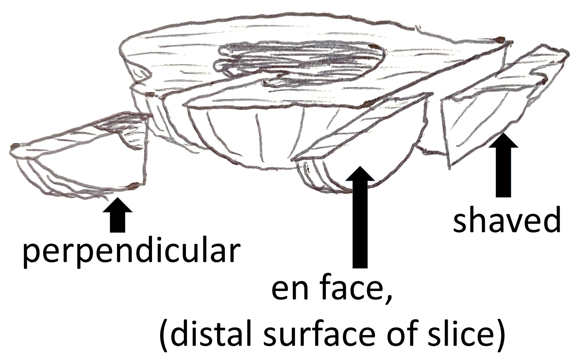
Gross processing or "grossing" is the process by which pathology specimens undergo examination with the bare eye to obtain diagnostic information, as well as cutting and tissue sampling in order to prepare material for subsequent microscopic ''examination.'' Responsibility Gross examination of surgical specimens is typically performed by a pathologist, or by a pathologists' assistant working within a pathology practice. Individuals trained in these fields are often able to gather diagnostically critical information in this stage of processing, including the stage and margin status of surgically removed tumors. Steps The initial step in any examination of a clinical specimen is confirmation of the identity of the patient and the anatomical site from which the specimen was obtained. Sufficient clinical data should be communicated by the clinical team to the pathology team in order to guide the appropriate diagnostic examination and interpretation of the specimen - if such informat ... [...More Info...] [...Related Items...] OR: [Wikipedia] [Google] [Baidu] |
Renal Cell Carcinoma
Renal cell carcinoma (RCC) is a kidney cancer that originates in the lining of the Proximal tubule, proximal convoluted tubule, a part of the very small tubes in the kidney that transport primary urine. RCC is the most common type of kidney cancer in adults, responsible for approximately 90–95% of cases. RCC occurrence shows a male predominance over women with a ratio of 1.5:1. RCC most commonly occurs between 6th and 7th decade of life. Initial treatment is most commonly either partial or complete removal of the affected kidney(s). Where the cancer has not metastasised (spread to other organs) or burrowed deeper into the tissues of the kidney, the five-year survival rate is 65–90%, but this is lowered considerably when the cancer has spread. The body is remarkably good at hiding the symptoms and as a result people with RCC often have advanced disease by the time it is discovered. The initial symptoms of RCC often include haematuria, blood in the urine (occurring in 40% of af ... [...More Info...] [...Related Items...] OR: [Wikipedia] [Google] [Baidu] |
Adenomas
An adenoma is a benign tumor of epithelial tissue with glandular origin, glandular characteristics, or both. Adenomas can grow from many glandular organs, including the adrenal glands, pituitary gland, thyroid, prostate, and others. Some adenomas grow from epithelial tissue in nonglandular areas but express glandular tissue structure (as can happen in familial polyposis coli). Although adenomas are benign, they should be treated as pre-cancerous. Over time adenomas may transform to become malignant, at which point they are called adenocarcinomas. Most adenomas do not transform. However, even though benign, they have the potential to cause serious health complications by compressing other structures ( mass effect) and by producing large amounts of hormones in an unregulated, non-feedback-dependent manner (causing paraneoplastic syndromes). Some adenomas are too small to be seen macroscopically but can still cause clinical symptoms. Histopathology Adenoma is a benign tumor o ... [...More Info...] [...Related Items...] OR: [Wikipedia] [Google] [Baidu] |
Renal
The kidneys are two reddish-brown bean-shaped organs found in vertebrates. They are located on the left and right in the retroperitoneal space, and in adult humans are about in length. They receive blood from the paired renal arteries; blood exits into the paired renal veins. Each kidney is attached to a ureter, a tube that carries excreted urine to the bladder. The kidney participates in the control of the volume of various body fluids, fluid osmolality, acid–base balance, various electrolyte concentrations, and removal of toxins. Filtration occurs in the glomerulus: one-fifth of the blood volume that enters the kidneys is filtered. Examples of substances reabsorbed are solute-free water, sodium, bicarbonate, glucose, and amino acids. Examples of substances secreted are hydrogen, ammonium, potassium and uric acid. The nephron is the structural and functional unit of the kidney. Each adult human kidney contains around 1 million nephrons, while a mouse kidney con ... [...More Info...] [...Related Items...] OR: [Wikipedia] [Google] [Baidu] |
Kidney
The kidneys are two reddish-brown bean-shaped organs found in vertebrates. They are located on the left and right in the retroperitoneal space, and in adult humans are about in length. They receive blood from the paired renal arteries; blood exits into the paired renal veins. Each kidney is attached to a ureter, a tube that carries excreted urine to the bladder. The kidney participates in the control of the volume of various body fluids, fluid osmolality, acid–base balance, various electrolyte concentrations, and removal of toxins. Filtration occurs in the glomerulus: one-fifth of the blood volume that enters the kidneys is filtered. Examples of substances reabsorbed are solute-free water, sodium, bicarbonate, glucose, and amino acids. Examples of substances secreted are hydrogen, ammonium, potassium and uric acid. The nephron is the structural and functional unit of the kidney. Each adult human kidney contains around 1 million nephrons, while a mouse kidney con ... [...More Info...] [...Related Items...] OR: [Wikipedia] [Google] [Baidu] |
Nephrectomy
A nephrectomy is the surgical removal of a kidney, performed to treat a number of kidney diseases including kidney cancer. It is also done to remove a normal healthy kidney from a living or deceased donor, which is part of a kidney transplant procedure. History The first recorded nephrectomy was performed in 1861 by Erastus B. Wolcott in Wisconsin. The patient had had a large tumor and the operation was initially successful, but the patient died fifteen days later. The first planned nephrectomy was performed by the German surgeon Gustav Simon on August 2, 1869, in Heidelberg. Simon practiced the operation beforehand in animal experiments. He proved that one healthy kidney can be sufficient for urine excretion in humans. Indications There are various indications for this procedure, including renal cell carcinoma, a non-functioning kidney (which may cause high blood pressure) and a congenitally small kidney (in which the kidney is swelling, causing it to press on nerves, which ... [...More Info...] [...Related Items...] OR: [Wikipedia] [Google] [Baidu] |
Carcinomas
Carcinoma is a malignancy that develops from epithelial cells. Specifically, a carcinoma is a cancer that begins in a tissue that lines the inner or outer surfaces of the body, and that arises from cells originating in the endodermal, mesodermal or ectodermal germ layer during embryogenesis. Carcinomas occur when the DNA of a cell is damaged or altered and the cell begins to grow uncontrollably and become malignant. It is from the el, καρκίνωμα, translit=karkinoma, lit=sore, ulcer, cancer (itself derived from meaning ''crab''). Classification As of 2004, no simple and comprehensive classification system has been devised and accepted within the scientific community. Traditionally, however, malignancies have generally been classified into various types using a combination of criteria, including: The cell type from which they start; specifically: * Epithelial cells ⇨ carcinoma * Non-hematopoietic mesenchymal cells ⇨ sarcoma * Hematopoietic cells ** Bone mar ... [...More Info...] [...Related Items...] OR: [Wikipedia] [Google] [Baidu] |


_Nephrectomy.jpg)

