|
Oligodendrogliomas
Oligodendrogliomas are a type of glioma that are believed to originate from the oligodendrocytes of the brain or from a glial precursor cell. They occur primarily in adults (9.4% of all primary brain and central nervous system tumors) but are also found in children (4% of all primary brain tumors). Signs and symptoms Oligodendroglioma arise mainly in the frontal lobe and in 50–80% of cases, the first symptom is the onset of seizure activity, without having any symptoms beforehand. Headaches combined with increased intracranial pressure are also a common symptom of oligodendroglioma. Depending on the location of the tumor, many different neurological deficits can be induced, including, but not limited to, visual loss, motor weakness and cognitive decline. A computed tomography (CT) or magnetic resonance imaging (MRI) scan is necessary to characterize tumor size, location, and hetero- or homogeneity. Final diagnosis relies on biopsy and histopathologic examination of the tumor ma ... [...More Info...] [...Related Items...] OR: [Wikipedia] [Google] [Baidu] |
Neuro-oncology
Neuro-oncology is the study of brain neoplasms, brain and Spinal neoplasms, spinal cord neoplasms, many of which are (at least eventually) very dangerous and life-threatening (astrocytoma, glioma, glioblastoma multiforme, ependymoma, pontine glioma, and brain stem tumors are among the many examples of these). Among the malignant brain cancers, gliomas of the brainstem and pons, glioblastoma multiforme, and Grading (tumors), high-grade (highly anaplastic) astrocytoma/oligodendroglioma are among the worst. In these cases, untreated survival usually amounts to only a few months, and survival with current radiation and chemotherapy treatments may extend that time from around a year to a year and a half, possibly two or more, depending on the patient's condition, immune function, treatments used, and the specific type of malignant brain neoplasm. Surgery may in some cases be curative, but, as a general rule, malignant brain cancers tend to regenerate and emerge from remission (medicine) ... [...More Info...] [...Related Items...] OR: [Wikipedia] [Google] [Baidu] |
Glioma
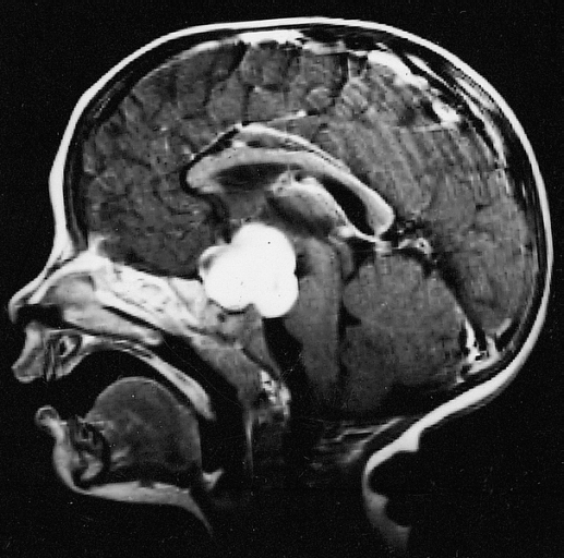
A glioma is a type of tumor that starts in the glial cells of the brain or the spine. Gliomas comprise about 30 percent of all brain tumors and central nervous system tumours, and 80 percent of all malignant brain tumours. Signs and symptoms Symptoms of gliomas depend on which part of the central nervous system is affected. A brain glioma can cause headaches, vomiting, seizures, and cranial nerve disorders as a result of increased intracranial pressure. A glioma of the optic nerve can cause visual loss. Spinal cord gliomas can cause pain, weakness, or numbness in the extremities. Gliomas do not usually metastasize by the bloodstream, but they can spread via the cerebrospinal fluid and cause "drop metastases" to the spinal cord. Complex visual hallucinations have been described as a symptom of low-grade glioma. A child who has a subacute disorder of the central nervous system that produces cranial nerve abnormalities (especially of cranial nerve VII and the lower bulbar nerv ... [...More Info...] [...Related Items...] OR: [Wikipedia] [Google] [Baidu] |
Micrograph
A micrograph or photomicrograph is a photograph or digital image taken through a microscope or similar device to show a magnified image of an object. This is opposed to a macrograph or photomacrograph, an image which is also taken on a microscope but is only slightly magnified, usually less than 10 times. Micrography is the practice or art of using microscopes to make photographs. A micrograph contains extensive details of microstructure. A wealth of information can be obtained from a simple micrograph like behavior of the material under different conditions, the phases found in the system, failure analysis, grain size estimation, elemental analysis and so on. Micrographs are widely used in all fields of microscopy. Types Photomicrograph A light micrograph or photomicrograph is a micrograph prepared using an optical microscope, a process referred to as ''photomicroscopy''. At a basic level, photomicroscopy may be performed simply by connecting a camera to a microscope, th ... [...More Info...] [...Related Items...] OR: [Wikipedia] [Google] [Baidu] |
Glial
Glia, also called glial cells (gliocytes) or neuroglia, are non-neuronal cells in the central nervous system (brain and spinal cord) and the peripheral nervous system that do not produce electrical impulses. They maintain homeostasis, form myelin in the peripheral nervous system, and provide support and protection for neurons. In the central nervous system, glial cells include oligodendrocytes, astrocytes, ependymal cells, and microglia, and in the peripheral nervous system they include Schwann cells and satellite cells. Function They have four main functions: *to surround neurons and hold them in place *to supply nutrients and oxygen to neurons *to insulate one neuron from another *to destroy pathogens and remove dead neurons. They also play a role in neurotransmission and synaptic connections, and in physiological processes such as breathing. While glia were thought to outnumber neurons by a ratio of 10:1, recent studies using newer methods and reappraisal of historical quant ... [...More Info...] [...Related Items...] OR: [Wikipedia] [Google] [Baidu] |
World Health Organization
The World Health Organization (WHO) is a specialized agency of the United Nations responsible for international public health. The WHO Constitution states its main objective as "the attainment by all peoples of the highest possible level of health". Headquartered in Geneva, Switzerland, it has six regional offices and 150 field offices worldwide. The WHO was established on 7 April 1948. The first meeting of the World Health Assembly (WHA), the agency's governing body, took place on 24 July of that year. The WHO incorporated the assets, personnel, and duties of the League of Nations' Health Organization and the , including the International Classification of Diseases (ICD). Its work began in earnest in 1951 after a significant infusion of financial and technical resources. The WHO's mandate seeks and includes: working worldwide to promote health, keeping the world safe, and serve the vulnerable. It advocates that a billion more people should have: universal health care coverag ... [...More Info...] [...Related Items...] OR: [Wikipedia] [Google] [Baidu] |
Ependymoma
An ependymoma is a tumor that arises from the ependyma, a tissue of the central nervous system. Usually, in pediatric cases the location is intracranial, while in adults it is spinal. The common location of intracranial ependymomas is the fourth ventricle. Rarely, ependymomas can occur in the pelvic cavity. Syringomyelia can be caused by an ependymoma. Ependymomas are also seen with neurofibromatosis type II. Signs and symptoms Source: * severe headache * visual loss (due to papilledema) * vomiting * bilateral Babinski sign * drowsiness (after several hours of the above symptoms) * gait change (rotation of feet when walking) * impaction/constipation * back flexibility Morphology Ependymomas are composed of cells with regular, round to oval nuclei. There is a variably dense fibrillary background. Tumor cells may form gland-like round or elongated structures that resemble the embryologic ependymal canal, with long, delicate processes extending into the lumen; more frequently pre ... [...More Info...] [...Related Items...] OR: [Wikipedia] [Google] [Baidu] |
Dysembryoplastic Neuroepithelial Tumor
Dysembryoplastic neuroepithelial tumour (DNT, DNET) is a type of brain tumor. Most commonly found in the temporal lobe, DNTs have been classified as benign tumours. These are glioneuronal tumours comprising both glial and neuron cells and often have ties to focal cortical dysplasia. Varying subclasses of DNTs have been presently identified, with dispute existing in the field on how to properly group these classes. The identification of possible genetic markers to these tumours is currently underway. With DNTs often causing epileptic seizures, surgical removal is a common treatment, providing high rates of success. Signs and symptoms Seizures and epilepsy are the strongest ties to dysembryoplastic neuroepithelial tumours. The most common symptom of DNTs are complex partial seizures. Simple DNTs more frequently manifest generalized seizures. In children, DNTs are considered to be the second leading cause of epilepsy. A headache is another common symptom. Diplopia may also be a r ... [...More Info...] [...Related Items...] OR: [Wikipedia] [Google] [Baidu] |
Central Neurocytoma
Central neurocytoma (CNC) is an extremely rare, ordinarily benign intraventricular brain tumour that typically forms from the neuronal cells of the septum pellucidum. The majority of central neurocytomas grow inwards into the ventricular system forming interventricular neurocytomas. This leads to two primary symptoms of CNCs, blurred vision and increased intracranial pressure. Treatment for a central neurocytoma typically involves surgical removal, with an approximate 1 in 5 chance of recurrence. Central neurocytomas are classified as a grade II tumor under the World Health Organization's classification of tumors of the nervous system. Signs and symptoms There is a wide range of symptoms that patients show. Symptoms can lie dormant, but come about due to Obstructive hydrocephalus. These symptoms include: * Intracranial pressure * Headache * Papilledema * Vomiting * Light headedness * Impaired mental activity * Gait instability In rare and extreme cases, more severe symptoms can ... [...More Info...] [...Related Items...] OR: [Wikipedia] [Google] [Baidu] |
Pilocytic Astrocytoma
Pilocytic astrocytoma (and its variant pilomyxoid astrocytoma) is a brain tumor that occurs most commonly in children and young adults (in the first 20 years of life). They usually arise in the cerebellum, near the brainstem, in the hypothalamic region, or the optic chiasm, but they may occur in any area where astrocytes are present, including the cerebral hemispheres and the spinal cord. These tumors are usually slow growing and benign, corresponding to WHO malignancy grade 1. Signs and symptoms Children affected by pilocytic astrocytoma can present with different symptoms that might include failure to thrive (lack of appropriate weight gain/ weight loss), headache, nausea, vomiting, irritability, torticollis (tilt neck or wry neck), difficulty to coordinate movements, and visual complaints (including nystagmus). The complaints may vary depending on the location and size of the neoplasm. The most common symptoms are associated with increased intracranial pressure due to the size ... [...More Info...] [...Related Items...] OR: [Wikipedia] [Google] [Baidu] |
Neuropathology
Neuropathology is the study of disease of nervous system tissue, usually in the form of either small surgical biopsies or whole-body autopsies. Neuropathologists usually work in a department of anatomic pathology, but work closely with the clinical disciplines of neurology, and neurosurgery, which often depend on neuropathology for a diagnosis. Neuropathology also relates to forensic pathology because brain disease or brain injury can be related to cause of death. Neuropathology should not be confused with neuropathy, which refers to disorders of the nerves themselves (usually in the peripheral nervous system) rather than the tissues. In neuropathology, the branches of the specializations of nervous system as well as the tissues come together into one field of study. Methodology The work of the neuropathologist consists largely of examining autopsy or biopsy tissue from the brain and spinal cord to aid in diagnosis of disease. Tissues are also observed through the eyes, muscles, ... [...More Info...] [...Related Items...] OR: [Wikipedia] [Google] [Baidu] |
Astrocytoma
Astrocytomas are a type of brain tumor. They originate in a particular kind of glial cells, star-shaped brain cells in the cerebrum called astrocytes. This type of tumor does not usually spread outside the brain and spinal cord and it does not usually affect other organs. Astrocytomas are the most common glioma and can occur in most parts of the brain and occasionally in the spinal cord. Within the astrocytomas, two broad classes are recognized in literature, those with: * Narrow zones of infiltration (mostly noninvasive tumors; e.g., pilocytic astrocytoma, subependymal giant cell astrocytoma, pleomorphic xanthoastrocytoma), that often are clearly outlined on diagnostic images * Diffuse zones of infiltration (e.g., high-grade astrocytoma, anaplastic astrocytoma, glioblastoma), that share various features, including the ability to arise at any location in the central nervous system, but with a preference for the cerebral hemispheres; they occur usually in adults, and have an intrins ... [...More Info...] [...Related Items...] OR: [Wikipedia] [Google] [Baidu] |


