|
Ocular Hypotony
Ocular hypotony, or ocular hypotension, or shortly hypotony, is the medical condition in which intraocular pressure (IOP) of the eye is very low. Description Normal IOP ranges between 10–20 mm Hg. The eye is considered hypotonous if the IOP is ≤5 mm Hg (some sources say IOP less than 6.5 mmHg). Types Ocular hypotony is divided into statistical and clinical types. If intraocular pressure is low (less than 6.5 mm Hg) it is called statistical hypotony, and if the reduced IOP causes a decrease in vision, it is called clinical. Causes Hypotony may occur either due to decreased production of aqueous humor or due to increased outflow. Hypotony has many causes including post-surgical wound leak from the eye, chronic inflammation within the eye including iridocyclitis, hypoperfusion, tractional ciliary body detachment or retinal detachment. Eye inflammation, medications including anti glaucoma drugs, or proliferative vitreoretinopathy causes decreased production. Increased outflow or ... [...More Info...] [...Related Items...] OR: [Wikipedia] [Google] [Baidu] |
Intraocular Pressure
Intraocular pressure (IOP) is the fluid pressure inside the eye. Tonometry is the method eye care professionals use to determine this. IOP is an important aspect in the evaluation of patients at risk of glaucoma. Most tonometers are calibrated to measure pressure in millimeters of mercury ( mmHg). Physiology Intraocular pressure is determined by the production and drainage of aqueous humour by the ciliary body and its drainage via the trabecular meshwork and uveoscleral outflow. The reason for this is because the vitreous humour in the posterior segment has a relatively fixed volume and thus does not affect intraocular pressure regulation. An important quantitative relationship (Goldmann's equation) is as follows: :P_o = \frac + P_v Where: * P_o is the IOP in millimeters of mercury (mmHg) * F the rate of aqueous humour formation in microliters per minute (μL/min) * U the resorption of aqueous humour through the uveoscleral route (μL/min) * C is the facility of outflow in micr ... [...More Info...] [...Related Items...] OR: [Wikipedia] [Google] [Baidu] |
Aqueous Humor
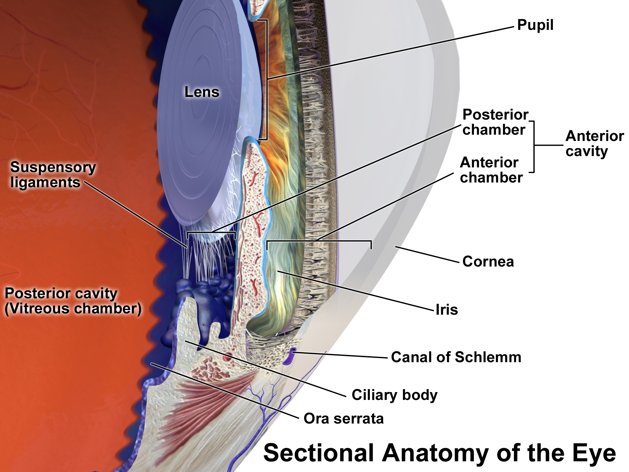
The aqueous humour is a transparent water-like fluid similar to plasma, but containing low protein concentrations. It is secreted from the ciliary body, a structure supporting the lens of the eyeball. It fills both the anterior and the posterior chambers of the eye, and is not to be confused with the vitreous humour, which is located in the space between the lens and the retina, also known as the posterior cavity or vitreous chamber. Blood cannot normally enter the eyeball. Structure Composition *Amino acids: transported by ciliary muscles *98% water *Electrolytes ( pH = 7.4 -one source gives 7.1) **Sodium = 142.09 **Potassium = 2.2 - 4.0 **Calcium = 1.8 **Magnesium = 1.1 **Chloride = 131.6 **HCO3- = 20.15 **Phosphate = 0.62 ** Osm = 304 *Ascorbic acid *Glutathione *Immunoglobulins Function *Maintains the intraocular pressure and inflates the globe of the eye. It is this hydrostatic pressure that keeps the eyeball in a roughly spherical shape and keeps the walls of the eyeball ... [...More Info...] [...Related Items...] OR: [Wikipedia] [Google] [Baidu] |
Iridocyclitis
Uveitis () is inflammation of the uvea, the pigmented layer of the eye between the inner retina and the outer fibrous layer composed of the sclera and cornea. The uvea consists of the middle layer of pigmented vascular structures of the eye and includes the iris, ciliary body, and choroid. Uveitis is described anatomically, by the part of the eye affected, as anterior, intermediate or posterior, or panuveitic if all parts are involved. Anterior uveitis ( iridocyclytis) is the most common, with the incidence of uveitis overall affecting approximately 1:4500, most commonly those between the ages of 20-60. Symptoms include eye pain, eye redness, floaters and blurred vision, and ophthalmic examination may show dilated ciliary blood vessels and the presence of cells in the anterior chamber. Uveitis may arise spontaneously, have a genetic component, or be associated with an autoimmune disease or infection. While the eye is a relatively protected environment, its immune mechanisms may ... [...More Info...] [...Related Items...] OR: [Wikipedia] [Google] [Baidu] |
Ciliary Body
The ciliary body is a part of the eye that includes the ciliary muscle, which controls the shape of the lens, and the ciliary epithelium, which produces the aqueous humor. The aqueous humor is produced in the non-pigmented portion of the ciliary body. The ciliary body is part of the uvea, the layer of tissue that delivers oxygen and nutrients to the eye tissues. The ciliary body joins the ora serrata of the choroid to the root of the iris.Cassin, B. and Solomon, S. ''Dictionary of Eye Terminology''. Gainesville, Florida: Triad Publishing Company, 1990. Structure The ciliary body is a ring-shaped thickening of tissue inside the eye that divides the posterior chamber from the vitreous body. It contains the ciliary muscle, vessels, and fibrous connective tissue. Folds on the inner ciliary epithelium are called ciliary processes, and these secrete aqueous humor into the posterior chamber. The aqueous humor then flows through the pupil into the anterior chamber. The ciliary bo ... [...More Info...] [...Related Items...] OR: [Wikipedia] [Google] [Baidu] |
Retinal Detachment
Retinal detachment is a disorder of the eye in which the retina peels away from its underlying layer of support tissue. Initial detachment may be localized, but without rapid treatment the entire retina may detach, leading to vision loss and blindness. It is a surgical emergency. The retina is a thin layer of light-sensitive tissue on the back wall of the eye. The optical system of the eye focuses light on the retina much like light is focused on the film in a camera. The retina translates that focused image into neural impulses and sends them to the brain via the optic nerve. Occasionally, posterior vitreous detachment, injury or trauma to the eye or head may cause a small tear in the retina. The tear allows vitreous fluid to seep through it under the retina, and peel it away like a bubble in wallpaper. Diagnosis Symptoms As the retina is responsible for vision, persons experiencing a retinal detachment have vision loss. This can be painful or painless. Imaging Ultraso ... [...More Info...] [...Related Items...] OR: [Wikipedia] [Google] [Baidu] |
Glaucoma Surgery
Glaucoma is a group of diseases affecting the optic nerve that results in vision loss and is frequently characterized by raised intraocular pressure (IOP). There are many glaucoma surgeries, and variations or combinations of those surgeries, that facilitate the escape of excess aqueous humor from the eye to lower intraocular pressure, and a few that lower IOP by decreasing the production of aqueous humor. Procedures that facilitate outflow of aqueous humor Laser trabeculoplasty A trabeculoplasty is a modification of the trabecular meshwork. Laser trabeculoplasty (LTP) is the application of a laser beam to burn areas of the trabecular meshwork, located near the base of the iris, to increase fluid outflow. LTP is used in the treatment of various open-angle glaucomas. The two types of laser trabeculoplasty are argon laser trabeculoplasty (ALT) and selective laser trabeculoplasty (SLT). As its name suggests, argon laser trabeculoplasty uses an argon laser to create tiny burns on the t ... [...More Info...] [...Related Items...] OR: [Wikipedia] [Google] [Baidu] |
Phthisis Bulbi
Phthisis bulbi is a shrunken, non-functional eye. It may result from severe eye disease, inflammation or injury, or it may represent a complication of eye surgery. Treatment options include insertion of a prosthesis, which may be preceded by enucleation of the eye. Symptoms The affected eye is shrunken, and has little to no vision. The intraocular pressure in the affected eye is very low or nonexistent. The layers in the eye may be fused together, thickened, or edematous. The eyelids may be glued shut. The eye may be soft when palpated. Under a microscope there may be deposits of calcium or bone, and the lens is often affected by cataract A cataract is a cloudy area in the lens of the eye that leads to a decrease in vision. Cataracts often develop slowly and can affect one or both eyes. Symptoms may include faded colors, blurry or double vision, halos around light, trouble w ...s. Causes It can be caused by injury, including burns to the eye, or long-term eye diseas ... [...More Info...] [...Related Items...] OR: [Wikipedia] [Google] [Baidu] |
Hypotony Maculopathy
Hypotony maculopathy is maculopathy due to very low intraocular pressure known as ocular hypotony. Maculopathy occurs either due to increased outflow of aqueous humor through angle of anterior chamber or less commonly, due to decreased aqueous humor secretion by ciliary body. Description Hypotony maculopathy is maculopathy due to ocular hypotony. Fundus examination may reveal abnormalities like chorioretinal folds, optic nerve head swelling (papilledema) and tortuosity of blood vessels. Causes Maculopathy occurs either due to increased outflow of aqueous humor through angle of anterior chamber or less commonly, due to decreased aqueous humor secretion by ciliary body. Chronic inflammation within the eye including iridocyclitis, medications including anti glaucoma drugs, or proliferative vitreoretinopathy causes decreased production. Increased outflow or aqueous loss may occur following a glaucoma surgery, trauma, post-surgical wound leak from the eye, cyclodialysis cleft, t ... [...More Info...] [...Related Items...] OR: [Wikipedia] [Google] [Baidu] |
Sodium Hyaluronate
Sodium hyaluronate is the sodium salt of hyaluronic acid, a glycosaminoglycan found in various connective tissue of humans. Chemistry Sodium hyaluronate is the sodium salt of hyaluronic acid. It is a glycosaminoglycan and long-chain polymer of disaccharide units of Na-glucuronate-N-acetylglucosamine. It can bind to specific receptors for which it has a high affinity. The polyanionic form, commonly referred to as hyaluronan, is a visco-elastic polymer found in the aqueous and vitreous humour of the eye and in the fluid of articulating joints. Natural occurrence Sodium hyaluronate, as hyaluronic acid, is distributed widely in the extracellular matrix of mammalian connective, epithelial, and neural tissues, as well as the corneal endothelium. Mechanism of action Sodium hyaluronate functions as a tissue lubricant and is thought to play an important role in modulating the interactions between adjacent tissues. It forms a viscoelastic solution in water. Mechanical protection fo ... [...More Info...] [...Related Items...] OR: [Wikipedia] [Google] [Baidu] |
Cycloplegia
Cycloplegia is paralysis of the ciliary muscle of the eye, resulting in a loss of accommodation. Because of the paralysis of the ciliary muscle, the curvature of the lens can no longer be adjusted to focus on nearby objects. This results in similar problems as those caused by presbyopia, in which the lens has lost elasticity and can also no longer focus on close-by objects. Cycloplegia with accompanying mydriasis (dilation of pupil) is usually due to topical application of muscarinic antagonists such as atropine and cyclopentolate. Belladonna alkaloids are used for testing the error of refraction and examination of eye. Management Cycloplegic drugs are generally muscarinic receptor blockers. These include atropine, cyclopentolate, homatropine, scopolamine and tropicamide. They are indicated for use in cycloplegic refraction (to paralyze the ciliary muscle in order to determine the true refractive error of the eye) and the treatment of uveitis. All cycloplegics are also mydriat ... [...More Info...] [...Related Items...] OR: [Wikipedia] [Google] [Baidu] |
Atropine
Atropine is a tropane alkaloid and anticholinergic medication used to treat certain types of nerve agent and pesticide poisonings as well as some types of slow heart rate, and to decrease saliva production during surgery. It is typically given intravenously or by injection into a muscle. Eye drops are also available which are used to treat uveitis and early amblyopia. The intravenous solution usually begins working within a minute and lasts half an hour to an hour. Large doses may be required to treat some poisonings. Common side effects include a dry mouth, large pupils, urinary retention, constipation, and a fast heart rate. It should generally not be used in people with angle closure glaucoma. While there is no evidence that its use during pregnancy causes birth defects, that has not been well studied. It is likely safe during breastfeeding. It is an antimuscarinic (a type of anticholinergic) that works by inhibiting the parasympathetic nervous system. Atropine occurs n ... [...More Info...] [...Related Items...] OR: [Wikipedia] [Google] [Baidu] |


