|
Occipitofrontalis Muscle
The occipitofrontalis muscle (epicranius muscle) is a muscle which covers parts of the skull. It consists of two parts or bellies: the occipital belly, near the occipital bone, and the frontal belly, near the frontal bone. It is supplied by the supraorbital artery, the supratrochlear artery, and the occipital artery. It is innervated by the facial nerve. In humans, the occipitofrontalis helps to create facial expressions. Structure The occipitofrontalis muscle consists of two parts or bellies: * the occipital belly, near the occipital bone. It originates on the lateral two-thirds of the highest nuchal line, and on the mastoid process of the temporal bone. It inserts into the epicranial aponeurosis. * the frontal belly, near the frontal bone. It originates from an intermediate tendon that connects to the occipital belly. It inserts in the fascia of the facial muscles and in the skin above the eyes and nose. Some sources consider the occipital and frontal bellies to be ... [...More Info...] [...Related Items...] OR: [Wikipedia] [Google] [Baidu] |
Epicranial Aponeurosis
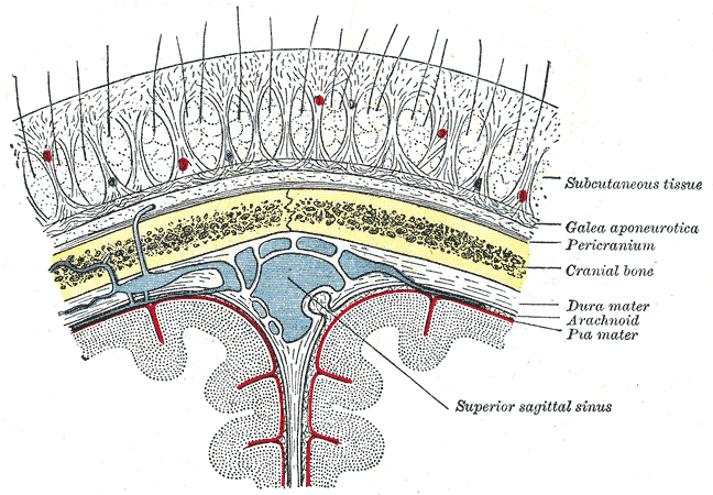
The epicranial aponeurosis (aponeurosis epicranialis, galea aponeurotica) is an aponeurosis (a tough layer of dense fibrous tissue). It covers the upper part of the skull in humans and many other animals. Structure In humans, the epicranial aponeurosis originates from the external occipital protuberance and highest nuchal lines of the occipital bone. It merges with the occipitofrontalis muscle. In front, it forms a short and narrow prolongation between its union with the frontalis muscle (the frontal part of the occipitofrontalis muscle). On either side, the epicranial aponeurosis attaches to the anterior auricular muscles and the superior auricular muscles. Here it is less aponeurotic, and is continued over the temporal fascia to the zygomatic arch as a layer of laminated areolar tissue. It is closely connected to the integument by the firm, dense, fibro-fatty layer which forms the superficial fascia of the scalp. It is attached to the pericranium by loose cellular tiss ... [...More Info...] [...Related Items...] OR: [Wikipedia] [Google] [Baidu] |
Temporal Bone
The temporal bones are situated at the sides and base of the skull, and lateral to the temporal lobes of the cerebral cortex. The temporal bones are overlaid by the sides of the head known as the temples, and house the structures of the ears. The lower seven cranial nerves and the major vessels to and from the brain traverse the temporal bone. Structure The temporal bone consists of four parts— the squamous, mastoid, petrous and tympanic parts. The squamous part is the largest and most superiorly positioned relative to the rest of the bone. The zygomatic process is a long, arched process projecting from the lower region of the squamous part and it articulates with the zygomatic bone. Posteroinferior to the squamous is the mastoid part. Fused with the squamous and mastoid parts and between the sphenoid and occipital bones lies the petrous part, which is shaped like a pyramid. The tympanic part is relatively small and lies inferior to the squamous part, anterior to the mast ... [...More Info...] [...Related Items...] OR: [Wikipedia] [Google] [Baidu] |
Saunders (imprint)
Saunders is an American academic publisher based in the United States. It is currently an imprint of Elsevier. Formerly independent, the W. B. Saunders company was acquired by CBS in 1968, who added it to their publishing division Holt, Rinehart & Winston. When CBS left the publishing field in 1986, it sold the academic publishing units to Harcourt Brace Jovanovich. Harcourt was acquired by Reed Elsevier in 2001. . . Retrieved May 2, 2015. W. B. Saunders published the Kinsey Reports and |
Atony
This glossary of medical terms is a list of definitions about medicine, its sub-disciplines, and related fields. A * Aarskog–Scott syndrome – (AAS) A rare, inherited (X-linked) disease characterized by short stature, facial abnormalities, skeletal and genital anomalies. *Abdomen – The part of the body between the chest and pelvis, which contains most of the tubelike organs of the digestive tract, as well as several solid organs. * Abdominal external oblique muscle – The largest, and outermost, of the three flat muscles of the lateral anterior abdominal wall. *Abdominal internal oblique muscle – A muscle of the abdominal wall, which lies below the external oblique and just above the transverse abdominal muscles. *Abductor pollicis brevis muscle – A muscle in the hand that abducts (straightens) the thumb. *Abductor pollicis longus muscle – One of the extrinsic muscles of the hand. Its major function is to abduct the thumb at the wrist. * Abscess – A collection ... [...More Info...] [...Related Items...] OR: [Wikipedia] [Google] [Baidu] |
Scalp
The scalp is the anatomical area bordered by the human face at the front, and by the neck at the sides and back. Structure The scalp is usually described as having five layers, which can conveniently be remembered as a mnemonic: * S: The skin on the head from which head hair grows. It contains numerous sebaceous glands and hair follicles. * C: Connective tissue. A dense subcutaneous layer of fat and fibrous tissue that lies beneath the skin, containing the nerves and vessels of the scalp. * A: The aponeurosis called epicranial aponeurosis (or galea aponeurotica) is the next layer. It is a tough layer of dense fibrous tissue which runs from the frontalis muscle anteriorly to the occipitalis posteriorly. * L: The loose areolar connective tissue layer provides an easy plane of separation between the upper three layers and the pericranium. In scalping the scalp is torn off through this layer. It also provides a plane of access in craniofacial surgery and neurosurgery. This layer i ... [...More Info...] [...Related Items...] OR: [Wikipedia] [Google] [Baidu] |
Edinburgh
Edinburgh ( ; gd, Dùn Èideann ) is the capital city of Scotland and one of its 32 Council areas of Scotland, council areas. Historically part of the county of Midlothian (interchangeably Edinburghshire before 1921), it is located in Lothian on the southern shore of the Firth of Forth. Edinburgh is Scotland's List of towns and cities in Scotland by population, second-most populous city, after Glasgow, and the List of cities in the United Kingdom, seventh-most populous city in the United Kingdom. Recognised as the capital of Scotland since at least the 15th century, Edinburgh is the seat of the Scottish Government, the Scottish Parliament and the Courts of Scotland, highest courts in Scotland. The city's Holyrood Palace, Palace of Holyroodhouse is the official residence of the Monarchy of the United Kingdom, British monarchy in Scotland. The city has long been a centre of education, particularly in the fields of medicine, Scots law, Scottish law, literature, philosophy, the sc ... [...More Info...] [...Related Items...] OR: [Wikipedia] [Google] [Baidu] |
Churchill Livingstone
Churchill Livingstone is an academic publisher. It was formed in 1971 from the merger of Longman's medical list, E & S Livingstone (Edinburgh, Scotland) and J & A Churchill (London, England) and was owned by Pearson. Harcourt acquired Churchill Livingstone in 1997. It is now integrated as an imprint in Elsevier's health science division after Elsevier acquired Harcourt in 2001. In the past it published a number of classic medical texts, including Sir William Osler's textbook '' The Principles and Practice of Medicine, Gray's Anatomy,'' and ''Myles In Greek mythology, Myles (; Ancient Greek: Μύλης means 'mill-man') was an ancient king of Laconia. He was the son of the King Lelex and possibly the naiad Queen Cleocharia, and brother of Polycaon. Myles was the father of Eurotas who begott ...' Textbook for Midwives.'' In the 1980s, in addition to new texts in all areas of clinical medicine, it published an extensive list of medical and nursing textbooks in low-cost editions ... [...More Info...] [...Related Items...] OR: [Wikipedia] [Google] [Baidu] |
Lambdoid Suture
The lambdoid suture (or lambdoidal suture) is a dense, fibrous connective tissue joint on the posterior aspect of the skull that connects the parietal bones with the occipital bone. It is continuous with the occipitomastoid suture. Structure The lambdoid suture is between the paired parietal bones and the occipital bone of the skull. It runs from the asterion on each side. Nerve supply The lambdoid suture may be supplied by a branch of the supraorbital nerve, a branch of the frontal branch of the trigeminal nerve. Clinical significance At birth, the bones of the skull do not meet. If certain bones of the skull grow too fast, then craniosynostosis (premature closure of the sutures) may occur. This can result in skull deformities. If the lambdoid suture closes too soon on one side, the skull will appear twisted and asymmetrical, a condition called "plagiocephaly". Plagiocephaly refers to the shape and not the condition. The condition is craniosynostosis. The lambdoid sut ... [...More Info...] [...Related Items...] OR: [Wikipedia] [Google] [Baidu] |
Supraorbital Nerve
The supraorbital nerve is one of two branches of the frontal nerve, itself a branch of the ophthalmic nerve. The other branch of the frontal nerve is the supratrochlear nerve. Structure The supraorbital nerve branches from the frontal nerve midway between the base and apex of the orbit. It travels anteriorly above the levator palpebrae superioris and exits the orbit through the supraorbital foramen (or notch) in the superior margin orbit. It exits the orbit lateral to the supratrochlear nerve. It then ascends onto the forehead beneath the corrugator supercilii and frontalis muscles and divides into a medial branch and lateral branch. Function The supraorbital nerve provides sensory innervation to the skin of the lateral forehead and upper eyelid, as well as the conjunctiva of the upper eyelid and mucosa of the frontal sinus The frontal sinuses are one of the four pairs of paranasal sinuses that are situated behind the brow ridges. Sinuses are mucosa-lined airspaces within the ... [...More Info...] [...Related Items...] OR: [Wikipedia] [Google] [Baidu] |
Artery
An artery (plural arteries) () is a blood vessel in humans and most animals that takes blood away from the heart to one or more parts of the body (tissues, lungs, brain etc.). Most arteries carry oxygenated blood; the two exceptions are the pulmonary and the umbilical arteries, which carry deoxygenated blood to the organs that oxygenate it (lungs and placenta, respectively). The effective arterial blood volume is that extracellular fluid which fills the arterial system. The arteries are part of the circulatory system, that is responsible for the delivery of oxygen and nutrients to all cells, as well as the removal of carbon dioxide and waste products, the maintenance of optimum blood pH, and the circulation of proteins and cells of the immune system. Arteries contrast with veins, which carry blood back towards the heart. Structure The anatomy of arteries can be separated into gross anatomy, at the macroscopic level, and microanatomy, which must be studied with a microscop ... [...More Info...] [...Related Items...] OR: [Wikipedia] [Google] [Baidu] |
Temporoparietalis Muscle
The temporoparietalis muscle is a distinct muscle of the head. It lies above the auricularis superior muscle. It lies just inferior to the epicranial aponeurosis of the occipitofrontalis muscle. The temporoparietalis muscle may be used in reconstructive ear surgery Otorhinolaryngology ( , abbreviated ORL and also known as otolaryngology, otolaryngology–head and neck surgery (ORL–H&N or OHNS), or ear, nose, and throat (ENT)) is a surgical subspeciality within medicine that deals with the surgical a .... References Muscles of the head and neck {{Anatomy-stub ... [...More Info...] [...Related Items...] OR: [Wikipedia] [Google] [Baidu] |
Terminologia Anatomica
''Terminologia Anatomica'' is the international standard for human anatomical terminology. It is developed by the Federative International Programme on Anatomical Terminology, a program of the International Federation of Associations of Anatomists (IFAA). The second edition was released in 2019 and approved and adopted by the IFAA General Assembly in 2020. ''Terminologia Anatomica'' supersedes the previous standard, ''Nomina Anatomica''. It contains terminology for about 7500 human anatomical structures. Categories of anatomical structures ''Terminologia Anatomica'' is divided into 16 chapters grouped into five parts. The official terms are in Latin. Although equivalent English-language terms are provided, as shown below, only the official Latin terms are used as the basis for creating lists of equivalent terms in other languages. Part I Chapter 1: General anatomy # General terms # Reference planes # Reference lines # Human body positions # Movements # Parts of human body ... [...More Info...] [...Related Items...] OR: [Wikipedia] [Google] [Baidu] |


