|
Oxaluria
Hyperoxaluria is an excessive urinary excretion of oxalate. Individuals with hyperoxaluria often have calcium oxalate kidney stones. It is sometimes called Bird's disease, after Golding Bird, who first described the condition. Causes Hyperoxaluria can be primary (as a result of a genetic defect) or secondary to another disease process. Type I primary hyperoxaluria (PH1) is associated mutations in the gene encoding AGXT, a key enzyme involved in oxalate metabolism. PH1 is an example of a protein mistargeting disease, wherein AGXT shows a trafficking defect. Instead of being trafficked to peroxisomes, it is targeted to mitochondria, where it is metabolically deficient despite being catalytically active. Type II is associated with GRHPR. Secondary hyperoxaluria can occur as a complication of jejunoileal bypass, or in a patient who has lost much of the ileum with an intact colon. In these cases, hyperoxaluria is caused by excessive gastrointestinal oxalate absorption. Excessive int ... [...More Info...] [...Related Items...] OR: [Wikipedia] [Google] [Baidu] |
Primary Hyperoxaluria
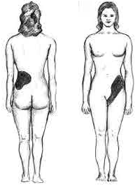
Primary hyperoxaluria is a rare condition (autosomal recessive), resulting in increased excretion of oxalate (up to 600 mg a day from normal 50 mg a day), with oxalate stones being common. Signs and symptoms Primary hyperoxaluria is an autosomal recessive disease, meaning both copies of the gene contain the mutation. Both parents must have one copy of this mutated gene to pass it on to their child, but they do not typically show signs or symptoms of the disease. A single kidney stone in children or recurrent stones in adults is often the first warning sign of primary hyperoxaluria. Other symptoms range from recurrent urinary tract infections, severe abdominal pain or pain in the side, blood in the urine, to chronic kidney disease and kidney failure. The age of symptom onset, progression and severity can vary greatly from one person to another, even among members of the same family. Some individuals may have mild cases that go undiagnosed well into adulthood; others may ... [...More Info...] [...Related Items...] OR: [Wikipedia] [Google] [Baidu] |
AGXT
Serine—pyruvate aminotransferase is an enzyme that in humans is encoded by the ''AGXT'' gene. This gene is expressed only in the liver and the encoded protein is localized mostly in the peroxisomes, where it is involved in glyoxylate detoxification. Mutations in this gene, some of which alter subcellular targeting, have been associated with Primary hyperoxaluria, type I primary hyperoxaluria. See also *Peroxisomal disorder References External links GeneReviews/NIH/NCBI/UW entry on Primary Hyperoxaluria Type 1* * Further reading * * * * * * * * * * * * * * * * * {{gene-2-stub ... [...More Info...] [...Related Items...] OR: [Wikipedia] [Google] [Baidu] |
Kidney Stone
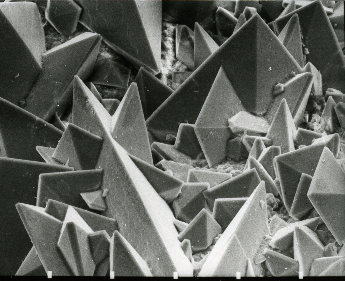
Kidney stone disease, also known as nephrolithiasis or urolithiasis, is a crystallopathy where a solid piece of material (kidney stone) develops in the urinary tract. Kidney stones typically form in the kidney and leave the body in the urine stream. A small stone may pass without causing symptoms. If a stone grows to more than , it can cause blockage of the ureter, resulting in sharp and severe pain in the lower back or abdomen. A stone may also result in blood in the urine, vomiting, or painful urination. About half of people who have had a kidney stone will have another within ten years. Most stones form by a combination of genetics and environmental factors. Risk factors include high urine calcium levels, obesity, certain foods, some medications, calcium supplements, hyperparathyroidism, gout and not drinking enough fluids. Stones form in the kidney when minerals in urine are at high concentration. The diagnosis is usually based on symptoms, urine testing, and medical i ... [...More Info...] [...Related Items...] OR: [Wikipedia] [Google] [Baidu] |
GRHPR
Glyoxylate reductase/hydroxypyruvate reductase is an enzyme that in humans is encoded by the ''GRHPR'' gene. This gene encodes an enzyme with hydroxypyruvate reductase, glyoxylate reductase, and D- glycerate dehydrogenase enzymatic activities. The enzyme has widespread tissue expression and has a role in metabolism. Type II hyperoxaluria Hyperoxaluria is an excessive urinary excretion of oxalate. Individuals with hyperoxaluria often have calcium oxalate kidney stones. It is sometimes called Bird's disease, after Golding Bird, who first described the condition. Causes Hyperoxalur ... is caused by mutations in this gene. GRHPR mutation analysis needs to pay attention to primer design, because allele dropout can cause false-positive result. References External links GeneReviews/NCBI/NIH/UW entry on Primary Hyperoxaluria Type 2 Further reading * * * * * * * * * * * * {{gene-9-stub ... [...More Info...] [...Related Items...] OR: [Wikipedia] [Google] [Baidu] |
Oxalate
Oxalate (IUPAC: ethanedioate) is an anion with the formula C2O42−. This dianion is colorless. It occurs naturally, including in some foods. It forms a variety of salts, for example sodium oxalate (Na2C2O4), and several esters such as dimethyl oxalate (C2O4(CH3)2). It is a conjugate base of oxalic acid. At neutral pH in aqueous solution, oxalic acid converts completely to oxalate. Relationship to oxalic acid The dissociation of protons from oxalic acid proceeds in a stepwise manner; as for other polyprotic acids, loss of a single proton results in the monovalent hydrogenoxalate anion . A salt with this anion is sometimes called an acid oxalate, monobasic oxalate, or hydrogen oxalate. The equilibrium constant ( ''K''a) for loss of the first proton is (p''K''a = 1.27). The loss of the second proton, which yields the oxalate ion, has an equilibrium constant of (p''K''a = 4.28). These values imply, in solutions with neutral pH, no oxalic acid and only trace am ... [...More Info...] [...Related Items...] OR: [Wikipedia] [Google] [Baidu] |
Golding Bird
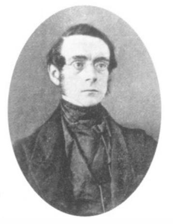
Golding Bird (9 December 1814 – 27 October 1854) was a British medical doctor and a Fellow of the Royal College of Physicians. He became a great authority on kidney diseases and published a comprehensive paper on urinary deposits in 1844. He was also notable for his work in related sciences, especially the medical uses of electricity and electrochemistry. From 1836, he lectured at Guy's Hospital, a well-known teaching hospital in London and now part of King's College London, and published a popular textbook on science for medical students called ''Elements of Natural Philosophy''. Having developed an interest in chemistry while still a child, largely through self-study, Bird was far enough advanced to deliver lectures to his fellow pupils at school. He later applied this knowledge to medicine and did much research on the chemistry of urine and of kidney stones. In 1842, he was the first to describe oxaluria, a condition which leads to the formation of a particul ... [...More Info...] [...Related Items...] OR: [Wikipedia] [Google] [Baidu] |
Jejunoileal Bypass
Jejunoileal bypass (JIB) was a surgical weight-loss procedure performed for the relief of morbid obesity from the 1950s through the 1970s in which all but 30 cm (12 in) to 45 cm (18 in) of the small bowel were detached and set to the side. Many complications that followed jejunoileal bypass operations were caused by bacterial overgrowth in the excluded blind loop. The arthritis-dermatitis syndrome was one of the common distressing disorders. The pathogenetic mechanism was thought to be an immune-complex-mediated process related to bypass enteritis. Problems Two variants of jejunoileal anastomosis were developed, the end-to-side and end-to end(Scott, Dean et al. 1973) anastomoses of the proximal jejunum to distal ileum. In both instances an extensive length of small intestine was bypassed, not excised, excluding it from the alimentary stream. In both these variants a total of only about 45 cm (18 in) of normally absorptive small intestine was retain ... [...More Info...] [...Related Items...] OR: [Wikipedia] [Google] [Baidu] |
Peroxisome
A peroxisome () is a membrane-bound organelle, a type of microbody, found in the cytoplasm of virtually all eukaryotic cells. Peroxisomes are oxidative organelles. Frequently, molecular oxygen serves as a co-substrate, from which hydrogen peroxide (H2O2) is then formed. Peroxisomes owe their name to hydrogen peroxide generating and scavenging activities. They perform key roles in lipid metabolism and the conversion of reactive oxygen species. Peroxisomes are involved in the catabolism of very long chain fatty acids, branched chain fatty acids, bile acid intermediates (in the liver), D-amino acids, and polyamines, the reduction of reactive oxygen species – specifically hydrogen peroxide – and the biosynthesis of plasmalogens, i.e., ether phospholipids critical for the normal function of mammalian brains and lungs. They also contain approximately 10% of the total activity of two enzymes (Glucose-6-phosphate dehydrogenase and 6-Phosphogluconate dehydrogenase) in the pentose ... [...More Info...] [...Related Items...] OR: [Wikipedia] [Google] [Baidu] |
Mitochondria
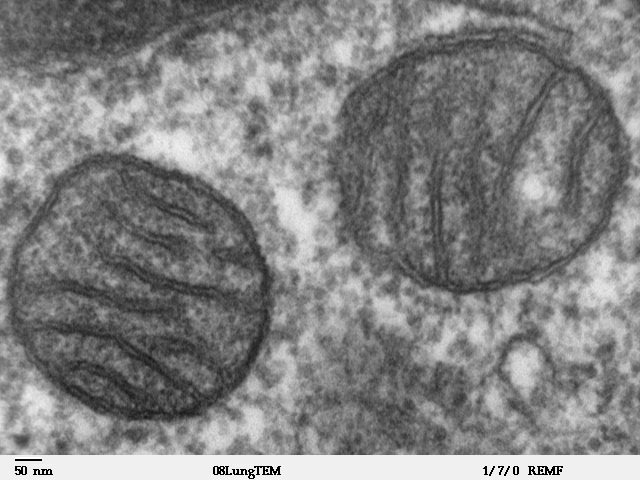
A mitochondrion (; ) is an organelle found in the Cell (biology), cells of most Eukaryotes, such as animals, plants and Fungus, fungi. Mitochondria have a double lipid bilayer, membrane structure and use aerobic respiration to generate adenosine triphosphate (ATP), which is used throughout the cell as a source of chemical energy. They were discovered by Albert von Kölliker in 1857 in the voluntary muscles of insects. The term ''mitochondrion'' was coined by Carl Benda in 1898. The mitochondrion is popularly nicknamed the "powerhouse of the cell", a phrase coined by Philip Siekevitz in a 1957 article of the same name. Some cells in some multicellular organisms lack mitochondria (for example, mature mammalian red blood cells). A large number of unicellular organisms, such as microsporidia, parabasalids and diplomonads, have reduced or transformed their mitochondria into mitosome, other structures. One eukaryote, ''Monocercomonoides'', is known to have completely lost its mitocho ... [...More Info...] [...Related Items...] OR: [Wikipedia] [Google] [Baidu] |
Ileum
The ileum () is the final section of the small intestine in most higher vertebrates, including mammals, reptiles, and birds. In fish, the divisions of the small intestine are not as clear and the terms posterior intestine or distal intestine may be used instead of ileum. Its main function is to absorb vitamin B12, bile salts, and whatever products of digestion that were not absorbed by the jejunum. The ileum follows the duodenum and jejunum and is separated from the cecum by the ileocecal valve (ICV). In humans, the ileum is about 2–4 m long, and the pH is usually between 7 and 8 (neutral or slightly basic). ''Ileum ''is derived from the Greek word ''eilein'', meaning "to twist up tightly". Structure The ileum is the third and final part of the small intestine. It follows the jejunum and ends at the ileocecal junction, where the terminal ileum communicates with the cecum of the large intestine through the ileocecal valve. The ileum, along with the jejunum, is suspended ... [...More Info...] [...Related Items...] OR: [Wikipedia] [Google] [Baidu] |


