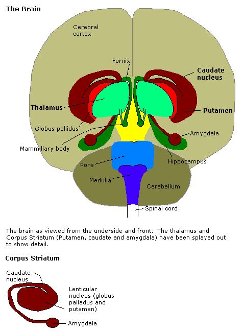
|
Orbitofrontal Cortex
The orbitofrontal cortex (OFC) is a prefrontal cortex region in the frontal lobes of the brain which is involved in the cognitive process of decision-making. In non-human primates it consists of the association cortex areas Brodmann area 11, 12 and 13; in humans it consists of Brodmann area 10, 11 and 47. The OFC is functionally related to the ventromedial prefrontal cortex.Phillips, LH., MacPherson, SE. & Della Sala, S. (2002). 'Age, cognition and emotion: the role of anatomical segregation in the frontal lobes: the role of anatomical segregation in the frontal lobes'. in J Grafman (ed.), Handbook of Neuropsychology: the frontal lobes. Elsevier Science, Amsterdam, pp. 73-98. Therefore, the region is distinguished due to the distinct neural connections and the distinct functions it performs.Barbas H, Ghashghaei H, Rempel-Clower N, Xiao D (2002) Anatomic basis of functional specialization in prefrontal cortices in primates. In: Handbook of Neuropsychology (Grafman J, ed), pp 1- ... [...More Info...] [...Related Items...] OR: [Wikipedia] [Google] [Baidu] |
Frontal Lobe
The frontal lobe is the largest of the four major lobes of the brain in mammals, and is located at the front of each cerebral hemisphere (in front of the parietal lobe and the temporal lobe). It is parted from the parietal lobe by a groove between tissues called the central sulcus and from the temporal lobe by a deeper groove called the lateral sulcus (Sylvian fissure). The most anterior rounded part of the frontal lobe (though not well-defined) is known as the frontal pole, one of the three poles of the cerebrum. The frontal lobe is covered by the frontal cortex. The frontal cortex includes the premotor cortex, and the primary motor cortex – parts of the motor cortex. The front part of the frontal cortex is covered by the prefrontal cortex. There are four principal gyri in the frontal lobe. The precentral gyrus is directly anterior to the central sulcus, running parallel to it and contains the primary motor cortex, which controls voluntary movements of specific body parts ... [...More Info...] [...Related Items...] OR: [Wikipedia] [Google] [Baidu] |
Orbit (anatomy)
In anatomy, the orbit is the cavity or socket of the skull in which the eye and its appendages are situated. "Orbit" can refer to the bony socket, or it can also be used to imply the contents. In the adult human, the volume of the orbit is , of which the eye occupies . The orbital contents comprise the eye, the orbital and retrobulbar fascia, extraocular muscles, cranial nerves II, III, IV, V, and VI, blood vessels, fat, the lacrimal gland with its sac and duct, the eyelids, medial and lateral palpebral ligaments, cheek ligaments, the suspensory ligament, septum, ciliary ganglion and short ciliary nerves. Structure The orbits are conical or four-sided pyramidal cavities, which open into the midline of the face and point back into the head. Each consists of a base, an apex and four walls."eye, human."Encyclopædia Britannica from Encyclopædia Britannica 2006 Ultimate Reference Suite DVD 2009 Openings There are two important foramina, or windows, two important fissu ... [...More Info...] [...Related Items...] OR: [Wikipedia] [Google] [Baidu] |
Parahippocampus
The parahippocampal gyrus (or hippocampal gyrus') is a grey matter cortical region of the brain that surrounds the hippocampus and is part of the limbic system. The region plays an important role in memory encoding and retrieval. It has been involved in some cases of hippocampal sclerosis. Asymmetry has been observed in schizophrenia. Structure The anterior part of the gyrus includes the perirhinal and entorhinal cortices. The term parahippocampal cortex is used to refer to an area that encompasses both the posterior parahippocampal gyrus and the medial portion of the fusiform gyrus. Function Scene recognition The parahippocampal place area (PPA) is a sub-region of the parahippocampal cortex that lies medially in the inferior temporo-occipital cortex. PPA plays an important role in the encoding and recognition of environmental scenes (rather than faces). fMRI studies indicate that this region of the brain becomes highly active when human subjects view topographical scene sti ... [...More Info...] [...Related Items...] OR: [Wikipedia] [Google] [Baidu] |
Amygdala
The amygdala (; plural: amygdalae or amygdalas; also '; Latin from Greek, , ', 'almond', 'tonsil') is one of two almond-shaped clusters of nuclei located deep and medially within the temporal lobes of the brain's cerebrum in complex vertebrates, including humans. Shown to perform a primary role in the processing of memory, decision making, and emotional responses (including fear, anxiety, and aggression), the amygdalae are considered part of the limbic system. The term "amygdala" was first introduced by Karl Friedrich Burdach in 1822. Structure The regions described as amygdala nuclei encompass several structures of the cerebrum with distinct connectional and functional characteristics in humans and other animals. Among these nuclei are the basolateral complex, the cortical nucleus, the medial nucleus, the central nucleus, and the intercalated cell clusters. The basolateral complex can be further subdivided into the lateral, the basal, and the accessory basal nucle ... [...More Info...] [...Related Items...] OR: [Wikipedia] [Google] [Baidu] |
Pyriform Cortex
The piriform cortex, or pyriform cortex, is a region in the brain, part of the rhinencephalon situated in the cerebrum. The function of the piriform cortex relates to the sense of smell. Structure The piriform cortex is part of the rhinencephalon situated in the cerebrum. In human anatomy, the piriform cortex has been described as consisting of the cortical amygdala, uncus, and anterior parahippocampal gyrus. More specifically, the human piriform cortex is located between the insula and the temporal lobe, anteriorly and laterally of the amygdala.Howard, J. D., Plailly, J., Grueschow, M., Haynes, J. D., & Gottfried, J. A. (2009). Odor quality coding and categorization in human posterior piriform cortex. Nature neuroscience, 12(7), 932-938. Supplementary material, p.4 Function The function of the piriform cortex relates to olfaction, which is the perception of smell. This has been particularly shown in humans for the posterior piriform cortex. The piriform cortex in rodents and ... [...More Info...] [...Related Items...] OR: [Wikipedia] [Google] [Baidu] |
Insular Cortex
The insular cortex (also insula and insular lobe) is a portion of the cerebral cortex folded deep within the lateral sulcus (the fissure separating the temporal lobe from the parietal and frontal lobes) within each hemisphere of the mammalian brain. The insulae are believed to be involved in consciousness and play a role in diverse functions usually linked to emotion or the regulation of the body's homeostasis. These functions include compassion, empathy, taste, perception, motor control, self-awareness, cognitive functioning, interpersonal experience, and awareness of homeostatic emotions such as hunger, pain and fatigue. In relation to these, it is involved in psychopathology. The insular cortex is divided into two parts: the anterior insula and the posterior insula in which more than a dozen field areas have been identified. The cortical area overlying the insula toward the lateral surface of the brain is the operculum (meaning ''lid''). The opercula are formed from parts o ... [...More Info...] [...Related Items...] OR: [Wikipedia] [Google] [Baidu] |
Internal Granular Layer (cerebral Cortex)
The internal granular layer of the cortex, also commonly referred to as the granular layer of the cortex, is the layer IV in the subdivision of the mammalian cortex into 6 layers. The adjective internal is used in opposition to the external granular layer of the cortex, the term granular refers to the granule cells found here. This layer receives the afferent connections from the thalamus and from other cortical regions and sends connections to the other layers. The line of Gennari The line of Gennari (also called the "band" or "stria" of Gennari) is a band of myelinated axons that runs parallel to the surface of the cerebral cortex on the banks of the calcarine fissure in the occipital lobe. This formation is visible to the ... (occipital stripe) is also present in this layer. Cerebral cortex {{neuroanatomy-stub ... [...More Info...] [...Related Items...] OR: [Wikipedia] [Google] [Baidu] |
Gyrus Rectus
The portion of the inferior frontal lobe immediately adjacent to the longitudinal fissure (and medial to the medial orbital gyrus and olfactory tract) is named the straight gyrus,(or gyrus rectus) and is continuous with the superior frontal gyrus on the medial surface. A specific function for the straight gyrus has not yet been brought to light; however, in males, greater activation of the straight gyrus within the medial orbitofrontal cortex while observing sexually visual pictures has been strongly linked to HSDD ( hypoactive sexual desire disorder). Additional images File:Straight gyrus animation small2.gif, Animation. Straight gyrus is depicted as red. File:Straight_gyrus_-_inferior_view.png, Basal surface of cerebrum. Straight gyrus is shown in red. File:Gray743 straight gyrus.png, Coronal section of human brain. Straight gyrus depicted as yellow in center bottom. File:Slide2STE.JPG, Cerebrum. Optic and olfactory nerves.Inferior view. Deep dissection. File:Slide2ZEB.JP ... [...More Info...] [...Related Items...] OR: [Wikipedia] [Google] [Baidu] |
Brodmann Area 10
Brodmann area 10 (BA10, frontopolar prefrontal cortex, rostrolateral prefrontal cortex, or anterior prefrontal cortex) is the anterior-most portion of the prefrontal cortex in the human brain. BA10 was originally defined broadly in terms of its cytoarchitectonic traits as they were observed in the brains of cadavers, but because modern functional imaging cannot precisely identify these boundaries, the terms anterior prefrontal cortex, rostral prefrontal cortex and frontopolar prefrontal cortex are used to refer to the area in the most anterior part of the frontal cortex that approximately covers BA10—simply to emphasize the fact that BA10 does not include ''all'' parts of the prefrontal cortex. BA10 is the largest cytoarchitectonic area in the human brain. It has been described as "one of the least well understood regions of the human brain". Present research suggests that it is involved in strategic processes in memory recall and various executive functions. During human evolu ... [...More Info...] [...Related Items...] OR: [Wikipedia] [Google] [Baidu] |
Brodmann Area 13
Brodmann area 13 is part of the Orbitofrontal cortex, a subdivision of the cerebral cortex as defined by cytoarchitecture. Location Brodmann area 13 is located in the posterior part of the Orbitofrontal cortex , and can be subdivided into areas 13a, 13b, 13m, 13l. Area 13a is anterior to the junction of olfactory tract and area 13b occupies a region just anterior to 13a along the olfactory sulcus. Area 13m is on the medial part of the middle orbital gyrus, whereas 13l is in the lateral part of the gyrus. Subregions Areas 13m and 13l are dysgranular regions of cortex. These areas are differentiated from the more anterior area 11 by a lack of continuous granular layer, and from the more posterior agranular Insular cortex. Area 13b is a thin and dysgranular cortical area, often characterized by crossing patterns of striations in layers III and V. Area 13a has an agranular structure. See also * Brodmann area A Brodmann area is a region of the cerebral cortex, in the human or o ... [...More Info...] [...Related Items...] OR: [Wikipedia] [Google] [Baidu] |
Brodmann Area 14
Brodmann Area 14 is one of Brodmann's subdivisions of the cerebral cortex in the brain. It was defined by Brodmann in the guenon monkey . While Brodmann, writing in 1909, argued that no equivalent structure existed in humans, later work demonstrated that area 14 has a clear homologue in the human ventromedial prefrontal cortex. Anatomy Brodmann areas were defined based on cytoarchitecture rather than function. Area 14 differs most clearly from Brodmann area 13-1905 in that it lacks a distinct internal granular layer (IV). Other differences are a less distinct external granular layer (II), a widening of the relatively cell-free zone of the external pyramidal layer (III); cells in the internal pyramidal layer (V) are denser and rounded; and the cells of the multiform layer (VI) assume a more distinct tangential orientation. Function According to one theory, Area 14 is believed to serve as association cortex for the visceral senses and olfaction along with Area 51. Its anatom ... [...More Info...] [...Related Items...] OR: [Wikipedia] [Google] [Baidu] |
Brodmann Area 11
Brodmann area 11 is one of Korbinian Brodmann, Brodmann's Cell biology, cytologically defined Brodmann area, regions of the brain. It is in the orbitofrontal cortex which is above the orbit (anatomy), eye sockets (orbitae). It is involved in decision making and processing rewards, planning, encoding new information into long-term memory, and reasoning. In humans Brodmann area 11, or BA11, is part of the frontal lobe, frontal cerebral cortex, cortex in the human brain. BA11 is the part of the orbitofrontal cortex that covers the medial portion of the ventral surface of the frontal lobe. Prefrontal area 11 of Brodmann-1909 is a subdivision of the frontal lobe in the human defined on the basis of cytoarchitecture. Defined and illustrated in Brodmann-1909, it included the areas subsequently illustrated in Brodmann-10 as prefrontal area 11 and rostral area 12. Area 11 is a subdivision of the cytoarchitecturally defined frontal region of cerebral cortex of the human. As illustrated in ... [...More Info...] [...Related Items...] OR: [Wikipedia] [Google] [Baidu] |


