|
Optic Disc
The optic disc or optic nerve head is the point of exit for ganglion cell axons leaving the eye. Because there are no rods or cones overlying the optic disc, it corresponds to a small blind spot in each eye. The ganglion cell axons form the optic nerve after they leave the eye. The optic disc represents the beginning of the optic nerve and is the point where the axons of retinal ganglion cells come together. The optic disc is also the entry point for the major blood vessels that supply the retina. The optic disc in a normal human eye carries 1–1.2 million afferent nerve fibers from the eye towards the brain. Structure The optic disc is placed 3 to 4 mm to the nasal side of the fovea. It is a vertical oval, with average dimensions of 1.76mm horizontally by 1.92mm vertically. There is a central depression, of variable size, called the optic cup. This depression can be a variety of shapes from a shallow indentation to a bean pot—this shape can be significant for dia ... [...More Info...] [...Related Items...] OR: [Wikipedia] [Google] [Baidu] |
Fovea Centralis
The fovea centralis is a small, central pit composed of closely packed cones in the eye. It is located in the center of the macula lutea of the retina. The fovea is responsible for sharp central vision (also called foveal vision), which is necessary in humans for activities for which visual detail is of primary importance, such as reading and driving. The fovea is surrounded by the ''parafovea'' belt and the ''perifovea'' outer region. The parafovea is the intermediate belt, where the ganglion cell layer is composed of more than five layers of cells, as well as the highest density of cones; the perifovea is the outermost region where the ganglion cell layer contains two to four layers of cells, and is where visual acuity is below the optimum. The perifovea contains an even more diminished density of cones, having 12 per 100 micrometres versus 50 per 100 micrometres in the most central fovea. That, in turn, is surrounded by a larger peripheral area, which delivers highly com ... [...More Info...] [...Related Items...] OR: [Wikipedia] [Google] [Baidu] |
Slit Lamp
A slit lamp is an instrument consisting of a high-intensity light source that can be focused to shine a thin sheet of light into the eye. It is used in conjunction with a biomicroscope. The lamp facilitates an examination of the anterior segment and posterior segment of the human eye, which includes the eyelid, sclera, conjunctiva, iris (anatomy), iris, natural crystalline lens, and cornea. The binocular slit-lamp examination provides a stereoscopic magnified view of the eye structures in detail, enabling anatomical diagnoses to be made for a variety of eye conditions. A second, hand-held lens is used to examine the retina. History Two conflicting trends emerged in the development of the slit lamp. One trend originated from clinical research and aimed to apply the increasingly complex and advanced technology of the time. [...More Info...] [...Related Items...] OR: [Wikipedia] [Google] [Baidu] |
SD-OCT Optic Disc Cross-Sections
OCT Biomicroscopy is the use of optical coherence tomography (OCT) in place of slit lamp biomicroscopy to examine the transparent axial tissues of the eye. Traditionally, ophthalmic biomicroscopy has been completed with a slit lamp biomicroscope that uses slit beam illumination and an optical microscope to enable stereoscopic, magnified, cross-sectional views of transparent tissues in the eye, with or without the aid of an additional lens. Like slit lamp biomicroscopy, OCT does not penetrate opaque tissues well but enables detailed, cross-sectional views of transparent tissues, often with greater detail than is possible with a slit lamp. Ultrasound biomicroscopy (UBM) is much better at imaging through opaque tissues since it uses high energy sound waves. Because of its limited depth of penetration, UBM's main use within ophthalmology has been to visualize anterior structures such as the angle and ciliary body. Both ultrasound and OCT biomicroscopy produce an objective image of ocul ... [...More Info...] [...Related Items...] OR: [Wikipedia] [Google] [Baidu] |
Schematic Diagram Of The Human Eye En
A schematic, or schematic diagram, is a designed representation of the elements of a system using abstract, graphic symbols rather than realistic pictures. A schematic usually omits all details that are not relevant to the key information the schematic is intended to convey, and may include oversimplified elements in order to make this essential meaning easier to grasp, as well as additional organization of the information. For example, a subway map intended for passengers may represent a subway station with a dot. The dot is not intended to resemble the actual station at all but aims to give the viewer information without unnecessary visual clutter. A schematic diagram of a chemical process uses symbols in place of detailed representations of the vessels, piping, valves, pumps, and other equipment that compose the system, thus emphasizing the functions of the individual elements and the interconnections among them and suppresses their physical details. In an electronic circuit ... [...More Info...] [...Related Items...] OR: [Wikipedia] [Google] [Baidu] |
Pre-eclampsia
Pre-eclampsia is a disorder of pregnancy characterized by the onset of high blood pressure and often a significant amount of protein in the urine. When it arises, the condition begins after 20 weeks of pregnancy. In severe cases of the disease there may be red blood cell breakdown, a low blood platelet count, impaired liver function, kidney dysfunction, swelling, shortness of breath due to fluid in the lungs, or visual disturbances. Pre-eclampsia increases the risk of undesirable outcomes for both the mother and the fetus. If left untreated, it may result in seizures at which point it is known as eclampsia. Risk factors for pre-eclampsia include obesity, prior hypertension, older age, and diabetes mellitus. It is also more frequent in a woman's first pregnancy and if she is carrying twins. The underlying mechanism involves abnormal formation of blood vessels in the placenta amongst other factors. Most cases are diagnosed before delivery. Commonly, pre-eclampsia continues i ... [...More Info...] [...Related Items...] OR: [Wikipedia] [Google] [Baidu] |
Optic Disc Drusen
Optic disc drusen (ODD) are globules of mucoproteins and mucopolysaccharides that progressively calcify in the optic disc.Golnik, K. (2006). Congenital anomalies and acquired abnormalities of the optic nerve, (Version 14.3). UptoDate (On-Line Serial) They are thought to be the remnants of the axonal transport system of degenerated retinal ganglion cells. ODD have also been referred to as congenitally elevated or anomalous discs, pseudopapilledema, pseudoneuritis, buried disc drusen, and disc hyaline bodies. Anatomy The optic nerve is a cable connection that transmits images from the retina to the brain. It consists of over one million retinal ganglion cell axons. The optic nerve head, or optic disc is the anterior end of the nerve that is in the eye and hence is visible with an ophthalmoscope. It is located nasally and slightly inferior to the macula of the eye. There is a blind spot at the optic disc because there are no rods or cones beneath it to detect light. The centr ... [...More Info...] [...Related Items...] OR: [Wikipedia] [Google] [Baidu] |
Intracranial Pressure
Intracranial pressure (ICP) is the pressure exerted by fluids such as cerebrospinal fluid (CSF) inside the skull and on the brain tissue. ICP is measured in millimeters of mercury ( mmHg) and at rest, is normally 7–15 mmHg for a supine adult. The body has various mechanisms by which it keeps the ICP stable, with CSF pressures varying by about 1 mmHg in normal adults through shifts in production and absorption of CSF. Changes in ICP are attributed to volume changes in one or more of the constituents contained in the cranium. CSF pressure has been shown to be influenced by abrupt changes in intrathoracic pressure during coughing (which is induced by contraction of the diaphragm and abdominal wall muscles, the latter of which also increases intra-abdominal pressure), the valsalva maneuver, and communication with the vasculature (venous and arterial systems). Intracranial hypertension (IH), also called increased ICP (IICP) or raised intracranial pressure (RICP), is elevation of ... [...More Info...] [...Related Items...] OR: [Wikipedia] [Google] [Baidu] |
Papilledema
Papilledema or papilloedema is optic disc swelling that is caused by increased intracranial pressure due to any cause. The swelling is usually bilateral and can occur over a period of hours to weeks. Unilateral presentation is extremely rare. In intracranial hypertension, the optic disc swelling most commonly occurs bilaterally. When papilledema is found on fundoscopy, further evaluation is warranted because vision loss can result if the underlying condition is not treated. Further evaluation with a CT or MRI of the brain and/or spine is usually performed. Recent research has shown that point-of-care ultrasound can be used to measure optic nerve sheath diameter for detection of increased intracranial pressure and shows good diagnostic test accuracy compared to CT. Thus, if there is a question of papilledema on fundoscopic examination or if the optic disc cannot be adequately visualized, ultrasound can be used to rapidly assess for increased intracranial pressure and help direct ... [...More Info...] [...Related Items...] OR: [Wikipedia] [Google] [Baidu] |
Optic Neuritis
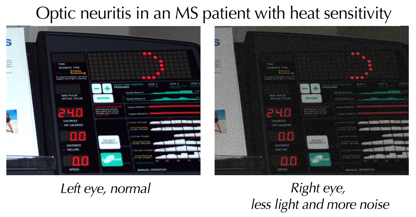
Optic neuritis describes any condition that causes inflammation of the optic nerve; it may be associated with demyelinating diseases, or infectious or inflammatory processes. It is also known as optic papillitis (when the head of the optic nerve is involved), neuroretinitis (when there is a combined involvement of the optic disc and surrounding retina in the macular area) and retrobulbar neuritis (when the posterior part of the nerve is involved). Prelaminar optic neuritis describes involvement of the non-myelinated axons in the retina. It is most often associated with multiple sclerosis, and it may lead to complete or partial loss of vision in one or both eyes. Other causes include: # Leber's hereditary optic neuropathy # Parainfectious optic neuritis (associated with viral infections such as measles, mumps, chickenpox, whooping cough and glandular fever) # Infectious optic neuritis (sinus related or associated with cat-scratch fever, tuberculosis, Lyme disease and cry ... [...More Info...] [...Related Items...] OR: [Wikipedia] [Google] [Baidu] |
Glaucoma
Glaucoma is a group of eye diseases that result in damage to the optic nerve (or retina) and cause vision loss. The most common type is open-angle (wide angle, chronic simple) glaucoma, in which the drainage angle for fluid within the eye remains open, with less common types including closed-angle (narrow angle, acute congestive) glaucoma and normal-tension glaucoma. Open-angle glaucoma develops slowly over time and there is no pain. Peripheral vision may begin to decrease, followed by central vision, resulting in blindness if not treated. Closed-angle glaucoma can present gradually or suddenly. The sudden presentation may involve severe eye pain, blurred vision, mid-dilated pupil, redness of the eye, and nausea. Vision loss from glaucoma, once it has occurred, is permanent. Eyes affected by glaucoma are referred to as being glaucomatous. Risk factors for glaucoma include increasing age, high pressure in the eye, a family history of glaucoma, and use of steroid medicatio ... [...More Info...] [...Related Items...] OR: [Wikipedia] [Google] [Baidu] |
Cup-to-disc Ratio
The optic cup is the white, cup-like area in the center of the optic disc. The ratio of the size of the optic cup to the optic disc (cup-to-disc ratio, or C/D) is one measure used in the diagnosis of glaucoma. Different C/Ds can be measured horizontally or vertically in the same patient. C/Ds vary widely in healthy individuals. However, larger vertical C/Ds, or C/Ds which are very different between the eyes, may raise suspicion of glaucoma. A C/D which enlarges vertically over months or years suggests glaucoma. Cup-to-disc ratio The ''cup-to-disc ratio'' (often notated ''CDR'') is a measurement used in ophthalmology and optometry to assess the progression of glaucoma. The optic disc is the anatomical location of the eye's "blind spot", the area where the optic nerve leave and blood vessels enter the retina. The optic disc can be flat or it can have a certain amount of normal cupping. But glaucoma, which is in most cases associated with an increase in intraocular pressure Int ... [...More Info...] [...Related Items...] OR: [Wikipedia] [Google] [Baidu] |


