|
Optic Chiasm
In neuroanatomy, the optic chiasm, or optic chiasma (; , ), is the part of the brain where the optic nerves cross. It is located at the bottom of the brain immediately inferior to the hypothalamus. The optic chiasm is found in all vertebrates, although in cyclostomes (lampreys and hagfishes), it is located within the brain. This article is about the optic chiasm of vertebrates, which is the best known nerve chiasm, but not every chiasm denotes a crossing of the body midline (e.g., in some invertebrates, see Chiasm (anatomy)). A midline crossing of nerves inside the brain is called a decussation (see Definition of types of crossings). Structure For the different types of optic chiasm, see In all vertebrates, the optic nerves of the left and the right eye meet in the body midline, ventral to the brain. In many vertebrates the left optic nerve crosses over the right one without fusing with it. In vertebrates with a large overlap of the visual fields of the two eyes, i ... [...More Info...] [...Related Items...] OR: [Wikipedia] [Google] [Baidu] |
Andreas Vesalius
Andreas Vesalius (Latinized from Andries van Wezel) () was a 16th-century anatomist, physician, and author of one of the most influential books on human anatomy, '' De Humani Corporis Fabrica Libri Septem'' (''On the fabric of the human body'' ''in seven books''). Vesalius is often referred to as the founder of modern human anatomy. He was born in Brussels, which was then part of the Habsburg Netherlands. He was a professor at the University of Padua (1537–1542) and later became Imperial physician at the court of Emperor Charles V. ''Andreas Vesalius'' is the Latinized form of the Dutch name Andries van Wesel. It was a common practice among European scholars in his time to Latinize their names. His name is also given as ''Andrea Vesalius'', ''André Vésale'', ''Andrea Vesalio'', ''Andreas Vesal'', ''Andrés Vesalio'' and ''Andre Vesale''. Early life and education Vesalius was born as Andries van Wesel to his father Anders van Wesel and mother Isabel Crabbe on 31 Decembe ... [...More Info...] [...Related Items...] OR: [Wikipedia] [Google] [Baidu] |
Amphibians
Amphibians are four-limbed and ectothermic vertebrates of the class Amphibia. All living amphibians belong to the group Lissamphibia. They inhabit a wide variety of habitats, with most species living within terrestrial, fossorial, arboreal or freshwater aquatic ecosystems. Thus amphibians typically start out as larvae living in water, but some species have developed behavioural adaptations to bypass this. The young generally undergo metamorphosis from larva with gills to an adult air-breathing form with lungs. Amphibians use their skin as a secondary respiratory surface and some small terrestrial salamanders and frogs lack lungs and rely entirely on their skin. They are superficially similar to reptiles like lizards but, along with mammals and birds, reptiles are amniotes and do not require water bodies in which to breed. With their complex reproductive needs and permeable skins, amphibians are often ecological indicators; in recent decades there has been a dramati ... [...More Info...] [...Related Items...] OR: [Wikipedia] [Google] [Baidu] |
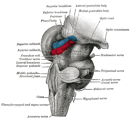
Superior Colliculus
In neuroanatomy, the superior colliculus () is a structure lying on the roof of the mammalian midbrain. In non-mammalian vertebrates, the homologous structure is known as the optic tectum, or optic lobe. The adjective form '' tectal'' is commonly used for both structures. In mammals, the superior colliculus forms a major component of the midbrain. It is a paired structure and together with the paired inferior colliculi forms the corpora quadrigemina. The superior colliculus is a layered structure, with a pattern that is similar to all mammals. The layers can be grouped into the superficial layers ( stratum opticum and above) and the deeper remaining layers. Neurons in the superficial layers receive direct input from the retina and respond almost exclusively to visual stimuli. Many neurons in the deeper layers also respond to other modalities, and some respond to stimuli in multiple modalities. The deeper layers also contain a population of motor-related neurons, capable of ac ... [...More Info...] [...Related Items...] OR: [Wikipedia] [Google] [Baidu] |
Mammals
Mammals () are a group of vertebrate animals constituting the class Mammalia (), characterized by the presence of mammary glands which in females produce milk for feeding (nursing) their young, a neocortex (a region of the brain), fur or hair, and three middle ear bones. These characteristics distinguish them from reptiles (including birds) from which they diverged in the Carboniferous, over 300 million years ago. Around 6,400 extant species of mammals have been described divided into 29 orders. The largest orders, in terms of number of species, are the rodents, bats, and Eulipotyphla ( hedgehogs, moles, shrews, and others). The next three are the Primates (including humans, apes, monkeys, and others), the Artiodactyla (cetaceans and even-toed ungulates), and the Carnivora (cats, dogs, seals, and others). In terms of cladistics, which reflects evolutionary history, mammals are the only living members of the Synapsida (synapsids); this clade, tog ... [...More Info...] [...Related Items...] OR: [Wikipedia] [Google] [Baidu] |
Optic Tectum
In neuroanatomy, the superior colliculus () is a structure lying on the roof of the mammalian midbrain. In non-mammalian vertebrates, the homologous structure is known as the optic tectum, or optic lobe. The adjective form '' tectal'' is commonly used for both structures. In mammals, the superior colliculus forms a major component of the midbrain. It is a paired structure and together with the paired inferior colliculi forms the corpora quadrigemina. The superior colliculus is a layered structure, with a pattern that is similar to all mammals. The layers can be grouped into the superficial layers (stratum opticum and above) and the deeper remaining layers. Neurons in the superficial layers receive direct input from the retina and respond almost exclusively to visual stimuli. Many neurons in the deeper layers also respond to other modalities, and some respond to stimuli in multiple modalities. The deeper layers also contain a population of motor-related neurons, capable of activa ... [...More Info...] [...Related Items...] OR: [Wikipedia] [Google] [Baidu] |
Optic Tracts
In neuroanatomy, the optic tract () is a part of the visual system in the brain. It is a continuation of the optic nerve that relays information from the optic chiasm to the ipsilateral lateral geniculate nucleus (LGN), pretectal nuclei, and superior colliculus. It is composed of two individual tracts, the left optic tract and the right optic tract, each of which conveys visual information exclusive to its respective contralateral half of the visual field. Each of these tracts is derived from a combination of temporal and nasal retinal fibers from each eye that corresponds to one half of the visual field. In more specific terms, the optic tract contains fibers from the ipsilateral temporal hemiretina and contralateral nasal hemiretina. Visual system The optic tract carries retinal information relating to the whole visual field. Specifically, the left optic tract corresponds to the right visual field, while the right optic tract corresponds to the left visual field. To form the r ... [...More Info...] [...Related Items...] OR: [Wikipedia] [Google] [Baidu] |
Stereopsis
Stereopsis () is the component of depth perception retrieved through binocular vision. Stereopsis is not the only contributor to depth perception, but it is a major one. Binocular vision happens because each eye receives a different image because they are in slightly different positions on one’s head (left and right eyes). These positional differences are referred to as "horizontal disparities" or, more generally, " binocular disparities". Disparities are processed in the visual cortex of the brain to yield depth perception. While binocular disparities are naturally present when viewing a real three-dimensional scene with two eyes, they can also be simulated by artificially presenting two different images separately to each eye using a method called stereoscopy. The perception of depth in such cases is also referred to as "stereoscopic depth". The perception of depth and three-dimensional structure is, however, possible with information visible from one eye alone, such as diff ... [...More Info...] [...Related Items...] OR: [Wikipedia] [Google] [Baidu] |
Binocular Vision
In biology, binocular vision is a type of vision in which an animal has two eyes capable of facing the same direction to perceive a single three-dimensional image of its surroundings. Binocular vision does not typically refer to vision where an animal has eyes on opposite sides of its head and shares no field of view between them, like in some animals. Neurological researcher Manfred Fahle has stated six specific advantages of having two eyes rather than just one: #It gives a creature a "spare eye" in case one is damaged. #It gives a wider field of view. For example, humans have a maximum horizontal field of view of approximately 190 degrees with two eyes, approximately 120 degrees of which makes up the binocular field of view (seen by both eyes) flanked by two uniocular fields (seen by only one eye) of approximately 40 degrees. #It can give stereopsis in which binocular disparity (or parallax) provided by the two eyes' different positions on the head gives precise depth per ... [...More Info...] [...Related Items...] OR: [Wikipedia] [Google] [Baidu] |
Retina
The retina (from la, rete "net") is the innermost, light-sensitive layer of tissue of the eye of most vertebrates and some molluscs. The optics of the eye create a focused two-dimensional image of the visual world on the retina, which then processes that image within the retina and sends nerve impulses along the optic nerve to the visual cortex to create visual perception. The retina serves a function which is in many ways analogous to that of the film or image sensor in a camera. The neural retina consists of several layers of neurons interconnected by synapses and is supported by an outer layer of pigmented epithelial cells. The primary light-sensing cells in the retina are the photoreceptor cells, which are of two types: rods and cones. Rods function mainly in dim light and provide monochromatic vision. Cones function in well-lit conditions and are responsible for the perception of colour through the use of a range of opsins, as well as high-acuity vision used f ... [...More Info...] [...Related Items...] OR: [Wikipedia] [Google] [Baidu] |
Stereopsis
Stereopsis () is the component of depth perception retrieved through binocular vision. Stereopsis is not the only contributor to depth perception, but it is a major one. Binocular vision happens because each eye receives a different image because they are in slightly different positions on one’s head (left and right eyes). These positional differences are referred to as "horizontal disparities" or, more generally, " binocular disparities". Disparities are processed in the visual cortex of the brain to yield depth perception. While binocular disparities are naturally present when viewing a real three-dimensional scene with two eyes, they can also be simulated by artificially presenting two different images separately to each eye using a method called stereoscopy. The perception of depth in such cases is also referred to as "stereoscopic depth". The perception of depth and three-dimensional structure is, however, possible with information visible from one eye alone, such as diff ... [...More Info...] [...Related Items...] OR: [Wikipedia] [Google] [Baidu] |
Depth Perception
Depth perception is the ability to perceive distance to objects in the world using the visual system and visual perception. It is a major factor in perceiving the world in three dimensions. Depth perception happens primarily due to stereopsis and accommodation of the eye. Depth sensation is the corresponding term for non-human animals, since although it is known that they can sense the distance of an object, it is not known whether they perceive it in the same way that humans do. Depth perception arises from a variety of depth cues. These are typically classified into binocular cues and monocular cues. Binocular cues are based on the receipt of sensory information in three dimensions from both eyes and monocular cues can be observed with just one eye. Binocular cues include retinal disparity, which exploits parallax and vergence. Stereopsis is made possible with binocular vision. Monocular cues include relative size (distant objects subtend smaller visual angles than ... [...More Info...] [...Related Items...] OR: [Wikipedia] [Google] [Baidu] |
Visual Field
The visual field is the "spatial array of visual sensations available to observation in introspectionist psychological experiments". Or simply, visual field can be defined as the entire area that can be seen when an eye is fixed straight at a point. The equivalent concept for optical instruments and image sensors is the field of view (FOV). In optometry, ophthalmology, and neurology, a visual field test is used to determine whether the visual field is affected by diseases that cause local scotoma or a more extensive loss of vision or a reduction in sensitivity (increase in threshold). Normal limits The normal (monocular) human visual field extends to approximately 60 degrees nasally (toward the nose, or inward) from the vertical meridian in each eye, to 107 degrees temporally (away from the nose, or outwards) from the vertical meridian, and approximately 70 degrees above and 80 below the horizontal meridian. The binocular visual field is the superimposition of the two mo ... [...More Info...] [...Related Items...] OR: [Wikipedia] [Google] [Baidu] |


.png)


