|
Omohyoid Muscle
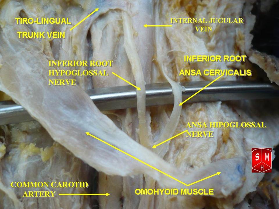
The omohyoid muscle is a muscle that depresses the hyoid. It is located in the front of the neck, and consists of two bellies separated by an intermediate tendon. The omohyoid muscle is proximally attached to the scapula and distally attached to the hyoid bone, stabilising it. Its superior belly serves as the most lateral member of the infrahyoid muscles, located lateral to both the sternothyroid muscles and the thyrohyoid muscles.Illustrated Anatomy of the Head and Neck, Fehrenbach and Herring, Elsevier, 2012, page 102 Structure The omohyoid muscle arises from the upper border of the scapula, inserting into the lower border of the body of the hyoid bone. It has two separate bellies, superior and inferior: * The ''inferior belly'' forms a flat, narrow fasciculus, which inclines forward and slightly upward across the lower part of the neck, being bound down to the clavicle by a fibrous expansion; it then passes behind the sternocleidomastoid, becomes tendinous and changes its ... [...More Info...] [...Related Items...] OR: [Wikipedia] [Google] [Baidu] |
Upper Border Of The Scapula
The scapula (plural scapulae or scapulas), also known as the shoulder blade, is the bone that connects the humerus (upper arm bone) with the clavicle (collar bone). Like their connected bones, the scapulae are paired, with each scapula on either side of the body being roughly a mirror image of the other. The name derives from the Classical Latin word for trowel or small shovel, which it was thought to resemble. In compound terms, the prefix omo- is used for the shoulder blade in medical terminology. This prefix is derived from ὦμος (ōmos), the Ancient Greek word for shoulder, and is cognate with the Latin , which in Latin signifies either the shoulder or the upper arm bone. The scapula forms the back of the shoulder girdle. In humans, it is a flat bone, roughly triangular in shape, placed on a posterolateral aspect of the thoracic cage. Structure The scapula is a thick, flat bone lying on the thoracic wall that provides an attachment for three groups of muscles: ... [...More Info...] [...Related Items...] OR: [Wikipedia] [Google] [Baidu] |
Clavicle
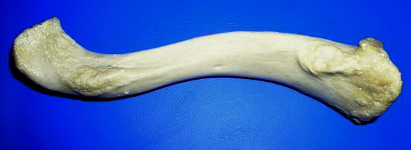
The clavicle, or collarbone, is a slender, S-shaped long bone approximately 6 inches (15 cm) long that serves as a strut between the shoulder blade and the sternum (breastbone). There are two clavicles, one on the left and one on the right. The clavicle is the only long bone in the body that lies horizontally. Together with the shoulder blade, it makes up the shoulder girdle. It is a palpable bone and, in people who have less fat in this region, the location of the bone is clearly visible. It receives its name from the Latin ''clavicula'' ("little key"), because the bone rotates along its axis like a key when the shoulder is abducted. The clavicle is the most commonly fractured bone. It can easily be fractured by impacts to the shoulder from the force of falling on outstretched arms or by a direct hit. Structure The collarbone is a thin doubly curved long bone that connects the arm to the trunk of the body. Located directly above the first rib, it acts as a strut t ... [...More Info...] [...Related Items...] OR: [Wikipedia] [Google] [Baidu] |
Muscular Triangle
The inferior carotid triangle (or muscular triangle), is bounded, in front, by the median line of the neck from the hyoid bone to the sternum; behind, by the anterior margin of the sternocleidomastoid; above, by the superior belly of the omohyoid. It is covered by the integument, superficial fascia, platysma, and deep fascia, ramifying in which are some of the branches of the supraclavicular nerves. Beneath these superficial structures are the sternohyoid and sternothyroid, which, together with the anterior margin of the sternocleidomastoid, conceal the lower part of the common carotid artery. This vessel is enclosed within its sheath, together with the internal jugular vein and vagus nerve; the vein lies lateral to the artery on the right side of the neck, but overlaps it below on the left side; the nerve lies between the artery and vein, on a plane posterior to both. In front of the sheath are a few descending filaments from the ansa cervicalis; behind the sheath are the infe ... [...More Info...] [...Related Items...] OR: [Wikipedia] [Google] [Baidu] |
Carotid Triangle
The carotid triangle (or superior carotid triangle) is a portion of the anterior triangle of the neck. Coverings and boundaries It is bounded: * Posteriorly by the anterior border of the Sternocleidomastoid; * Anteroinferiorly, by the superior belly of the Omohyoid muscle. * Superiorly by the posterior belly of the digastric muscle. It is (covered) by the integument, superficial fascia, Platysma and deep fascia; ramifying in which are branches of the facial and cutaneous cervical nerves. Its floor is formed by parts of the * Thyrohyoid membrane, *Hyoglossus, and the *Constrictores pharyngis medius and inferior. Arteries The external and internal carotids lie side by side, the external being the more anterior of the two. The following branches of the external carotid are also met with in this space: * the superior thyroid artery, running forward and downward; * the lingual artery, directly forward; * the facial artery, forward and upward; * the occipital artery, backward ... [...More Info...] [...Related Items...] OR: [Wikipedia] [Google] [Baidu] |
Anterior Triangle
The anterior triangle is a region of the neck. Structure The triangle is inverted with its apex inferior to its base which is under the chin. Investing fascia covers the roof of the triangle while visceral fascia covers the floor. Anatomy Muscles: * Suprahyoid muscles - Digastric (Ant and Post Belly), mylohyoid, geniohyoid and Stylohyoid. * Infrahyoid muscles - Omohyoid, Sternohyoid, Sternothyroid, and Thyrohyoid. Nerve supply 2 Bellies of Digastric * Anterior: Mylohyoid nerve * Posterior: Facial nerve Stylohyoid: by the facial nerve, by a branch from that to the posterior belly of digastric. Mylohyoid: by its own nerve, a branch of the inferior alveolar ( from the mandibular division of trigeminal nerve), which arises just before the parent nerve enters the mandibular foramen, pierces the sphenomandibular ligament, and runs forward on the inferior surface of the mylohyoid, supplying it and the anterior belly of the digastric. Geniohyoid: by a branch from the hypoglossal n ... [...More Info...] [...Related Items...] OR: [Wikipedia] [Google] [Baidu] |
Subclavian Triangle
The subclavian triangle (or supraclavicular triangle, omoclavicular triangle, Ho's triangle), the smaller division of the posterior triangle, is bounded, above, by the inferior belly of the omohyoideus; below, by the clavicle; its base is formed by the posterior border of the sternocleidomastoideus. Its floor is formed by the first rib with the first digitation of the serratus anterior. The size of the subclavian triangle varies with the extent of attachment of the clavicular portions of the Sternocleidomastoideus and Trapezius, and also with the height at which the Omohyoideus crosses the neck. Its height also varies according to the position of the arm, being diminished by raising the limb, on account of the ascent of the clavicle, and increased by drawing the arm downward, when that bone is depressed. This space is covered by the integument, the superficial and deep fasciæ and the platysma, and crossed by the supraclavicular nerves. Just above the level of the clavicl ... [...More Info...] [...Related Items...] OR: [Wikipedia] [Google] [Baidu] |
Occipital Triangle
The occipital bone () is a cranial dermal bone and the main bone of the occiput (back and lower part of the skull). It is trapezoidal in shape and curved on itself like a shallow dish. The occipital bone overlies the occipital lobes of the cerebrum. At the base of skull in the occipital bone, there is a large oval opening called the foramen magnum, which allows the passage of the spinal cord. Like the other cranial bones, it is classed as a flat bone. Due to its many attachments and features, the occipital bone is described in terms of separate parts. From its front to the back is the basilar part, also called the basioccipital, at the sides of the foramen magnum are the lateral parts, also called the exoccipitals, and the back is named as the squamous part. The basilar part is a thick, somewhat quadrilateral piece in front of the foramen magnum and directed towards the pharynx. The squamous part is the curved, expanded plate behind the foramen magnum and is the largest part o ... [...More Info...] [...Related Items...] OR: [Wikipedia] [Google] [Baidu] |
Posterior Triangle Of The Neck
Posterior may refer to: * Posterior (anatomy), the end of an organism opposite to its head ** Buttocks, as a euphemism * Posterior horn (other) * Posterior probability The posterior probability is a type of conditional probability that results from updating the prior probability with information summarized by the likelihood via an application of Bayes' rule. From an epistemological perspective, the posterior p ..., the conditional probability that is assigned when the relevant evidence is taken into account * Posterior tense, a relative future tense {{disambiguation ... [...More Info...] [...Related Items...] OR: [Wikipedia] [Google] [Baidu] |
Superior Root Of Ansa Cervicalis
The ansa cervicalis (or ansa hypoglossi in older literature) is a loop of nerves that are part of the cervical plexus. It lies superficial to the internal jugular vein in the carotid triangle. Its name means "handle of the neck" in Latin. Branches from the ansa cervicalis innervate most of the infrahyoid muscles, including the sternothyroid muscle, sternohyoid muscle and the omohyoid muscle. Note that the thyrohyoid muscle, which is also an infrahyoid muscle and the geniohyoid muscle which is a suprahyoid muscle are innervated by cervical spinal nerve 1 via the hypoglossal nerve. Roots Two roots make up the ansa cervicalis, a superior root, and an inferior root. The superior root of the ansa cervicalis is formed from cervical spinal nerve 1 of the cervical plexus. These nerve fibers travel in the hypoglossal nerve before separating in the carotid triangle to form the superior root. The superior root goes around the occipital artery and then descends on the carotid sheath. It ... [...More Info...] [...Related Items...] OR: [Wikipedia] [Google] [Baidu] |
Ansa Cervicalis
The ansa cervicalis (or ansa hypoglossi in older literature) is a loop of nerves that are part of the cervical plexus. It lies superficial to the internal jugular vein in the carotid triangle. Its name means "handle of the neck" in Latin. Branches from the ansa cervicalis innervate most of the infrahyoid muscles, including the sternothyroid muscle, sternohyoid muscle and the omohyoid muscle. Note that the thyrohyoid muscle, which is also an infrahyoid muscle and the geniohyoid muscle which is a suprahyoid muscle are innervated by cervical spinal nerve 1 via the hypoglossal nerve. Roots Two roots make up the ansa cervicalis, a superior root, and an inferior root. The superior root of the ansa cervicalis is formed from cervical spinal nerve 1 of the cervical plexus. These nerve fibers travel in the hypoglossal nerve before separating in the carotid triangle to form the superior root. The superior root goes around the occipital artery and then descends on the caroti ... [...More Info...] [...Related Items...] OR: [Wikipedia] [Google] [Baidu] |
Cervical Plexus
The cervical plexus is a plexus of the anterior rami of the first four cervical spinal nerves which arise from C1 to C4 cervical segment in the neck. They are located laterally to the transverse processes between prevertebral muscles from the medial side and vertebral (m. scalenus, m. levator scapulae, m. splenius cervicis) from lateral side. There is anastomosis with accessory nerve, hypoglossal nerve and sympathetic trunk. It is located in the neck, deep to the sternocleidomastoid muscle. Nerves formed from the cervical plexus innervate the back of the head, as well as some neck muscles. The branches of the cervical plexus emerge from the posterior triangle at the nerve point, a point which lies midway on the posterior border of the sternocleidomastoid. Branches The cervical plexus has two types of branches: cutaneous and muscular. *Cutaneous (4 branches): ** Lesser occipital nerve - innervates the skin and the scalp posterosuperior to the auricle (C2) ** Great auricu ... [...More Info...] [...Related Items...] OR: [Wikipedia] [Google] [Baidu] |
Scapular Notch
The suprascapular notch (or ''scapular notch'') is a notch in the superior border of the scapula, just medial to the base of the coracoid process. It forms the entrance site into the suprascapular canal. Structure This notch is converted into a foramen by the suprascapular ligament, and serves for the passage of the suprascapular nerve; sometimes the ligament is ossified. The suprascapular vessels varies in number as well as in their course as they run at the suprascapular notch site. The suprascapular artery pass above the suprascapular ligament in most cases. The suprascapular vein been found to pass above the suprascapular ligament as well as passing through the suprascapular notch. Types Two main classification systems exists with others being modified approaches of the same principle. Typing based on subjective observation of the suprascapular notch shape. Introduced by and modified by . There are six basic types of scapular notch: * Type I: Notch is absent. The sup ... [...More Info...] [...Related Items...] OR: [Wikipedia] [Google] [Baidu] |


