|
Occipitalis
The occipitalis muscle (occipital belly) is a muscle which covers parts of the skull. Some sources consider the occipital muscle to be a distinct muscle. However, Terminologia Anatomica currently classifies it as part of the occipitofrontalis muscle along with the frontalis muscle. The occipitalis muscle is thin and quadrilateral in form. It arises from tendinous fibers from the lateral two-thirds of the superior nuchal line of the occipital bone and from the mastoid process of the temporal and ends in the epicranial aponeurosis. The occipitalis muscle is innervated by the facial nerve and its function is to move the scalp back. The muscles receives blood from the occipital artery. Additional image File:Occipitalis muscle animation small.gif, Position of occipitalis muscle (shown in red). See also * Occipitofrontalis muscle The occipitofrontalis muscle (epicranius muscle) is a muscle which covers parts of the skull. It consists of two parts or bellies: the occipital belly, ... [...More Info...] [...Related Items...] OR: [Wikipedia] [Google] [Baidu] |
Posterior Auricular Nerve
The posterior auricular nerve is a nerve of the head. It is a branch of the facial nerve (CN VII). It communicates with branches from the vagus nerve, the great auricular nerve, and the lesser occipital nerve. Its auricular branch supplies the posterior auricular muscle, the intrinsic muscles of the auricle, and gives sensation to the auricle. Its occipital branch supplies the occipitalis muscle. Structure The posterior auricular nerve arises from the facial nerve (CN VII). It is the first branch outside of the skull. This origin is close to the stylomastoid foramen. It runs upward in front of the mastoid process. It is joined by a branch from the auricular branch of the vagus nerve (CN X). It communicates with the posterior branch of the great auricular nerve, as well as with the lesser occipital nerve. As it ascends between the external acoustic meatus and mastoid process it divides into auricular and occipital branches. * The ''auricular branch'' travels to the posteri ... [...More Info...] [...Related Items...] OR: [Wikipedia] [Google] [Baidu] |
Mastoid Process
The mastoid part of the temporal bone is the posterior (back) part of the temporal bone, one of the bones of the skull. Its rough surface gives attachment to various muscles (via tendons) and it has openings for blood vessels. From its borders, the mastoid part articulates with two other bones. Etymology The word "mastoid" is derived from the Greek word for "breast", a reference to the shape of this bone. Surfaces Outer surface Its outer surface is rough and gives attachment to the occipitalis and posterior auricular muscles. It is perforated by numerous foramina (holes); for example, the mastoid foramen is situated near the posterior border and transmits a vein to the transverse sinus and a small branch of the occipital artery to the dura mater. The position and size of this foramen are very variable; it is not always present; sometimes it is situated in the occipital bone, or in the suture between the temporal and the occipital. Mastoid process The mastoid process is ... [...More Info...] [...Related Items...] OR: [Wikipedia] [Google] [Baidu] |
Frontalis Muscle
The frontalis muscle () is a muscle which covers parts of the forehead of the skull. Some sources consider the frontalis muscle to be a distinct muscle. However, Terminologia Anatomica currently classifies it as part of the occipitofrontalis muscle along with the occipitalis muscle. In humans, the frontalis muscle only serves for facial expressions. The frontalis muscle is supplied by the facial nerve and receives blood from the supraorbital and supratrochlear arteries. Structure The frontalis muscle is thin, of a quadrilateral form, and intimately adherent to the superficial fascia. It is broader than the occipitalis and its fibers are longer and paler in color. It is located on the front of the head. The muscle has no bony attachments. Its medial fibers are continuous with those of the procerus; its intermediate fibers blend with the corrugator and orbicularis oculi muscles, thus attached to the skin of the eyebrows; and its lateral fibers are also blended with the latter mus ... [...More Info...] [...Related Items...] OR: [Wikipedia] [Google] [Baidu] |
Superior Nuchal Line
The nuchal lines are four curved lines on the external surface of the occipital bone: * The upper, often faintly marked, is named the highest nuchal line, but is sometimes referred to as the Mempin line or linea suprema, and it attaches to the epicranial aponeurosis. * Below the highest nuchal line is the superior nuchal line. To it is attached, the splenius capitis muscle, the trapezius muscle, and the occipitalis. * From the external occipital protuberance a ridge or crest, the external occipital crest also called the median nuchal line, often faintly marked, descends to the foramen magnum, and affords attachment to the nuchal ligament. * Running from the middle of this line is the inferior nuchal line. Attached are the obliquus capitis superior muscle, rectus capitis posterior major muscle, and rectus capitis posterior minor muscle The rectus capitis posterior minor (or rectus capitis posticus minor, both being Latin for ''lesser posterior straight muscle of the head'') arises ... [...More Info...] [...Related Items...] OR: [Wikipedia] [Google] [Baidu] |
Human Skull
The skull is a bone protective cavity for the brain. The skull is composed of four types of bone i.e., cranial bones, facial bones, ear ossicles and hyoid bone. However two parts are more prominent: the cranium and the mandible. In humans, these two parts are the neurocranium and the viscerocranium ( facial skeleton) that includes the mandible as its largest bone. The skull forms the anterior-most portion of the skeleton and is a product of cephalisation—housing the brain, and several sensory structures such as the eyes, ears, nose, and mouth. In humans these sensory structures are part of the facial skeleton. Functions of the skull include protection of the brain, fixing the distance between the eyes to allow stereoscopic vision, and fixing the position of the ears to enable sound localisation of the direction and distance of sounds. In some animals, such as horned ungulates (mammals with hooves), the skull also has a defensive function by providing the mount (on the front ... [...More Info...] [...Related Items...] OR: [Wikipedia] [Google] [Baidu] |
Occipital Artery
The occipital artery arises from the external carotid artery opposite the facial artery. Its path is below the posterior belly of digastric to the occipital region. This artery supplies blood to the back of the scalp and sternocleidomastoid muscles, and deep muscles in the back and neck. Structure At its origin, it is covered by the posterior belly of the digastricus and the stylohyoideus, and the hypoglossal nerve winds around it from behind forward; higher up, it crosses the internal carotid artery, the internal jugular vein, and the vagus and accessory nerves. It next ascends to the interval between the transverse process of the atlas and the mastoid process of the temporal bone, and passes horizontally backward, grooving the surface of the latter bone, being covered by the sternocleidomastoideus, splenius capitis, longissimus capitis, and digastricus, and resting upon the rectus capitis lateralis, the obliquus superior, and semispinalis capitis. It then changes its course and ... [...More Info...] [...Related Items...] OR: [Wikipedia] [Google] [Baidu] |
Superior Nuchal Line
The nuchal lines are four curved lines on the external surface of the occipital bone: * The upper, often faintly marked, is named the highest nuchal line, but is sometimes referred to as the Mempin line or linea suprema, and it attaches to the epicranial aponeurosis. * Below the highest nuchal line is the superior nuchal line. To it is attached, the splenius capitis muscle, the trapezius muscle, and the occipitalis. * From the external occipital protuberance a ridge or crest, the external occipital crest also called the median nuchal line, often faintly marked, descends to the foramen magnum, and affords attachment to the nuchal ligament. * Running from the middle of this line is the inferior nuchal line. Attached are the obliquus capitis superior muscle, rectus capitis posterior major muscle, and rectus capitis posterior minor muscle The rectus capitis posterior minor (or rectus capitis posticus minor, both being Latin for ''lesser posterior straight muscle of the head'') arises ... [...More Info...] [...Related Items...] OR: [Wikipedia] [Google] [Baidu] |
Occipitofrontalis Muscle
The occipitofrontalis muscle (epicranius muscle) is a muscle which covers parts of the skull. It consists of two parts or bellies: the occipital belly, near the occipital bone, and the frontal belly, near the frontal bone. It is supplied by the supraorbital artery, the supratrochlear artery, and the occipital artery. It is innervated by the facial nerve. In humans, the occipitofrontalis helps to create facial expressions. Structure The occipitofrontalis muscle consists of two parts or bellies: * the occipital belly, near the occipital bone. It originates on the lateral two-thirds of the highest nuchal line, and on the mastoid process of the temporal bone. It inserts into the epicranial aponeurosis. * the frontal belly, near the frontal bone. It originates from an intermediate tendon that connects to the occipital belly. It inserts in the fascia of the facial muscles and in the skin above the eyes and nose. Some sources consider the occipital and frontal bellies to be ... [...More Info...] [...Related Items...] OR: [Wikipedia] [Google] [Baidu] |
Occipital Artery
The occipital artery arises from the external carotid artery opposite the facial artery. Its path is below the posterior belly of digastric to the occipital region. This artery supplies blood to the back of the scalp and sternocleidomastoid muscles, and deep muscles in the back and neck. Structure At its origin, it is covered by the posterior belly of the digastricus and the stylohyoideus, and the hypoglossal nerve winds around it from behind forward; higher up, it crosses the internal carotid artery, the internal jugular vein, and the vagus and accessory nerves. It next ascends to the interval between the transverse process of the atlas and the mastoid process of the temporal bone, and passes horizontally backward, grooving the surface of the latter bone, being covered by the sternocleidomastoideus, splenius capitis, longissimus capitis, and digastricus, and resting upon the rectus capitis lateralis, the obliquus superior, and semispinalis capitis. It then changes its course and ... [...More Info...] [...Related Items...] OR: [Wikipedia] [Google] [Baidu] |
Scalp
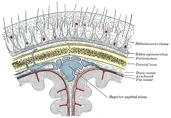
The scalp is the anatomical area bordered by the human face at the front, and by the neck at the sides and back. Structure The scalp is usually described as having five layers, which can conveniently be remembered as a mnemonic: * S: The skin on the head from which head hair grows. It contains numerous sebaceous glands and hair follicles. * C: Connective tissue. A dense subcutaneous layer of fat and fibrous tissue that lies beneath the skin, containing the nerves and vessels of the scalp. * A: The aponeurosis called epicranial aponeurosis (or galea aponeurotica) is the next layer. It is a tough layer of dense fibrous tissue which runs from the frontalis muscle anteriorly to the occipitalis posteriorly. * L: The loose areolar connective tissue layer provides an easy plane of separation between the upper three layers and the pericranium. In scalping the scalp is torn off through this layer. It also provides a plane of access in craniofacial surgery and neurosurgery. This layer i ... [...More Info...] [...Related Items...] OR: [Wikipedia] [Google] [Baidu] |
Occipital Bone
The occipital bone () is a neurocranium, cranial dermal bone and the main bone of the occiput (back and lower part of the skull). It is trapezoidal in shape and curved on itself like a shallow dish. The occipital bone overlies the occipital lobes of the cerebrum. At the base of skull in the occipital bone, there is a large oval opening called the foramen magnum, which allows the passage of the spinal cord. Like the other cranial bones, it is classed as a flat bone. Due to its many attachments and features, the occipital bone is described in terms of separate parts. From its front to the back is the basilar part of occipital bone, basilar part, also called the basioccipital, at the sides of the foramen magnum are the lateral parts of occipital bone, lateral parts, also called the exoccipitals, and the back is named as the squamous part of occipital bone, squamous part. The basilar part is a thick, somewhat quadrilateral piece in front of the foramen magnum and directed towards the ... [...More Info...] [...Related Items...] OR: [Wikipedia] [Google] [Baidu] |


