|
Occipital
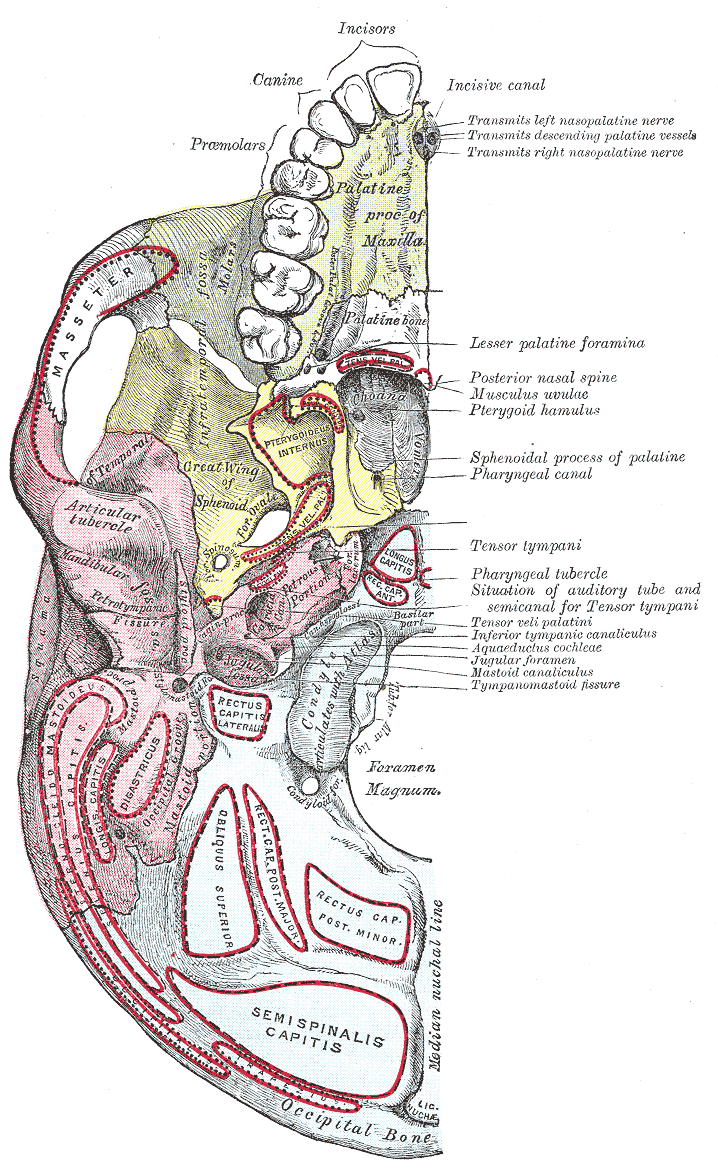
The occipital bone () is a cranial dermal bone and the main bone of the occiput (back and lower part of the skull). It is trapezoidal in shape and curved on itself like a shallow dish. The occipital bone overlies the occipital lobes of the cerebrum. At the base of skull in the occipital bone, there is a large oval opening called the foramen magnum, which allows the passage of the spinal cord. Like the other cranial bones, it is classed as a flat bone. Due to its many attachments and features, the occipital bone is described in terms of separate parts. From its front to the back is the basilar part, also called the basioccipital, at the sides of the foramen magnum are the lateral parts, also called the exoccipitals, and the back is named as the squamous part. The basilar part is a thick, somewhat quadrilateral piece in front of the foramen magnum and directed towards the pharynx. The squamous part is the curved, expanded plate behind the foramen magnum and is the largest part o ... [...More Info...] [...Related Items...] OR: [Wikipedia] [Google] [Baidu] |
Occipital Lobe
The occipital lobe is one of the four major lobes of the cerebral cortex in the brain of mammals. The name derives from its position at the back of the head, from the Latin ''ob'', "behind", and ''caput'', "head". The occipital lobe is the visual processing center of the mammalian brain containing most of the anatomical region of the visual cortex. The primary visual cortex is Brodmann area 17, commonly called V1 (visual one). Human V1 is located on the medial side of the occipital lobe within the calcarine sulcus; the full extent of V1 often continues onto the occipital pole. V1 is often also called striate cortex because it can be identified by a large stripe of myelin, the Stria of Gennari. Visually driven regions outside V1 are called extrastriate cortex. There are many extrastriate regions, and these are specialized for different visual tasks, such as visuospatial processing, color differentiation, and motion perception. Bilateral lesions of the occipital lobe can lead ... [...More Info...] [...Related Items...] OR: [Wikipedia] [Google] [Baidu] |
Occipital Condyles
The occipital condyles are undersurface protuberances of the occipital bone in vertebrates, which function in articulation with the superior facets of the atlas vertebra. The condyles are oval or reniform (kidney-shaped) in shape, and their anterior extremities, directed forward and medialward, are closer together than their posterior, and encroach on the basilar portion of the bone; the posterior extremities extend back to the level of the middle of the foramen magnum. The articular surfaces of the condyles are convex from before backward and from side to side, and look downward and lateralward. To their margins are attached the capsules of the atlanto-occipital joints, and on the medial side of each is a rough impression or tubercle for the alar ligament. At the base of either condyle the bone is tunnelled by a short canal, the hypoglossal canal. Clinical significance Fracture of an occipital condyle may occur in isolation, or as part of a more extended basilar skull fracture ... [...More Info...] [...Related Items...] OR: [Wikipedia] [Google] [Baidu] |
Lateral Parts Of Occipital Bone
The lateral parts of the occipital bone (also called the exoccipitals) are situated at the sides of the foramen magnum; on their under surfaces are the condyles for articulation with the superior facets of the atlas. Description The condyles are oval or reniform (kidney-shaped) in shape, and their anterior extremities, directed forward and medialward, are closer together than their posterior, and encroach on the basilar portion of the bone; the posterior extremities extend back to the level of the middle of the foramen magnum. The articular surfaces of the condyles are convex from before backward and from side to side, and look downward and lateralward. To their margins are attached the capsules of the atlantoöccipital articulations, and on the medial side of each is a rough impression or tubercle for the alar ligament. At the base of either condyle the bone is tunnelled by a short canal, the hypoglossal canal (anterior condyloid foramen). This begins on the cranial surface o ... [...More Info...] [...Related Items...] OR: [Wikipedia] [Google] [Baidu] |
Basilar Part Of Occipital Bone
The basilar part of the occipital bone (also basioccipital) extends forward and upward from the foramen magnum, and presents in front an area more or less quadrilateral in outline. In the young skull this area is rough and uneven, and is joined to the body of the sphenoid by a plate of cartilage. By the twenty-fifth year this cartilaginous plate is ossified, and the occipital and sphenoid form a continuous bone. Surfaces On its ''lower surface'', about 1 cm. in front of the foramen magnum, is the pharyngeal tubercle which gives attachment to the fibrous raphe of the pharynx. On either side of the middle line the longus capitis and rectus capitis anterior are inserted, and immediately in front of the foramen magnum the anterior atlantooccipital membrane is attached. The ''upper surface'', which constitutes the lower half of the clivus, presents a broad, shallow groove which inclines upward and forward from the foramen magnum; it supports the medulla oblongata, and ... [...More Info...] [...Related Items...] OR: [Wikipedia] [Google] [Baidu] |
Foramen Magnum
The foramen magnum ( la, great hole) is a large, oval-shaped opening in the occipital bone of the skull. It is one of the several oval or circular openings (foramina) in the base of the skull. The spinal cord, an extension of the medulla oblongata, passes through the foramen magnum as it exits the cranial cavity. Apart from the transmission of the medulla oblongata and its membranes, the foramen magnum transmits the vertebral arteries, the anterior and posterior spinal arteries, the tectorial membranes and alar ligaments. It also transmits the accessory nerve into the skull. The foramen magnum is a very important feature in bipedal mammals. One of the attributes of a biped's foramen magnum is a forward shift of the anterior border of the cerebellar tentorium; this is caused by the shortening of the cranial base. Studies on the foramen magnum position have shown a connection to the functional influences of both posture and locomotion. The forward shift of the foramen magnum i ... [...More Info...] [...Related Items...] OR: [Wikipedia] [Google] [Baidu] |
Nuchal Plane
The squamous part of occipital bone is situated above and behind the foramen magnum, and is curved from above downward and from side to side. External surface The external surface is convex and presents midway between the summit of the bone and the foramen magnum a prominence, the external occipital protuberance and inion. Extending lateralward from this on either side are two curved lines, one a little above the other. The upper, often faintly marked, is named the highest nuchal line, and to it the epicranial aponeurosis is attached. The lower is termed the superior nuchal line. That area of the squamous part, which lies above the highest nuchal lines is named the occipital plane ''(planum occipitale)'' and is covered by the ''occipitalis muscle''. That below, termed the nuchal plane, is rough and irregular for the attachment of several muscles. From the external occipital protuberance, an often faintly marked ridge or crest, the median nuchal line, descends to the foramen ma ... [...More Info...] [...Related Items...] OR: [Wikipedia] [Google] [Baidu] |
Occipital Plane
The squamous part of occipital bone is situated above and behind the foramen magnum, and is curved from above downward and from side to side. External surface The external surface is convex and presents midway between the summit of the bone and the foramen magnum a prominence, the external occipital protuberance and inion. Extending lateralward from this on either side are two curved lines, one a little above the other. The upper, often faintly marked, is named the highest nuchal line, and to it the epicranial aponeurosis is attached. The lower is termed the superior nuchal line. That area of the squamous part, which lies above the highest nuchal lines is named the occipital plane ''(planum occipitale)'' and is covered by the ''occipitalis muscle''. That below, termed the nuchal plane, is rough and irregular for the attachment of several muscles. From the external occipital protuberance, an often faintly marked ridge or crest, the median nuchal line, descends to the foramen ma ... [...More Info...] [...Related Items...] OR: [Wikipedia] [Google] [Baidu] |
Squamous Part Of Occipital Bone
The squamous part of occipital bone is situated above and behind the foramen magnum, and is curved from above downward and from side to side. External surface The external surface is convex and presents midway between the summit of the bone and the foramen magnum a prominence, the external occipital protuberance and external occipital protuberance, inion. Extending lateralward from this on either side are two curved lines, one a little above the other. The upper, often faintly marked, is named the nuchal lines, highest nuchal line, and to it the epicranial aponeurosis is attached. The lower is termed the nuchal lines, superior nuchal line. That area of the squamous part, which lies above the highest nuchal lines is named the occipital plane ''(planum occipitale)'' and is covered by the ''occipitalis muscle''. That below, termed the nuchal plane, is rough and irregular for the attachment of several muscles. From the external occipital protuberance, an often faintly marked ridge ... [...More Info...] [...Related Items...] OR: [Wikipedia] [Google] [Baidu] |
External Occipital Protuberance
Near the middle of the squamous part of occipital bone is the external occipital protuberance, the highest point of which is referred to as the inion. The inion is the most prominent projection of the protuberance which is located at the posterioinferior (rear lower) part of the human skull. The nuchal ligament and trapezius muscle attach to it. The inion (ἰνίον, iníon, Greek for the occipital bone) is used as a landmark in the 10-20 system in electroencephalography (EEG) recording. Extending laterally from it on either side is the superior nuchal line, and above it is the faintly marked highest nuchal line. A study of 16th-century Anatolian remains showed that the external occipital protuberance statistically tends to be less pronounced in female remains. Additional images See also * Internal occipital protuberance * Occipital bun The occipital bone () is a cranial dermal bone and the main bone of the occiput (back and lower part of the skull). It is trapezoidal in ... [...More Info...] [...Related Items...] OR: [Wikipedia] [Google] [Baidu] |
Base Of Skull
The base of skull, also known as the cranial base or the cranial floor, is the most inferior area of the skull. It is composed of the endocranium and the lower parts of the calvaria. Structure Structures found at the base of the skull are for example: Bones There are five bones that make up the base of the skull: *Ethmoid bone *Sphenoid bone *Occipital bone *Frontal bone *Temporal bone Sinuses *Occipital sinus *Superior sagittal sinus *Superior petrosal sinus Foramina of the skull * Foramen cecum *Optic foramen *Foramen lacerum *Foramen rotundum *Foramen magnum * Foramen ovale *Jugular foramen *Internal auditory meatus *Mastoid foramen *Sphenoidal emissary foramen *Foramen spinosum Sutures *Frontoethmoidal suture *Sphenofrontal suture *Sphenopetrosal suture *Sphenoethmoidal suture * Petrosquamous suture *Sphenosquamosal suture Other *Sphenoidal lingula *Subarcuate fossa *Dorsum sellae *Jugular process *Petro-occipital fissure *Condylar canal *Jugular tubercle *Tuberc ... [...More Info...] [...Related Items...] OR: [Wikipedia] [Google] [Baidu] |
Nuchal Line
The nuchal lines are four curved lines on the external surface of the occipital bone: * The upper, often faintly marked, is named the highest nuchal line, but is sometimes referred to as the Mempin line or linea suprema, and it attaches to the epicranial aponeurosis. * Below the highest nuchal line is the superior nuchal line. To it is attached, the splenius capitis muscle, the trapezius muscle, and the occipitalis. * From the external occipital protuberance a ridge or crest, the external occipital crest also called the median nuchal line, often faintly marked, descends to the foramen magnum, and affords attachment to the nuchal ligament. * Running from the middle of this line is the inferior nuchal line. Attached are the obliquus capitis superior muscle, rectus capitis posterior major muscle, and rectus capitis posterior minor muscle The rectus capitis posterior minor (or rectus capitis posticus minor, both being Latin for ''lesser posterior straight muscle of the head'') arises ... [...More Info...] [...Related Items...] OR: [Wikipedia] [Google] [Baidu] |
Nuchal Ligament
The nuchal ligament is a ligament at the back of the neck that is continuous with the supraspinous ligament. Structure The nuchal ligament extends from the external occipital protuberance on the skull and median nuchal line to the spinous process of the seventh cervical vertebra in the lower part of the neck. From the anterior border of the nuchal ligament, a fibrous lamina is given off. This is attached to the posterior tubercle of the atlas, and to the spinous processes of the cervical vertebrae, and forms a septum between the muscles on either side of the neck. The trapezius and splenius capitis muscle attach to the nuchal ligament. Function It is a tendon-like structure that has developed independently in humans and other animals well adapted for running. In some four-legged animals, particularly ungulates, the nuchal ligament serves to sustain the weight of the head. Clinical significance In Chiari malformation treatment, decompression and duraplasty with a harvested n ... [...More Info...] [...Related Items...] OR: [Wikipedia] [Google] [Baidu] |


