|
Nuclear Atypia
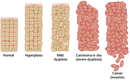
Nuclear atypia refers to abnormal appearance of cell nuclei. It is a term used in cytopathology and histopathology. Atypical nuclei are often pleomorphic. Nuclear atypia can be seen in reactive changes, pre-neoplastic changes and malignancy. Severe nuclear atypia is, in most cases, considered an indicator of malignancy. See also * Arias-Stella reaction *NC ratio The nuclear-cytoplasmic ratio (also variously known as the nucleus:cytoplasm ratio, nucleus-cytoplasm ratio, N:C ratio, or N/C) is a measurement used in cell biology. It is a ratio of the size (i.e., volume) of the cell nucleus, nucleus of a cell ... * Nuclear pleomorphism {{pathology-stub Pathology ... [...More Info...] [...Related Items...] OR: [Wikipedia] [Google] [Baidu] |
Glioblastoma With Extreme Nuclear Enlargement - Very High Mag
Glioblastoma, previously known as glioblastoma multiforme (GBM), is one of the most aggressive types of cancer that begin within the brain. Initially, signs and symptoms of glioblastoma are nonspecific. They may include headaches, personality changes, nausea, and symptoms similar to those of a stroke. Symptoms often worsen rapidly and may progress to unconsciousness. The cause of most cases of glioblastoma is not known. Uncommon risk factors include genetic disorders, such as neurofibromatosis and Li–Fraumeni syndrome, and previous radiation therapy. Glioblastomas represent 15% of all brain tumors. They can either start from normal brain cells or develop from an existing low-grade astrocytoma. The diagnosis typically is made by a combination of a CT scan, MRI scan, and tissue biopsy. There is no known method of preventing the cancer. Treatment usually involves surgery, after which chemotherapy and radiation therapy are used. The medication temozolomide is frequently used as ... [...More Info...] [...Related Items...] OR: [Wikipedia] [Google] [Baidu] |
Cytopathology Of Reactive Urothelial Changes
Cytopathology (from Greek , ''kytos'', "a hollow"; , ''pathos'', "fate, harm"; and , ''-logia'') is a branch of pathology that studies and diagnoses diseases on the cellular level. The discipline was founded by George Nicolas Papanicolaou in 1928. Cytopathology is generally used on samples of free cells or tissue fragments, in contrast to histopathology, which studies whole tissues. Cytopathology is frequently, less precisely, called "cytology", which means "the study of cells". Cytopathology is commonly used to investigate diseases involving a wide range of body sites, often to aid in the diagnosis of cancer but also in the diagnosis of some infectious diseases and other inflammatory conditions. For example, a common application of cytopathology is the Pap smear, a screening tool used to detect precancerous cervical lesions that may lead to cervical cancer. Cytopathologic tests are sometimes called smear tests because the samples may be smeared across a glass microscope slide ... [...More Info...] [...Related Items...] OR: [Wikipedia] [Google] [Baidu] |
Cell Nucleus
The cell nucleus (pl. nuclei; from Latin or , meaning ''kernel'' or ''seed'') is a membrane-bound organelle found in eukaryotic cells. Eukaryotic cells usually have a single nucleus, but a few cell types, such as mammalian red blood cells, have no nuclei, and a few others including osteoclasts have many. The main structures making up the nucleus are the nuclear envelope, a double membrane that encloses the entire organelle and isolates its contents from the cellular cytoplasm; and the nuclear matrix, a network within the nucleus that adds mechanical support. The cell nucleus contains nearly all of the cell's genome. Nuclear DNA is often organized into multiple chromosomes – long stands of DNA dotted with various proteins, such as histones, that protect and organize the DNA. The genes within these chromosomes are structured in such a way to promote cell function. The nucleus maintains the integrity of genes and controls the activities of the cell by regulating gene expres ... [...More Info...] [...Related Items...] OR: [Wikipedia] [Google] [Baidu] |
Cytopathology
Cytopathology (from Greek , ''kytos'', "a hollow"; , ''pathos'', "fate, harm"; and , '' -logia'') is a branch of pathology that studies and diagnoses diseases on the cellular level. The discipline was founded by George Nicolas Papanicolaou in 1928. Cytopathology is generally used on samples of free cells or tissue fragments, in contrast to histopathology, which studies whole tissues. Cytopathology is frequently, less precisely, called "cytology", which means "the study of cells". Cytopathology is commonly used to investigate diseases involving a wide range of body sites, often to aid in the diagnosis of cancer but also in the diagnosis of some infectious diseases and other inflammatory conditions. For example, a common application of cytopathology is the Pap smear, a screening tool used to detect precancerous cervical lesions that may lead to cervical cancer. Cytopathologic tests are sometimes called smear tests because the samples may be smeared across a glass microscope slid ... [...More Info...] [...Related Items...] OR: [Wikipedia] [Google] [Baidu] |
Histopathology
Histopathology (compound of three Greek words: ''histos'' "tissue", πάθος ''pathos'' "suffering", and -λογία '' -logia'' "study of") refers to the microscopic examination of tissue in order to study the manifestations of disease. Specifically, in clinical medicine, histopathology refers to the examination of a biopsy or surgical specimen by a pathologist, after the specimen has been processed and histological sections have been placed onto glass slides. In contrast, cytopathology examines free cells or tissue micro-fragments (as "cell blocks"). Collection of tissues Histopathological examination of tissues starts with surgery, biopsy, or autopsy. The tissue is removed from the body or plant, and then, often following expert dissection in the fresh state, placed in a fixative which stabilizes the tissues to prevent decay. The most common fixative is 10% neutral buffered formalin (corresponding to 3.7% w/v formaldehyde in neutral buffered water, such as phosphate buf ... [...More Info...] [...Related Items...] OR: [Wikipedia] [Google] [Baidu] |
Pleomorphism (cytology)
Pleomorphism is a term used in histology and cytopathology to describe variability in the size, shape and staining of cells and/or their nuclei. Several key determinants of cell and nuclear size, like ploidy and the regulation of cellular metabolism, are commonly disrupted in tumors. Therefore, cellular and nuclear pleomorphism is one of the earliest hallmarks of cancer progression and a feature characteristic of malignant neoplasms and dysplasia. Certain benign cell types may also exhibit pleomorphism, e.g. neuroendocrine cells, Arias-Stella reaction. A rare type of rhabdomyosarcoma that is found in adults is known as pleomorphic rhabdomyosarcoma. Despite the prevalence of pleomorphism in human pathology, its role in disease progression is unclear. In epithelial tissue, pleomorphism in cellular size can induce packing defects and disperse aberrant cells. But the consequence of atypical cell and nuclear morphology in other tissues is unknown. See also *Anaplasia *Cell growt ... [...More Info...] [...Related Items...] OR: [Wikipedia] [Google] [Baidu] |
Precancerous Condition
A precancerous condition is a condition, tumor or lesion involving abnormal cells which are associated with an increased risk of developing into cancer. Clinically, precancerous conditions encompass a variety of abnormal tissues with an increased risk of developing into cancer. Some of the most common precancerous conditions include certain colon polyps, which can progress into colon cancer, monoclonal gammopathy of undetermined significance, which can progress into multiple myeloma or myelodysplastic syndrome. and cervical dysplasia, which can progress into cervical cancer. Bronchial premalignant lesions can progress to squamous cell carcinoma of the lung. Pathologically, precancerous tissue can range from benign neoplasias, which are tumors which don't invade neighboring normal tissues or spread to distant organs, to dysplasia, a collection of highly abnormal cells which, in some cases, has an increased risk of progressing to anaplasia and invasive cancer which is life-threat ... [...More Info...] [...Related Items...] OR: [Wikipedia] [Google] [Baidu] |
Malignancy
Malignancy () is the tendency of a medical condition to become progressively worse. Malignancy is most familiar as a characterization of cancer. A ''malignant'' tumor contrasts with a non-cancerous ''benign'' tumor in that a malignancy is not self-limited in its growth, is capable of invading into adjacent tissues, and may be capable of spreading to distant tissues. A benign tumor has none of those properties. Malignancy in cancers is characterized by anaplasia, invasiveness, and metastasis. Malignant tumors are also characterized by genome instability, so that cancers, as assessed by whole genome sequencing, frequently have between 10,000 and 100,000 mutations in their entire genomes. Cancers usually show tumour heterogeneity, containing multiple subclones. They also frequently have reduced expression of DNA repair enzymes due to epigenetic methylation of DNA repair genes or altered microRNAs that control DNA repair gene expression. Tumours can be detected through the visual ... [...More Info...] [...Related Items...] OR: [Wikipedia] [Google] [Baidu] |
Arias-Stella Reaction
Arias-Stella reaction, also Arias-Stella phenomenon, is a benign change in the endometrium associated with the presence of chorionic tissue. Arias-Stella reaction is due to progesterone primarily. Cytologically, it looks like a malignancy and, historically, it was diagnosed as endometrial cancer. Significance It is significant only because it can be misdiagnosed as a cancer. It may be seen in a completely normal pregnancy. Diagnosis It is characterized by nuclear enlargement and may also have any of the following: an irregular nuclear membrane, granular chromatin, centronuclear vacuolization, and pseudonuclear inclusions. Five subtypes are recognized: #Minimal atypia. #Early secretory pattern. #Secretory or hypersecretory pattern. #Regenerative, proliferative or nonsecretory pattern. #Monstrous cell pattern. History It was first described by Javier Arias Stella, a Peruvian pathologist, in 1954. See also * Choriocarcinoma * Chorioangioma * Herpes * Nuclear atypia Nucl ... [...More Info...] [...Related Items...] OR: [Wikipedia] [Google] [Baidu] |
NC Ratio
The nuclear-cytoplasmic ratio (also variously known as the nucleus:cytoplasm ratio, nucleus-cytoplasm ratio, N:C ratio, or N/C) is a measurement used in cell biology. It is a ratio of the size (i.e., volume) of the nucleus of a cell to the size of the cytoplasm of that cell. The N:C ratio indicates the maturity of a cell, because as a cell matures the size of its nucleus generally decreases. For example, "blast" forms of erythrocytes, leukocytes, and megakaryocytes start with an N:C ratio of 4:1, which decreases as they mature to 2:1 or even 1:1 (with exceptions for mature thrombocytes and erythrocytes, which are anuclear cells, and mature lymphocytes, which only decrease to a 3:1 ratio and often retain the original 4:1 ratio). An increased N:C ratio is commonly associated with precancerous dysplasia as well as with malignant cells. See also * *Cytopathology *Nuclear atypia Nuclear atypia refers to abnormal appearance of cell nuclei. It is a term used in cytopathology and his ... [...More Info...] [...Related Items...] OR: [Wikipedia] [Google] [Baidu] |
Nuclear Pleomorphism
Pleomorphism is a term used in histology and cytopathology to describe variability in the size, shape and staining of cells and/or their nuclei. Several key determinants of cell and nuclear size, like ploidy and the regulation of cellular metabolism, are commonly disrupted in tumors. Therefore, cellular and nuclear pleomorphism is one of the earliest hallmarks of cancer progression and a feature characteristic of malignant neoplasms and dysplasia. Certain benign cell types may also exhibit pleomorphism, e.g. neuroendocrine cells, Arias-Stella reaction. A rare type of rhabdomyosarcoma that is found in adults is known as pleomorphic rhabdomyosarcoma. Despite the prevalence of pleomorphism in human pathology, its role in disease progression is unclear. In epithelial tissue, pleomorphism in cellular size can induce packing defects and disperse aberrant cells. But the consequence of atypical cell and nuclear morphology in other tissues is unknown. See also *Anaplasia *Cell growt ... [...More Info...] [...Related Items...] OR: [Wikipedia] [Google] [Baidu] |


.jpg)
