|
Nodal Cilia
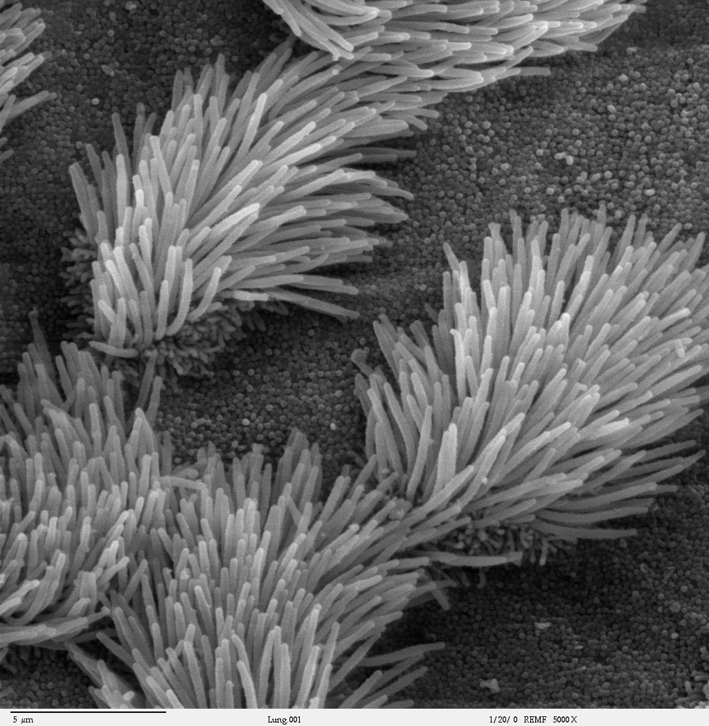
The cilium, plural cilia (), is a membrane-bound organelle found on most types of eukaryotic cell, and certain microorganisms known as ciliates. Cilia are absent in bacteria and archaea. The cilium has the shape of a slender threadlike projection that extends from the surface of the much larger cell body. Eukaryotic flagella found on sperm cells and many protozoans have a similar structure to motile cilia that enables swimming through liquids; they are longer than cilia and have a different undulating motion. There are two major classes of cilia: ''motile'' and ''non-motile'' cilia, each with a subtype, giving four types in all. A cell will typically have one primary cilium or many motile cilia. The structure of the cilium core called the axoneme determines the cilium class. Most motile cilia have a central pair of single microtubules surrounded by nine pairs of double microtubules called a 9+2 axoneme. Most non-motile cilia have a 9+0 axoneme that lacks the central pair of m ... [...More Info...] [...Related Items...] OR: [Wikipedia] [Google] [Baidu] |
9+2 Axoneme
9 (nine) is the natural number following and preceding . Evolution of the Arabic digit In the beginning, various Indians wrote a digit 9 similar in shape to the modern closing question mark without the bottom dot. The Kshatrapa, Andhra and Gupta started curving the bottom vertical line coming up with a -look-alike. The Nagari continued the bottom stroke to make a circle and enclose the 3-look-alike, in much the same way that the sign @ encircles a lowercase ''a''. As time went on, the enclosing circle became bigger and its line continued beyond the circle downwards, as the 3-look-alike became smaller. Soon, all that was left of the 3-look-alike was a squiggle. The Arabs simply connected that squiggle to the downward stroke at the middle and subsequent European change was purely cosmetic. While the shape of the glyph for the digit 9 has an ascender in most modern typefaces, in typefaces with text figures the character usually has a descender, as, for example, in . The mod ... [...More Info...] [...Related Items...] OR: [Wikipedia] [Google] [Baidu] |
Fallopian Tube
The fallopian tubes, also known as uterine tubes, oviducts or salpinges (singular salpinx), are paired tubes in the human female that stretch from the uterus to the ovaries. The fallopian tubes are part of the female reproductive system. In other mammals they are only called oviducts. Each tube is a muscular hollow organ that is on average between 10 and 14 cm in length, with an external diameter of 1 cm. It has four described parts: the intramural part, isthmus, ampulla, and infundibulum with associated fimbriae. Each tube has two openings a proximal opening nearest and opening to the uterus, and a distal opening furthest and opening to the abdomen. The fallopian tubes are held in place by the mesosalpinx, a part of the broad ligament mesentery that wraps around the tubes. Another part of the broad ligament, the mesovarium suspends the ovaries in place. An egg cell is transported from an ovary to a fallopian tube where it may be fertilized in the ampulla of the tu ... [...More Info...] [...Related Items...] OR: [Wikipedia] [Google] [Baidu] |
Brain
A brain is an organ that serves as the center of the nervous system in all vertebrate and most invertebrate animals. It is located in the head, usually close to the sensory organs for senses such as vision. It is the most complex organ in a vertebrate's body. In a human, the cerebral cortex contains approximately 14–16 billion neurons, and the estimated number of neurons in the cerebellum is 55–70 billion. Each neuron is connected by synapses to several thousand other neurons. These neurons typically communicate with one another by means of long fibers called axons, which carry trains of signal pulses called action potentials to distant parts of the brain or body targeting specific recipient cells. Physiologically, brains exert centralized control over a body's other organs. They act on the rest of the body both by generating patterns of muscle activity and by driving the secretion of chemicals called hormones. This centralized control allows rapid and coordinated respon ... [...More Info...] [...Related Items...] OR: [Wikipedia] [Google] [Baidu] |
Ventricular System
The ventricular system is a set of four interconnected cavities known as cerebral ventricles in the brain. Within each ventricle is a region of choroid plexus which produces the circulating cerebrospinal fluid (CSF). The ventricular system is continuous with the central canal of the spinal cord from the fourth ventricle, allowing for the flow of CSF to circulate. All of the ventricular system and the central canal of the spinal cord are lined with ependyma, a specialised form of epithelium connected by tight junctions that make up the blood–cerebrospinal fluid barrier. Structure The system comprises four ventricles: * lateral ventricles right and left (one for each hemisphere) * third ventricle * fourth ventricle There are several foramina, openings acting as channels, that connect the ventricles. The interventricular foramina (also called the foramina of Monro) connect the lateral ventricles to the third ventricle through which the cerebrospinal fluid can flow. Ventric ... [...More Info...] [...Related Items...] OR: [Wikipedia] [Google] [Baidu] |
Cerebrospinal Fluid
Cerebrospinal fluid (CSF) is a clear, colorless body fluid found within the tissue that surrounds the brain and spinal cord of all vertebrates. CSF is produced by specialised ependymal cells in the choroid plexus of the ventricles of the brain, and absorbed in the arachnoid granulations. There is about 125 mL of CSF at any one time, and about 500 mL is generated every day. CSF acts as a shock absorber, cushion or buffer, providing basic mechanical and immunological protection to the brain inside the skull. CSF also serves a vital function in the cerebral autoregulation of cerebral blood flow. CSF occupies the subarachnoid space (between the arachnoid mater and the pia mater) and the ventricular system around and inside the brain and spinal cord. It fills the ventricles of the brain, cisterns, and sulci, as well as the central canal of the spinal cord. There is also a connection from the subarachnoid space to the bony labyrinth of the inner ear via the perilymphat ... [...More Info...] [...Related Items...] OR: [Wikipedia] [Google] [Baidu] |
Ependymal Cells
The ependyma is the thin neuroepithelial ( simple columnar ciliated epithelium) lining of the ventricular system of the brain and the central canal of the spinal cord. The ependyma is one of the four types of neuroglia in the central nervous system (CNS). It is involved in the production of cerebrospinal fluid (CSF), and is shown to serve as a reservoir for neuroregeneration. Structure The ependyma is made up of ependymal cells called ependymocytes, a type of glial cell. These cells line the ventricles in the brain and the central canal of the spinal cord, which become filled with cerebrospinal fluid. These are nervous tissue cells with simple columnar shape, much like that of some mucosal epithelial cells. Early monociliated ependymal cells are differentiated to multiciliated ependymal cells for their function in circulating cerebrospinal fluid. The basal membranes of these cells are characterized by tentacle-like extensions that attach to astrocytes. The apical side is cove ... [...More Info...] [...Related Items...] OR: [Wikipedia] [Google] [Baidu] |
Chemoreceptor
A chemoreceptor, also known as chemosensor, is a specialized sensory receptor which transduces a chemical substance (endogenous or induced) to generate a biological signal. This signal may be in the form of an action potential, if the chemoreceptor is a neuron, or in the form of a neurotransmitter that can activate a nerve fiber if the chemoreceptor is a specialized cell, such as taste receptors, or an internal peripheral chemoreceptor, such as the carotid bodies. In physiology, a chemoreceptor detects changes in the normal environment, such as an increase in blood levels of carbon dioxide (hypercapnia) or a decrease in blood levels of oxygen (hypoxia), and transmits that information to the central nervous system which engages body responses to restore homeostasis. In bacteria, chemoreceptors are essential in the mediation of chemotaxis. Cellular chemoreceptors In prokaryotes Bacteria utilize complex long helical proteins as chemoreceptors, permitting signals to travel long di ... [...More Info...] [...Related Items...] OR: [Wikipedia] [Google] [Baidu] |
Mechanosensation
Mechanosensation is the transduction of mechanical stimuli into neural signals. Mechanosensation provides the basis for the senses of light touch, hearing, proprioception, and pain. Mechanoreceptors found in the skin, called cutaneous mechanoreceptors, are responsible for the sense of touch. Tiny cells in the inner ear, called hair cells, are responsible for hearing and balance. States of neuropathic pain, such as hyperalgesia and allodynia, are also directly related to mechanosensation. A wide array of elements are involved in the process of mechanosensation, many of which are still not fully understood. Cutaneous mechanoreceptors Cutaneous mechanoreceptors are physiologically classified with respect to conduction velocity, which is directly related to the diameter and myelination of the axon. Rapidly adapting and slowly adapting mechanoreceptors Mechanoreceptors that possess a large diameter and high myelination are called ''low-threshold mechanoreceptors''. Fibers that respond ... [...More Info...] [...Related Items...] OR: [Wikipedia] [Google] [Baidu] |
Mucociliary Clearance
Mucociliary clearance (MCC), mucociliary transport, or the mucociliary escalator, describes the self-clearing mechanism of the airways in the respiratory system. It is one of the two protective processes for the lungs in removing inhaled particles including pathogens before they can reach the delicate tissue of the lungs. The other clearance mechanism is provided by the cough reflex. Mucociliary clearance has a major role in pulmonary hygiene. MCC effectiveness relies on the correct properties of the airway surface liquid produced, both of the periciliary sol layer and the overlying mucus gel layer, and of the number and quality of the cilia present in the lining of the airways. An important factor is the rate of mucin secretion. The ion channels CFTR and ENaC work together to maintain the necessary hydration of the airway surface liquid. Any disturbance in the closely regulated functioning of the cilia can cause a disease. Disturbances in the structural formation of the c ... [...More Info...] [...Related Items...] OR: [Wikipedia] [Google] [Baidu] |
Respiratory Epithelium
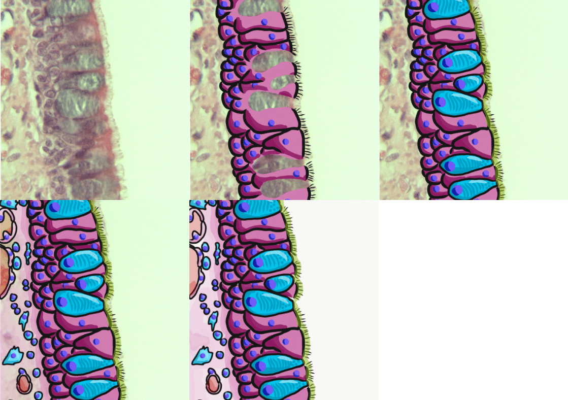
Respiratory epithelium, or airway epithelium, is a type of ciliated columnar epithelium found lining most of the respiratory tract as respiratory mucosa, where it serves to moisten and protect the airways. It is not present in the vocal cords of the larynx, or the oropharynx and laryngopharynx, where instead the epithelium is stratified squamous. It also functions as a barrier to potential pathogens and foreign particles, preventing infection and tissue injury by the secretion of mucus and the action of mucociliary clearance. Structure The respiratory epithelium lining the upper respiratory airways is classified as ciliated pseudostratified columnar epithelium. This designation is due to the arrangement of the multiple cell types composing the respiratory epithelium. While all cells make contact with the basement membrane and are, therefore, a single layer of cells, their nuclei are not aligned in the same plane. Hence, it appears as though several layers of cells are presen ... [...More Info...] [...Related Items...] OR: [Wikipedia] [Google] [Baidu] |
Hair Cell
Hair cells are the sensory receptors of both the auditory system and the vestibular system in the ears of all vertebrates, and in the lateral line organ of fishes. Through mechanotransduction, hair cells detect movement in their environment. In mammals, the auditory hair cells are located within the spiral organ of Corti on the thin basilar membrane in the cochlea of the inner ear. They derive their name from the tufts of stereocilia called ''hair bundles'' that protrude from the apical surface of the cell into the fluid-filled cochlear duct. The stereocilia number from 50-100 in each cell while being tightly packed together and decrease in size the further away they are located from the kinocilium. The hair bundles are arranged as stiff columns that move at their base in response to stimuli applied to the tips. Mammalian cochlear hair cells are of two anatomically and functionally distinct types, known as outer, and inner hair cells. Damage to these hair cells results in ... [...More Info...] [...Related Items...] OR: [Wikipedia] [Google] [Baidu] |


