|
Neurogliaform Cells
Neurogliaform cells (NGF) are inhibitory ( GABAergic) interneurons found in the cortex and the hippocampus. NGF cells represent approximately 10% of the total hippocampal inhibitory interneuron population. In terms of morphology, they are comparatively small and have an unusually high presynaptic bouton density. Almost all NGF neurons express neuropeptide Y (NPY) and are commonly positive for other signalling and non-signalling peptides, including reelin, α-actinin 2, COUP-TFII, and neuronal nitric oxide synthase (nNOS). However, populations of NGF cells which do not express NPY have been reported in both cortical layer I and the striatum. Developmentally, in the cortex, all NGF cells are derived from caudal ganglionic eminence (CGE) but NGF cells of the hippocampus have their origins within both the CGE and the medial ganglionic eminence (MGE). Functionally, NGF cells are GABAergic and their function in the mature brain is inhibition. However, they are suspected to sign ... [...More Info...] [...Related Items...] OR: [Wikipedia] [Google] [Baidu] |
Nitric Oxide Synthase
Nitric oxide synthases () (NOSs) are a family of enzymes catalyzing the production of nitric oxide (NO) from L-arginine. NO is an important cellular signaling molecule. It helps modulate vascular tone, insulin secretion, airway tone, and peristalsis, and is involved in angiogenesis and neural development. It may function as a retrograde neurotransmitter. Nitric oxide is mediated in mammals by the calcium-calmodulin controlled isoenzymes eNOS ( endothelial NOS) and nNOS (neuronal NOS). The inducible isoform, iNOS, involved in immune response, binds calmodulin at physiologically relevant concentrations, and produces NO as an immune defense mechanism, as NO is a free radical with an unpaired electron. It is the proximate cause of septic shock and may function in autoimmune disease. NOS catalyzes the reaction: * 2 L-arginine + 3 NADPH + 3 H+ + 4 O2 \rightleftharpoons 2 citrulline +2 nitric oxide + 4 H2O + 3 NADP+ NOS isoforms catalyze other leak and side reactions, such as superox ... [...More Info...] [...Related Items...] OR: [Wikipedia] [Google] [Baidu] |
Brain
A brain is an organ that serves as the center of the nervous system in all vertebrate and most invertebrate animals. It is located in the head, usually close to the sensory organs for senses such as vision. It is the most complex organ in a vertebrate's body. In a human, the cerebral cortex contains approximately 14–16 billion neurons, and the estimated number of neurons in the cerebellum is 55–70 billion. Each neuron is connected by synapses to several thousand other neurons. These neurons typically communicate with one another by means of long fibers called axons, which carry trains of signal pulses called action potentials to distant parts of the brain or body targeting specific recipient cells. Physiologically, brains exert centralized control over a body's other organs. They act on the rest of the body both by generating patterns of muscle activity and by driving the secretion of chemicals called hormones. This centralized control allows rapid and coordinated respon ... [...More Info...] [...Related Items...] OR: [Wikipedia] [Google] [Baidu] |
Hippocampus (brain)
The hippocampus (via Latin from Ancient Greek, Greek , 'seahorse') is a major component of the brain of humans and other vertebrates. Humans and other mammals have two hippocampi, one in each cerebral hemisphere, side of the brain. The hippocampus is part of the limbic system, and plays important roles in the Memory consolidation, consolidation of information from short-term memory to long-term memory, and in spatial memory that enables navigation. The hippocampus is located in the allocortex, with neural projections into the neocortex in humans, as well as primates. The hippocampus, as the Pallium (neuroanatomy)#In humans, medial pallium, is a structure found in all vertebrates. In humans, it contains two main interlocking parts: the hippocampus proper (also called ''Ammon's horn''), and the dentate gyrus. In Alzheimer's disease (and other forms of dementia), the hippocampus is one of the first regions of the brain to suffer damage; short-term memory, short-term memory loss and ... [...More Info...] [...Related Items...] OR: [Wikipedia] [Google] [Baidu] |
Chemical Synapse
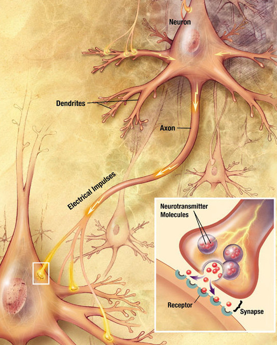
Chemical synapses are biological junctions through which neurons' signals can be sent to each other and to non-neuronal cells such as those in muscles or glands. Chemical synapses allow neurons to form circuits within the central nervous system. They are crucial to the biological computations that underlie perception and thought. They allow the nervous system to connect to and control other systems of the body. At a chemical synapse, one neuron releases neurotransmitter molecules into a small space (the synaptic cleft) that is adjacent to another neuron. The neurotransmitters are contained within small sacs called synaptic vesicles, and are released into the synaptic cleft by exocytosis. These molecules then bind to neurotransmitter receptors on the postsynaptic cell. Finally, the neurotransmitters are cleared from the synapse through one of several potential mechanisms including enzymatic degradation or re-uptake by specific transporters either on the presynaptic cell o ... [...More Info...] [...Related Items...] OR: [Wikipedia] [Google] [Baidu] |
Ganglionic Eminence
The ganglionic eminence (GE) is a transitory structure in the development of the nervous system that guides cell and axon migration. It is present in the embryonic and fetal stages of neural development found between the thalamus and caudate nucleus.Encha-Razavi & Sonigo. (2003). Features of the developing brain. ''Child's Nervous System''. pp. 426-428 The eminence is divided into three regions of the ventral ventricular zone of the telencephalon (a lateral, medial and caudal eminence), where they facilitate tangential cell migration during embryonic development. Tangential migration does not involve interactions with radial glial cells; instead the interneurons migrate perpendicularly through the radial glial cells to reach their final location. The characteristics and function of the cells that follow the tangential migration pathway seem to be closely related to the location and precise timing of their production, and the GEs contribute significantly to building up the GABAerg ... [...More Info...] [...Related Items...] OR: [Wikipedia] [Google] [Baidu] |
Striatum
The striatum, or corpus striatum (also called the striate nucleus), is a nucleus (a cluster of neurons) in the subcortical basal ganglia of the forebrain. The striatum is a critical component of the motor and reward systems; receives glutamatergic and dopaminergic inputs from different sources; and serves as the primary input to the rest of the basal ganglia. Functionally, the striatum coordinates multiple aspects of cognition, including both motor and action planning, decision-making, motivation, reinforcement, and reward perception. The striatum is made up of the caudate nucleus and the lentiform nucleus. The lentiform nucleus is made up of the larger putamen, and the smaller globus pallidus. Strictly speaking the globus pallidus is part of the striatum. It is common practice, however, to implicitly exclude the globus pallidus when referring to striatal structures. In primates, the striatum is divided into a ventral striatum, and a dorsal striatum, subdivisions that are ... [...More Info...] [...Related Items...] OR: [Wikipedia] [Google] [Baidu] |
COUP-TFII
COUP-TFII (COUP transcription factor 2), also known as NR2F2 (nuclear receptor subfamily 2, group F, member 2) is a protein that in humans is encoded by the ''NR2F2'' gene. The COUP acronym stands for chicken ovalbumin upstream promoter. Function COUP-TFII plays a critical role in controlling the development of a number of tissues and organs including heart, blood vessels, muscles and limbs. The glucocorticoid receptor (GR) stimulates COUP-TFII-induced transactivation while COUP-TFII represses the GR transcriptional activity. COUP-TFII interacts with GATA2 to inhibit adipocyte differentiation. Structure and ligands The structure of COUP-TF2 LBD is known. Retinoic acid, although not at physiological concentrations, activate this receptor. Interactions COUP-TFII has been shown to interact with: * HDAC1 * Lck * V-erbA-related gene. * Nucleolin Nucleolin is a protein that in humans is encoded by the ''NCL'' gene. Gene The human NCL gene is located on chromosome 2 and ... [...More Info...] [...Related Items...] OR: [Wikipedia] [Google] [Baidu] |
Interneuron
Interneurons (also called internuncial neurons, relay neurons, association neurons, connector neurons, intermediate neurons or local circuit neurons) are neurons that connect two brain regions, i.e. not direct motor neurons or sensory neurons. Interneurons are the central nodes of neural circuits, enabling communication between sensory or motor neurons and the central nervous system (CNS). They play vital roles in reflexes, neuronal oscillations, and neurogenesis in the adult mammalian brain. Interneurons can be further broken down into two groups: local interneurons and relay interneurons. Local interneurons have short axons and form circuits with nearby neurons to analyze small pieces of information. Relay interneurons have long axons and connect circuits of neurons in one region of the brain with those in other regions. However, interneurons are generally considered to operate mainly within local brain areas. The interaction between interneurons allow the brain to perform c ... [...More Info...] [...Related Items...] OR: [Wikipedia] [Google] [Baidu] |
Actinin Alpha 2
Alpha-actinin-2 is a protein which in humans is encoded by the ''ACTN2'' gene. This gene encodes an alpha-actinin isoform that is expressed in both skeletal and cardiac muscles and functions to anchor myofibrillar actin thin filaments and titin to Z-discs. Structure Alpha-actinin-2 is a 103.8 kDa protein composed of 894 amino acids. Each molecule is rod-shaped (35 nm in length) and it homodimerizes in an anti-parallel fashion. Each monomer has an N-terminal actin-binding region composed of two calponin homology domains, two C-terminal EF hand domains, and four tandem spectrin-like repeats form the rod domain in the central region of the molecule. The high-resolution crystal structure of human alpha-actinin 2 at 3.5 Å was recently resolved. Alpha actinins belong to the spectrin gene superfamily which represents a diverse group of actin-binding cytoskeletal proteins, including spectrin, dystrophin, utrophin and fimbrin. Skeletal, cardiac, and smooth muscle isoforms are local ... [...More Info...] [...Related Items...] OR: [Wikipedia] [Google] [Baidu] |
Reelin
Reelin, encoded by the ''RELN'' gene, is a large secreted extracellular matrix glycoprotein that helps regulate processes of neuronal migration and positioning in the developing brain by controlling cell–cell interactions. Besides this important role in early development, reelin continues to work in the adult brain. It modulates synaptic plasticity by enhancing the induction and maintenance of long-term potentiation. It also stimulates dendrite and dendritic spine development and regulates the continuing migration of neuroblasts generated in adult neurogenesis sites like the subventricular and subgranular zones. It is found not only in the brain but also in the liver, thyroid gland, adrenal gland, Fallopian tube, breast and in comparatively lower levels across a range of anatomical regions. Reelin has been suggested to be implicated in pathogenesis of several brain diseases. The expression of the protein has been found to be significantly lower in schizophrenia and psycho ... [...More Info...] [...Related Items...] OR: [Wikipedia] [Google] [Baidu] |


