|
Nephrocalcinosis
Nephrocalcinosis, once known as Albright's calcinosis after Fuller Albright, is a term originally used to describe deposition of calcium salts in the renal parenchyma due to hyperparathyroidism. The term nephrocalcinosis is used to describe the deposition of both calcium oxalate and calcium phosphate. It may cause acute kidney injury. It is now more commonly used to describe diffuse, fine, renal parenchymal calcification in radiology. It is caused by multiple different conditions and is determined progressive kidney dysfunction. These outlines eventually come together to form a dense mass. During its early stages, nephrocalcinosis is visible on x-ray, and appears as a fine granular mottling over the renal outlines. It is most commonly seen as an incidental finding with medullary sponge kidney on an abdominal x-ray. It may be severe enough to cause (as well as be caused by) renal tubular acidosis or even end stage kidney disease, due to disruption of the kidney tissue by the deposi ... [...More Info...] [...Related Items...] OR: [Wikipedia] [Google] [Baidu] |
Renal Tubular Acidosis
Renal tubular acidosis (RTA) is a medical condition that involves an accumulation of acid in the body due to a failure of the kidneys to appropriately acidify the urine. In renal physiology, when blood is filtered by the kidney, the filtrate passes through the tubules of the nephron, allowing for exchange of salts, acid equivalents, and other solutes before it drains into the bladder as urine. The metabolic acidosis that results from RTA may be caused either by insufficient secretion of hydrogen ions (which are acidic) into the latter portions of the nephron (the distal tubule) or by failure to reabsorb sufficient bicarbonate ions (which are alkaline) from the filtrate in the early portion of the nephron (the proximal tubule). Although a metabolic acidosis also occurs in those with chronic kidney disease, the term RTA is reserved for individuals with poor urinary acidification in otherwise well-functioning kidneys. Several different types of RTA exist, which all have different ... [...More Info...] [...Related Items...] OR: [Wikipedia] [Google] [Baidu] |
Phosphate Nephropathy
Phosphate nephropathy or nephrocalcinosis is an adverse renal condition that arises with a formation of phosphate crystals within the kidney's tubules. This renal insufficiency is associated with the use of oral sodium phosphate (OSP) such as C.B. Fleet's Phospho soda and Salix's Visocol, for bowel cleansing prior to a colonoscopy. According to the U.S. Food and Drug Administration (FDA), the potential risk factors of this complication include pre-existing kidney disease, increased age, female gender, dehydration, comorbidities such as diabetes mellitus and hypertension, and concurrent treatment with hypertensive medications (ACE inhibitors and angiotensin receptor blockers) and medications that affect renal perfusion (Nonsteroidal anti-inflammatory drug or NSAIDs and diuretics). This complication can be diagnosed with renal tests and biomarkers in laboratories including histochemical staining of renal biopsy specimens, the measure of creatinine level, GFR level, and u ... [...More Info...] [...Related Items...] OR: [Wikipedia] [Google] [Baidu] |
Hypercalciuria
Hypercalciuria is the condition of elevated calcium in the urine. Chronic hypercalciuria may lead to impairment of renal function, nephrocalcinosis, and chronic kidney disease. Patients with hypercalciuria have kidneys that put out higher levels of calcium than normal. Calcium may come from one of two paths: through the gut where higher than normal levels of calcium are absorbed by the body or from the bones. A bone density scan (DSX) may be performed to determine if calcium is obtained from the bones. Hypercalciuria in patients can be due to genetic causes. See also * Kidney stones * Nephrocalcinosis * Dent's disease * Hypercalcaemia, elevated calcium level in the blood * Vegan nutrition *Hyperparathyroidism *Vitamin D toxicity Vitamin D toxicity, or hypervitaminosis D is the toxic state of an excess of vitamin D. The normal range for blood concentration is 20 to 50 nanograms per milliliter (ng/mL). However, the toxic state is known to be a value of 100 ng/ml or more in ... ... [...More Info...] [...Related Items...] OR: [Wikipedia] [Google] [Baidu] |
Nephrolithiasis
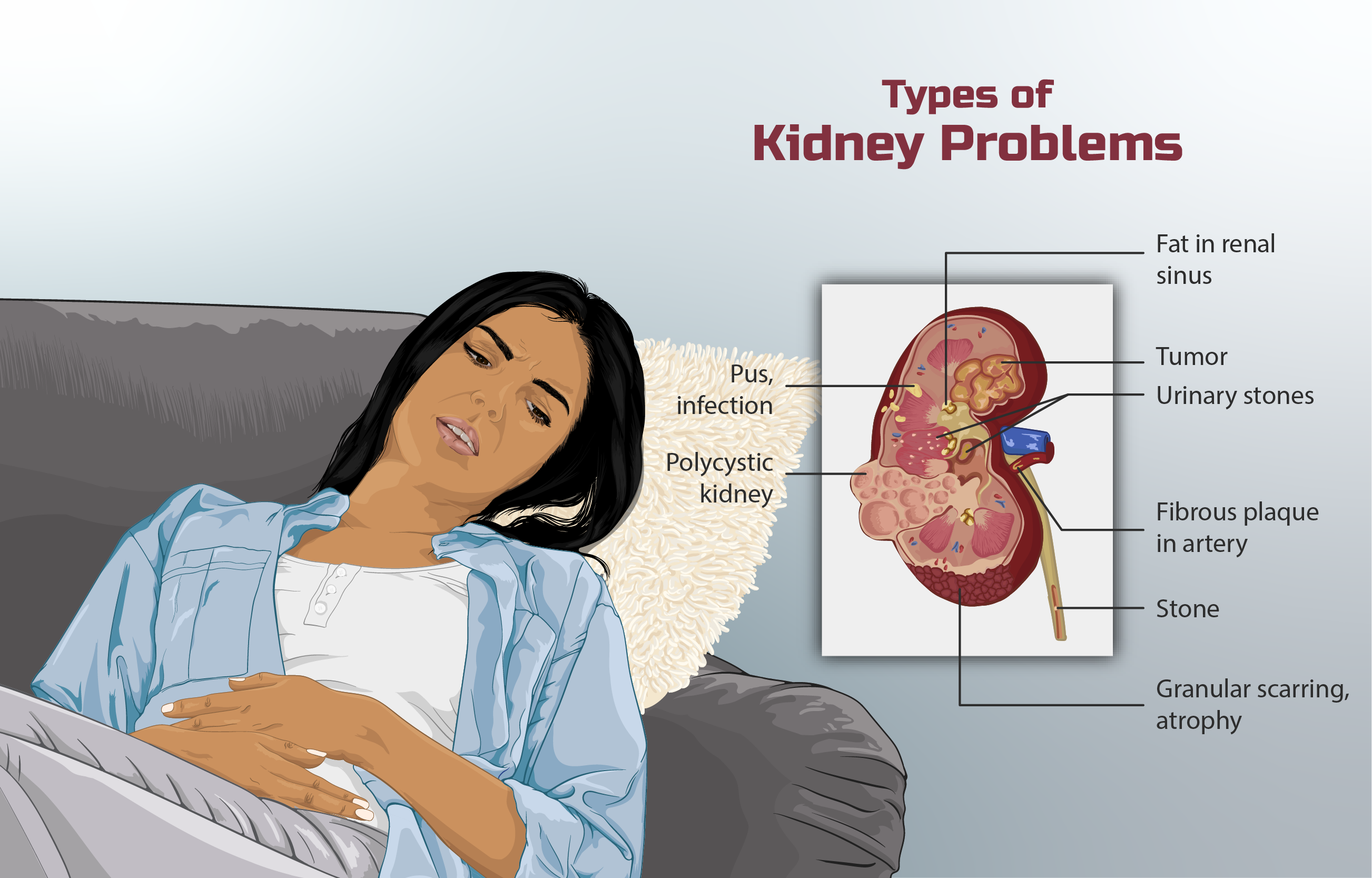
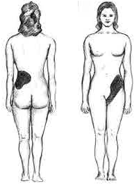
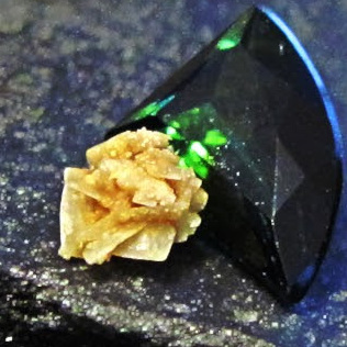
Kidney stone disease, also known as nephrolithiasis or urolithiasis, is a crystallopathy where a solid piece of material (kidney stone) develops in the urinary tract. Kidney stones typically form in the kidney and leave the body in the urine stream. A small stone may pass without causing symptoms. If a stone grows to more than , it can cause blockage of the ureter, resulting in sharp and severe pain in the lower back or abdomen. A stone may also result in blood in the urine, vomiting, or painful urination. About half of people who have had a kidney stone will have another within ten years. Most stones form by a combination of genetics and environmental factors. Risk factors include high urine calcium levels, obesity, certain foods, some medications, calcium supplements, hyperparathyroidism, gout and not drinking enough fluids. Stones form in the kidney when minerals in urine are at high concentration. The diagnosis is usually based on symptoms, urine testing, and medical ... [...More Info...] [...Related Items...] OR: [Wikipedia] [Google] [Baidu] |
Kidney Stone Disease
Kidney stone disease, also known as nephrolithiasis or urolithiasis, is a crystallopathy where a solid piece of material (kidney stone) develops in the urinary tract. Kidney stones typically form in the kidney and leave the body in the urine stream. A small stone may pass without causing symptoms. If a stone grows to more than , it can cause blockage of the ureter, resulting in sharp and severe pain in the lower back or abdomen. A stone may also result in blood in the urine, vomiting, or painful urination. About half of people who have had a kidney stone will have another within ten years. Most stones form by a combination of genetics and environmental factors. Risk factors include high urine calcium levels, obesity, certain foods, some medications, calcium supplements, hyperparathyroidism, gout and not drinking enough fluids. Stones form in the kidney when minerals in urine are at high concentration. The diagnosis is usually based on symptoms, urine testing, and medical i ... [...More Info...] [...Related Items...] OR: [Wikipedia] [Google] [Baidu] |
Fuller Albright
Fuller Albright (January 12, 1900 – December 8, 1969) was an American endocrinologist who made numerous contributions to his field, especially to the area of calcium metabolism. Albright made great strides and contributions to the understanding of disorders associated with calcium and phosphate abnormalities in the body. He also was a published author and in his books he detailed his findings. Early life Albright was born on January 12, 1900, in Buffalo, New York. He was the third child of John J. Albright, a prominent businessman and philanthropist who constructed the Albright Art Gallery in Buffalo, and Susan Fuller, a Smith College graduate who was Albright's second wife. John J. Albright had three children from his first marriage and six from his marriage to Fuller. His maternal grandparents were Eben and Nancy Fuller, of Lancaster, Massachusetts. His paternal grandparents were Joseph Jacob and Elizabeth S. Albright, both from Pennsylvania. The family was descended from ... [...More Info...] [...Related Items...] OR: [Wikipedia] [Google] [Baidu] |
Sarcoidosis
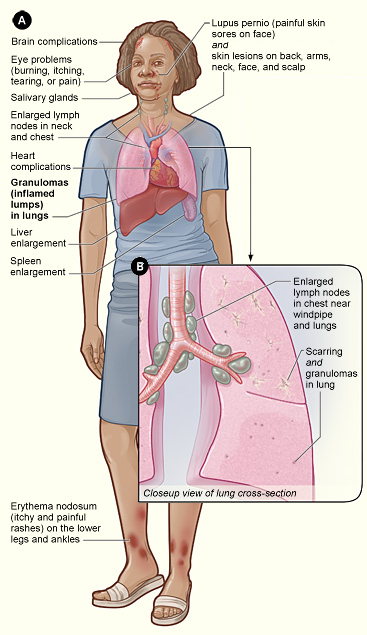
Sarcoidosis (also known as ''Besnier-Boeck-Schaumann disease'') is a disease involving abnormal collections of inflammatory cells that form lumps known as granulomata. The disease usually begins in the lungs, skin, or lymph nodes. Less commonly affected are the eyes, liver, heart, and brain. Any organ can be affected though. The signs and symptoms depend on the organ involved. Often, no, or only mild, symptoms are seen. When it affects the lungs, wheezing, coughing, shortness of breath, or chest pain may occur. Some may have Löfgren syndrome with fever, large lymph nodes, arthritis, and a rash known as erythema nodosum. The cause of sarcoidosis is unknown. Some believe it may be due to an immune reaction to a trigger such as an infection or chemicals in those who are genetically predisposed. Those with affected family members are at greater risk. Diagnosis is partly based on signs and symptoms, which may be supported by biopsy. Findings that make it likely include large lymph n ... [...More Info...] [...Related Items...] OR: [Wikipedia] [Google] [Baidu] |
Hyperoxaluria
Hyperoxaluria is an excessive urinary excretion of oxalate. Individuals with hyperoxaluria often have calcium oxalate kidney stones. It is sometimes called Bird's disease, after Golding Bird, who first described the condition. Causes Hyperoxaluria can be primary (as a result of a genetic defect) or secondary to another disease process. Type I primary hyperoxaluria (PH1) is associated mutations in the gene encoding AGXT, a key enzyme involved in oxalate metabolism. PH1 is an example of a protein mistargeting disease, wherein AGXT shows a trafficking defect. Instead of being trafficked to peroxisomes, it is targeted to mitochondria, where it is metabolically deficient despite being catalytically active. Type II is associated with GRHPR. Secondary hyperoxaluria can occur as a complication of jejunoileal bypass, or in a patient who has lost much of the ileum with an intact colon. In these cases, hyperoxaluria is caused by excessive gastrointestinal oxalate absorption. Excessive int ... [...More Info...] [...Related Items...] OR: [Wikipedia] [Google] [Baidu] |
Hypercalcaemia
Hypercalcemia, also spelled hypercalcaemia, is a high calcium (Ca2+) level in the blood serum. The normal range is 2.1–2.6 mmol/L (8.8–10.7 mg/dL, 4.3–5.2 mEq/L), with levels greater than 2.6 mmol/L defined as hypercalcemia. Those with a mild increase that has developed slowly typically have no symptoms. In those with greater levels or rapid onset, symptoms may include abdominal pain, bone pain, confusion, depression, weakness, kidney stones or an abnormal heart rhythm including cardiac arrest. Most outpatient cases are due to primary hyperparathyroidism and inpatient cases due to cancer. Other causes of hypercalcemia include sarcoidosis, tuberculosis, Paget disease, multiple endocrine neoplasia (MEN), vitamin D toxicity, familial hypocalciuric hypercalcaemia and certain medications such as lithium and hydrochlorothiazide. Diagnosis should generally include either a corrected calcium or ionized calcium level and be confirmed after a week. Specific changes ... [...More Info...] [...Related Items...] OR: [Wikipedia] [Google] [Baidu] |
Vitamin D
Vitamin D is a group of fat-soluble secosteroids responsible for increasing intestinal absorption of calcium, magnesium, and phosphate, and many other biological effects. In humans, the most important compounds in this group are vitamin D3 (cholecalciferol) and vitamin D2 (ergocalciferol). The major natural source of the vitamin is synthesis of cholecalciferol in the lower layers of epidermis of the skin through a chemical reaction that is dependent on sun exposure (specifically UVB radiation). Cholecalciferol and ergocalciferol can be ingested from the diet and supplements. Only a few foods, such as the flesh of fatty fish, naturally contain significant amounts of vitamin D. In the U.S. and other countries, cow's milk and plant-derived milk substitutes are fortified with vitamin D, as are many breakfast cereals. Mushrooms exposed to ultraviolet light contribute useful amounts of vitamin D2. Dietary recommendations typically assume that all of a person's vitamin D is taken ... [...More Info...] [...Related Items...] OR: [Wikipedia] [Google] [Baidu] |
Kidney Failure
Kidney failure, also known as end-stage kidney disease, is a medical condition in which the kidneys can no longer adequately filter waste products from the blood, functioning at less than 15% of normal levels. Kidney failure is classified as either acute kidney failure, which develops rapidly and may resolve; and chronic kidney failure, which develops slowly and can often be irreversible. Symptoms may include leg swelling, feeling tired, vomiting, loss of appetite, and confusion. Complications of acute and chronic failure include uremia, high blood potassium, and volume overload. Complications of chronic failure also include heart disease, high blood pressure, and anemia. Causes of acute kidney failure include low blood pressure, blockage of the urinary tract, certain medications, muscle breakdown, and hemolytic uremic syndrome. Causes of chronic kidney failure include diabetes, high blood pressure, nephrotic syndrome, and polycystic kidney disease. Diagnosis of acute failure ... [...More Info...] [...Related Items...] OR: [Wikipedia] [Google] [Baidu] |
Medullary Nephrocalcinoses
Medulla or Medullary may refer to: Science * Medulla oblongata, a part of the brain stem * Renal medulla, a part of the kidney * Adrenal medulla, a part of the adrenal gland * Medulla of ovary, a stroma in the center of the ovary * Medulla of the thymus, a part of the lobes of the thymus * Medulla of lymph node * Medulla (hair), the innermost layer of the hair shaft * Medulla, a part of the optic lobe of arthropods * Medulla (lichenology), a layer of the internal structure of a lichen * Pith, or medulla, a tissue in the stems of vascular plants Other uses * ''Medúlla'', a 2004 album by Björk * Medulla, Florida, a place in the U.S. * Las Médulas, a gold mining site in León, Spain See also * *Medullary cavity The medullary cavity (''medulla'', innermost part) is the central cavity of bone shafts where red bone marrow and/or yellow bone marrow (adipose tissue) is stored; hence, the medullary cavity is also known as the marrow cavity. Located in the m ..., the central ca ... [...More Info...] [...Related Items...] OR: [Wikipedia] [Google] [Baidu] |


