|
Nucleus Dentatus
The dentate nucleus is a cluster of neurons, or nerve cells, in the central nervous system that has a dentate – tooth-like or serrated – edge. It is located within the deep white matter of each cerebellar hemisphere, and it is the largest single structure linking the cerebellum to the rest of the brain.Sultan, F., Hamodeh, S., & Baizer, J. S. (2010). THE HUMAN DENTATE NUCLEUS: A COMPLEX SHAPE UNTANGLED. rticle Neuroscience, 167(4), 965–968. It is the largest and most lateral, or farthest from the midline, of the four pairs of deep cerebellar nuclei, the others being the globose and emboliform nuclei, which together are referred to as the interposed nucleus, and the fastigial nucleus. The dentate nucleus is responsible for the planning, initiation and control of voluntary movements. The dorsal region of the dentate nucleus contains output channels involved in motor function, which is the movement of skeletal muscle, while the ventral region contains output channels involved i ... [...More Info...] [...Related Items...] OR: [Wikipedia] [Google] [Baidu] |
Cerebellum
The cerebellum (Latin for "little brain") is a major feature of the hindbrain of all vertebrates. Although usually smaller than the cerebrum, in some animals such as the mormyrid fishes it may be as large as or even larger. In humans, the cerebellum plays an important role in motor control. It may also be involved in some cognition, cognitive functions such as attention and language as well as emotion, emotional control such as regulating fear and pleasure responses, but its movement-related functions are the most solidly established. The human cerebellum does not initiate movement, but contributes to Motor coordination, coordination, precision, and accurate timing: it receives input from sensory systems of the spinal cord and from other parts of the brain, and integrates these inputs to fine-tune motor activity. Cerebellar damage produces disorders in Fine motor skill, fine movement, Equilibrioception, equilibrium, Human positions, posture, and motor learning in humans. Anatomica ... [...More Info...] [...Related Items...] OR: [Wikipedia] [Google] [Baidu] |
Vestibular Nuclei
The vestibular nuclei (VN) are the cranial nuclei for the vestibular nerve located in the brainstem. In Terminologia Anatomica they are grouped in both the pons and the medulla in the brainstem. Structure Path The fibers of the vestibular nerve enter the medulla oblongata on the medial side of those of the cochlear, and pass between the inferior peduncle and the spinal tract of the trigeminal nerve. They then divide into ascending and descending fibers. The latter end by arborizing around the cells of the medial nucleus, which is situated in the area acustica of the rhomboid fossa. The ascending fibers either end in the same manner or in the lateral nucleus, which is situated lateral to the area acustica and farther from the ventricular floor. Some of the axons of the cells of the lateral nucleus, and possibly also of the medial nucleus, are continued upward through the inferior peduncle to the roof nuclei of the opposite side of the cerebellum, to which also other fibers of th ... [...More Info...] [...Related Items...] OR: [Wikipedia] [Google] [Baidu] |
Medullary Laminae Of Thalamus
Medullary laminae of thalamus are layers of myelinated fibres that appear on cross sections of the thalamus. They also are commonly referred to as laminae medullares thalami or medullary layers of thalamus. The specific layers are: *Lamina medullaris lateralis (external medullary lamina) - separates ventral and lateral thalamus from the subthalamus and thalamic reticular nucleus *Lamina medullaris medialis (internal medullary lamina) - positioned between the dorsomedial and ventral nuclei of thalamus, encloses the intralaminar nuclei (centromedian nucleus In the anatomy of the brain, the centromedian nucleus, also known as the centrum medianum, (CM or Cm-Pf) is a part of the intralaminar thalamic nuclei (ITN) in the thalamus. There are two centromedian nuclei arranged bilaterally. In humans, it c ..., paracentral, and central lateral) External links * References Thalamus {{neuroanatomy-stub ... [...More Info...] [...Related Items...] OR: [Wikipedia] [Google] [Baidu] |
Frontal Lobe
The frontal lobe is the largest of the four major lobes of the brain in mammals, and is located at the front of each cerebral hemisphere (in front of the parietal lobe and the temporal lobe). It is parted from the parietal lobe by a groove between tissues called the central sulcus and from the temporal lobe by a deeper groove called the lateral sulcus (Sylvian fissure). The most anterior rounded part of the frontal lobe (though not well-defined) is known as the frontal pole, one of the three poles of the cerebrum. The frontal lobe is covered by the frontal cortex. The frontal cortex includes the premotor cortex, and the primary motor cortex – parts of the motor cortex. The front part of the frontal cortex is covered by the prefrontal cortex. There are four principal gyri in the frontal lobe. The precentral gyrus is directly anterior to the central sulcus, running parallel to it and contains the primary motor cortex, which controls voluntary movements of specific body parts ... [...More Info...] [...Related Items...] OR: [Wikipedia] [Google] [Baidu] |
Posterior Parietal
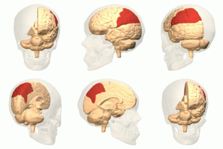
The parietal lobe is one of the four major lobes of the cerebral cortex in the brain of mammals. The parietal lobe is positioned above the temporal lobe and behind the frontal lobe and central sulcus. The parietal lobe integrates sensory information among various modalities, including spatial sense and navigation (proprioception), the main sensory receptive area for the sense of touch in the somatosensory cortex which is just posterior to the central sulcus in the postcentral gyrus, and the dorsal stream of the visual system. The major sensory inputs from the skin (touch, temperature, and pain receptors), relay through the thalamus to the parietal lobe. Several areas of the parietal lobe are important in language processing. The somatosensory cortex can be illustrated as a distorted figure – the cortical homunculus (Latin: "little man") in which the body parts are rendered according to how much of the somatosensory cortex is devoted to them. The superior parietal lobule and in ... [...More Info...] [...Related Items...] OR: [Wikipedia] [Google] [Baidu] |
Prefrontal Cortex
In mammalian brain anatomy, the prefrontal cortex (PFC) covers the front part of the frontal lobe of the cerebral cortex. The PFC contains the Brodmann areas BA8, BA9, BA10, BA11, BA12, BA13, BA14, BA24, BA25, BA32, BA44, BA45, BA46, and BA47. The basic activity of this brain region is considered to be orchestration of thoughts and actions in accordance with internal goals. Many authors have indicated an integral link between a person's will to live, personality, and the functions of the prefrontal cortex. This brain region has been implicated in executive functions, such as planning, decision making, short-term memory, personality expression, moderating social behavior and controlling certain aspects of speech and language. Executive function relates to abilities to differentiate among conflicting thoughts, determine good and bad, better and best, same and different, future consequences of current activities, working toward a defined goal, prediction of outcomes, e ... [...More Info...] [...Related Items...] OR: [Wikipedia] [Google] [Baidu] |
Cerebral Cortex
The cerebral cortex, also known as the cerebral mantle, is the outer layer of neural tissue of the cerebrum of the brain in humans and other mammals. The cerebral cortex mostly consists of the six-layered neocortex, with just 10% consisting of allocortex. It is separated into two cortices, by the longitudinal fissure that divides the cerebrum into the left and right cerebral hemispheres. The two hemispheres are joined beneath the cortex by the corpus callosum. The cerebral cortex is the largest site of neural integration in the central nervous system. It plays a key role in attention, perception, awareness, thought, memory, language, and consciousness. The cerebral cortex is part of the brain responsible for cognition. In most mammals, apart from small mammals that have small brains, the cerebral cortex is folded, providing a greater surface area in the confined volume of the cranium. Apart from minimising brain and cranial volume, cortical folding is crucial for the brain ... [...More Info...] [...Related Items...] OR: [Wikipedia] [Google] [Baidu] |
Premotor
The premotor cortex is an area of the motor cortex lying within the frontal lobe of the brain just anterior to the primary motor cortex. It occupies part of Brodmann's area 6. It has been studied mainly in primates, including monkeys and humans. The functions of the premotor cortex are diverse and not fully understood. It projects directly to the spinal cord and therefore may play a role in the direct control of behavior, with a relative emphasis on the trunk muscles of the body. It may also play a role in planning movement, in the spatial guidance of movement, in the sensory guidance of movement, in understanding the actions of others, and in using abstract rules to perform specific tasks. Different subregions of the premotor cortex have different properties and presumably emphasize different functions. Nerve signals generated in the premotor cortex cause much more complex patterns of movement than the discrete patterns generated in the primary motor cortex. Structure The premo ... [...More Info...] [...Related Items...] OR: [Wikipedia] [Google] [Baidu] |
Primary Motor Cortex
The primary motor cortex (Brodmann area 4) is a brain region that in humans is located in the dorsal portion of the frontal lobe. It is the primary region of the motor system and works in association with other motor areas including premotor cortex, the supplementary motor area, posterior parietal cortex, and several subcortical brain regions, to plan and execute voluntary movements. Primary motor cortex is defined anatomically as the region of cortex that contains large neurons known as Betz cells, which, along with other cortical neurons, send long axons down the spinal cord to synapse onto the interneuron circuitry of the spinal cord and also directly onto the alpha motor neurons in the spinal cord which connect to the muscles. At the primary motor cortex, motor representation is orderly arranged (in an inverted fashion) from the toe (at the top of the cerebral hemisphere) to mouth (at the bottom) along a fold in the cortex called the central sulcus. However, some body parts ... [...More Info...] [...Related Items...] OR: [Wikipedia] [Google] [Baidu] |
Somata
The soma (pl. ''somata'' or ''somas''), perikaryon (pl. ''perikarya''), neurocyton, or cell body is the bulbous, non-process portion of a neuron or other brain cell type, containing the cell nucleus. The word 'soma' comes from the Greek '' σῶμα'', meaning 'body'. Although it is often used to refer to neurons, it can also refer to other cell types as well, including astrocytes, oligodendrocytes, and microglia. There are many different specialized types of neurons, and their sizes vary from as small as about 5 micrometres to over 10 millimetres for some of the smallest and largest neurons of invertebrates, respectively. The soma of a neuron (i.e., the main part of the neuron in which the dendrites branch off of) contains many organelles, including granules called Nissl granules, which are composed largely of rough endoplasmic reticulum and free polyribosomes. The cell nucleus is a key feature of the soma. The nucleus is the source of most of the RNA that is produced in neuron ... [...More Info...] [...Related Items...] OR: [Wikipedia] [Google] [Baidu] |
Dendrite
Dendrites (from Greek δένδρον ''déndron'', "tree"), also dendrons, are branched protoplasmic extensions of a nerve cell that propagate the electrochemical stimulation received from other neural cells to the cell body, or soma, of the neuron from which the dendrites project. Electrical stimulation is transmitted onto dendrites by upstream neurons (usually via their axons) via synapses which are located at various points throughout the dendritic tree. Dendrites play a critical role in integrating these synaptic inputs and in determining the extent to which action potentials are produced by the neuron. Dendritic arborization, also known as dendritic branching, is a multi-step biological process by which neurons form new dendritic trees and branches to create new synapses. The morphology of dendrites such as branch density and grouping patterns are highly correlated to the function of the neuron. Malformation of dendrites is also tightly correlated to impaired nervous syste ... [...More Info...] [...Related Items...] OR: [Wikipedia] [Google] [Baidu] |
Pontine Nuclei
Pontine may refer to: * Having to do with the pons, a structure located in the brain stem (from ''pons'', "bridge") * Pontine Marshes, a region of Italy near Rome * Pontine Islands The Pontine Islands (, also ; it, Isole Ponziane ) are an archipelago in the Tyrrhenian Sea off the coast of Lazio region, Italy. The islands were collectively named after the largest island in the group, Ponza. The other islands in the archipe ..., islands of Italy near Circeo See also {{disambig ... [...More Info...] [...Related Items...] OR: [Wikipedia] [Google] [Baidu] |


