|
Nevus Cell
Nevus cells are a variant of melanocytes. They are larger than typical melanocytes, do not have dendrites, and have more abundant cytoplasm with coarse granules. They are usually located at the dermoepidermal junction or in the dermis of the skin. Dermal nevus cells can be further classified: type A (epithelioid) dermal nevus cells mature into type B (lymphocytoid) dermal nevus cells which mature further into type C (neuroid) dermal nevus cells, through a process involving downwards migration. Nevus cells are the primary component of a melanocytic nevus. Nevus cells can also be found in lymph nodes and the thymus. See also *List of human cell types derived from the germ layers This is a list of cells in humans derived from the three embryonic germ layers – ectoderm, mesoderm, and endoderm. Cells derived from ectoderm Surface ectoderm Skin * Trichocyte * Keratinocyte Anterior pituitary * Gonadotrope * Corti ... References Skin anatomy {{anatomy-stub ... [...More Info...] [...Related Items...] OR: [Wikipedia] [Google] [Baidu] |
Melanocytes
Melanocytes are melanin-producing neural crest-derived cells located in the bottom layer (the stratum basale) of the skin's epidermis, the middle layer of the eye (the uvea), the inner ear, vaginal epithelium, meninges, bones, and heart. Melanin is a dark pigment primarily responsible for skin color. Once synthesized, melanin is contained in special organelles called melanosomes which can be transported to nearby keratinocytes to induce pigmentation. Thus darker skin tones have more melanosomes present than lighter skin tones. Functionally, melanin serves as protection against UV radiation. Melanocytes also have a role in the immune system. Function Through a process called melanogenesis, melanocytes produce melanin, which is a pigment found in the skin, eyes, hair, nasal cavity, and inner ear. This melanogenesis leads to a long-lasting pigmentation, which is in contrast to the pigmentation that originates from oxidation of already-existing melanin. There a ... [...More Info...] [...Related Items...] OR: [Wikipedia] [Google] [Baidu] |
Dendrite (non-neuronal)
{{unreferenced, date=December 2011 A dendrite is a branching projection of the cytoplasm of a cell. While the term is most commonly used to refer to the branching projections of neurons, it can also be used to refer to features of other types of cells that, while having a similar appearance, are actually quite distinct structures. Non-neuronal cells that have dendrites: *Dendritic cells, part of the mammalian immune system *Melanocytes, pigment-producing cells located in the skin *Merkel cells, receptor-cells in the skin associated with the sense of touch * Corneal keratocytes, specialized fibroblasts A fibroblast is a type of biological cell that synthesizes the extracellular matrix and collagen, produces the structural framework ( stroma) for animal tissues, and plays a critical role in wound healing. Fibroblasts are the most common cells ... residing in the stroma. Cell biology ... [...More Info...] [...Related Items...] OR: [Wikipedia] [Google] [Baidu] |
Cytoplasm
In cell biology, the cytoplasm is all of the material within a eukaryotic cell, enclosed by the cell membrane, except for the cell nucleus. The material inside the nucleus and contained within the nuclear membrane is termed the nucleoplasm. The main components of the cytoplasm are cytosol (a gel-like substance), the organelles (the cell's internal sub-structures), and various cytoplasmic inclusions. The cytoplasm is about 80% water and is usually colorless. The submicroscopic ground cell substance or cytoplasmic matrix which remains after exclusion of the cell organelles and particles is groundplasm. It is the hyaloplasm of light microscopy, a highly complex, polyphasic system in which all resolvable cytoplasmic elements are suspended, including the larger organelles such as the ribosomes, mitochondria, the plant plastids, lipid droplets, and vacuoles. Most cellular activities take place within the cytoplasm, such as many metabolic pathways including glycolysis, ... [...More Info...] [...Related Items...] OR: [Wikipedia] [Google] [Baidu] |
Dermoepidermal Junction
The dermoepidermal junction or dermal-epidermal junction (DEJ) is the area of tissue that joins the epidermal and the dermal layers of the skin. The basal cells in the stratum basale of the epidermis connect to the basement membrane by the anchoring filaments of hemidesmosomes; the cells of the papillary layer of the dermis are attached to the basement membrane by anchoring fibrils, which consist of type VII collagen. Stevens–Johnson syndrome and toxic epidermal necrolysis Toxic epidermal necrolysis (TEN) is a type of severe skin reaction. Together with Stevens–Johnson syndrome (SJS) it forms a spectrum of disease, with TEN being more severe. Early symptoms include fever and flu-like symptoms. A few days later ... are diseases where there is a breakdown of the dermoepidermal junction. References Skin anatomy {{dermatology-stub ... [...More Info...] [...Related Items...] OR: [Wikipedia] [Google] [Baidu] |
Dermis
The dermis or corium is a layer of skin between the epidermis (with which it makes up the cutis) and subcutaneous tissues, that primarily consists of dense irregular connective tissue and cushions the body from stress and strain. It is divided into two layers, the superficial area adjacent to the epidermis called the papillary region and a deep thicker area known as the reticular dermis.James, William; Berger, Timothy; Elston, Dirk (2005). ''Andrews' Diseases of the Skin: Clinical Dermatology'' (10th ed.). Saunders. Pages 1, 11–12. . The dermis is tightly connected to the epidermis through a basement membrane. Structural components of the dermis are collagen, elastic fibers, and extrafibrillar matrix.Marks, James G; Miller, Jeffery (2006). ''Lookingbill and Marks' Principles of Dermatology'' (4th ed.). Elsevier Inc. Page 8–9. . It also contains mechanoreceptors that provide the sense of touch and thermoreceptors that provide the sense of heat. In addition, hair foll ... [...More Info...] [...Related Items...] OR: [Wikipedia] [Google] [Baidu] |
Melanocytic Nevus
A melanocytic nevus (also known as nevocytic nevus, nevus-cell nevus and commonly as a mole) is a type of melanocytic tumor that contains nevus cells. Some sources equate the term mole with "melanocytic nevus", but there are also sources that equate the term mole with any nevus form. The majority of moles appear during the first two decades of a person's life, with about one in every 100 babies being born with moles. Acquired moles are a form of benign neoplasm, while congenital moles, or congenital nevi, are considered a minor malformation or hamartoma and may be at a higher risk for melanoma. A mole can be either subdermal (under the skin) or a pigmented growth on the skin, formed mostly of a type of cell known as a melanocyte. The high concentration of the body's pigmenting agent, melanin, is responsible for their dark color. Moles are a member of the family of skin lesions known as nevi and can occur in all mammalian species, but have been documented most extensively in human ... [...More Info...] [...Related Items...] OR: [Wikipedia] [Google] [Baidu] |
Lymph Nodes
A lymph node, or lymph gland, is a kidney-shaped organ of the lymphatic system and the adaptive immune system. A large number of lymph nodes are linked throughout the body by the lymphatic vessels. They are major sites of lymphocytes that include B and T cells. Lymph nodes are important for the proper functioning of the immune system, acting as filters for foreign particles including cancer cells, but have no detoxification function. In the lymphatic system a lymph node is a secondary lymphoid organ. A lymph node is enclosed in a fibrous capsule and is made up of an outer cortex and an inner medulla. Lymph nodes become inflamed or enlarged in various diseases, which may range from trivial throat infections to life-threatening cancers. The condition of lymph nodes is very important in cancer staging, which decides the treatment to be used and determines the prognosis. Lymphadenopathy refers to glands that are enlarged or swollen. When inflamed or enlarged, lymph nodes can ... [...More Info...] [...Related Items...] OR: [Wikipedia] [Google] [Baidu] |
Thymus
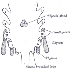
The thymus is a specialized primary lymphoid organ of the immune system. Within the thymus, thymus cell lymphocytes or ''T cells'' mature. T cells are critical to the adaptive immune system, where the body adapts to specific foreign invaders. The thymus is located in the upper front part of the chest, in the anterior superior mediastinum, behind the sternum, and in front of the heart. It is made up of two lobes, each consisting of a central medulla and an outer cortex, surrounded by a capsule. The thymus is made up of immature T cells called thymocytes, as well as lining cells called epithelial cells which help the thymocytes develop. T cells that successfully develop react appropriately with MHC immune receptors of the body (called ''positive selection'') and not against proteins of the body (called ''negative selection''). The thymus is largest and most active during the neonatal and pre-adolescent periods. By the early teens, the thymus begins to decrease in size and a ... [...More Info...] [...Related Items...] OR: [Wikipedia] [Google] [Baidu] |
List Of Human Cell Types Derived From The Germ Layers
This is a list of cells in humans derived from the three embryonic germ layers – ectoderm, mesoderm, and endoderm. Cells derived from ectoderm Surface ectoderm Skin * Trichocyte * Keratinocyte Anterior pituitary * Gonadotrope * Corticotrope * Thyrotrope * Somatotrope * Lactotroph Tooth enamel * Ameloblast Neural crest Peripheral nervous system * Neuron * Glia ** Schwann cell ** Satellite glial cell Neuroendocrine system * Chromaffin cell * Glomus cell Skin * Melanocyte ** Nevus cell * Merkel cell Teeth * Odontoblast * Cementoblast Eyes * Corneal keratocyte Neural tube Central nervous system * Neuron * Glia ** Astrocyte ** Ependymocytes ** Muller glia (retina) ** Oligodendrocyte ** Oligodendrocyte progenitor cell ** Pituicyte (posterior pituitary) Pineal gland * Pinealocyte Cells derived from mesoderm Paraxial mesoderm Mesenchymal stem cell =Osteochondroprogenitor cell= * Bone (Osteoblast → Osteocyte) * Cartilage (Chondroblast → Chond ... [...More Info...] [...Related Items...] OR: [Wikipedia] [Google] [Baidu] |


.jpg)
