|
Muscle Biopsy
In medicine, a muscle biopsy is a procedure in which a piece of muscle tissue is removed from an organism and examined microscopically. A muscle biopsy can lead to the discovery of problems with the nervous system, connective tissue, vascular system, or musculoskeletal system. Indications In humans with weakness and low muscle tone, a muscle biopsy can help distinguish between myopathies (where the pathology is in the muscle tissue itself) and neuropathies (where the pathology is at the nerves innervating those muscles). Muscle biopsies can also help to distinguish among various types of myopathies, by microscopic analysis for differing characteristics when exposed to a variety of chemical reactions and stains. However, in some cases the muscle biopsy alone is inadequate to distinguish between certain myopathies. For example, a muscle biopsy showing the nucleus pathologically located in the center of the muscle cell would indicate "centronuclear myopathy", but research has shown ... [...More Info...] [...Related Items...] OR: [Wikipedia] [Google] [Baidu] |
Micrograph
A micrograph or photomicrograph is a photograph or digital image taken through a microscope or similar device to show a magnified image of an object. This is opposed to a macrograph or photomacrograph, an image which is also taken on a microscope but is only slightly magnified, usually less than 10 times. Micrography is the practice or art of using microscopes to make photographs. A micrograph contains extensive details of microstructure. A wealth of information can be obtained from a simple micrograph like behavior of the material under different conditions, the phases found in the system, failure analysis, grain size estimation, elemental analysis and so on. Micrographs are widely used in all fields of microscopy. Types Photomicrograph A light micrograph or photomicrograph is a micrograph prepared using an optical microscope, a process referred to as ''photomicroscopy''. At a basic level, photomicroscopy may be performed simply by connecting a camera to a microscope, th ... [...More Info...] [...Related Items...] OR: [Wikipedia] [Google] [Baidu] |
Needle Aspiration Biopsy
Fine-needle aspiration (FNA) is a diagnostic procedure used to investigate lumps or masses. In this technique, a thin (23–25 gauge (0.52 to 0.64 mm outer diameter)), hollow needle is inserted into the mass for sampling of cells that, after being stained, are examined under a microscope (biopsy). The sampling and biopsy considered together are called fine-needle aspiration biopsy (FNAB) or fine-needle aspiration cytology (FNAC) (the latter to emphasize that any aspiration biopsy involves cytopathology, not histopathology). Fine-needle aspiration biopsies are very safe minor surgical procedures. Often, a major surgical (excisional or open) biopsy can be avoided by performing a needle aspiration biopsy instead, eliminating the need for hospitalization. In 1981, the first fine-needle aspiration biopsy in the United States was done at Maimonides Medical Center. Today, this procedure is widely used in the diagnosis of cancer and inflammatory conditions. Aspiration is safer and ... [...More Info...] [...Related Items...] OR: [Wikipedia] [Google] [Baidu] |
Amyotrophic Lateral Sclerosis
Amyotrophic lateral sclerosis (ALS), also known as motor neuron disease (MND) or Lou Gehrig's disease, is a neurodegenerative disease that results in the progressive loss of motor neurons that control voluntary muscles. ALS is the most common type of motor neuron diseases. Early symptoms of ALS include stiff muscles, muscle twitches, and gradual increasing weakness and muscle wasting. ''Limb-onset ALS'' begins with weakness in the arms or legs, while ''bulbar-onset ALS'' begins with difficulty speaking or swallowing. Half of the people with ALS develop at least mild difficulties with thinking and behavior, and about 15% develop frontotemporal dementia. Most people experience pain. The affected muscles are responsible for chewing food, speaking, and walking. Motor neuron loss continues until the ability to eat, speak, move, and finally the ability to breathe is lost. ALS eventually causes paralysis and early death, usually from respiratory failure. Most cases of ALS (a ... [...More Info...] [...Related Items...] OR: [Wikipedia] [Google] [Baidu] |
Dermatomyositis
Dermatomyositis (DM) is a long-term inflammatory disorder which affects skin and the muscles. Its symptoms are generally a skin rash and worsening muscle weakness over time. These may occur suddenly or develop over months. Other symptoms may include weight loss, fever, lung inflammation, or light sensitivity. Complications may include calcium deposits in muscles or skin. The cause is unknown. Theories include that it is an autoimmune disease or a result of a viral infection. Dermatomyositis may develop as a paraneoplastic syndrome associated with several forms of malignancy. It is a type of inflammatory myopathy. Diagnosis is typically based on some combination of symptoms, blood tests, electromyography, and muscle biopsies. While no cure for the condition is known, treatments generally improve symptoms. Treatments may include medication, physical therapy, exercise, heat therapy, orthotics and assistive devices, and rest. Medications in the corticosteroids family are typic ... [...More Info...] [...Related Items...] OR: [Wikipedia] [Google] [Baidu] |
Polymyositis
Polymyositis (PM) is a type of chronic inflammation of the muscles (inflammatory myopathy) related to dermatomyositis and inclusion body myositis. Its name means "inflammation of many muscles" ('' poly-'' + '' myos-'' + ''-itis''). The inflammation of polymyositis is mainly found in the endomysial layer of skeletal muscle, whereas dermatomyositis is characterized primarily by inflammation of the perimysial layer of skeletal muscles. Signs and symptoms The hallmark of polymyositis is weakness and/or loss of muscle mass in the proximal musculature, as well as flexion of the neck and torso. These symptoms can be associated with marked pain in these areas as well. The hip extensors are often severely affected, leading to particular difficulty in climbing stairs and rising from a seated position. The skin involvement of dermatomyositis is absent in polymyositis. Dysphagia (difficulty swallowing) or other problems with esophageal motility occur in as many as 1/3 of patients. Low grad ... [...More Info...] [...Related Items...] OR: [Wikipedia] [Google] [Baidu] |
Myasthenia Gravis
Myasthenia gravis (MG) is a long-term neuromuscular junction disease that leads to varying degrees of skeletal muscle weakness. The most commonly affected muscles are those of the eyes, face, and swallowing. It can result in double vision, drooping eyelids, trouble talking, and trouble walking. Onset can be sudden. Those affected often have a large thymus or develop a thymoma. Myasthenia gravis is an autoimmune disease of the neuro-muscular junction which results from antibodies that block or destroy nicotinic acetylcholine receptors (AChR) at the junction between the nerve and muscle. This prevents nerve impulses from triggering muscle contractions. Most cases are due to immunoglobulin G1 (IgG1) and IgG3 antibodies that attack AChR in the postsynaptic membrane, causing complement-mediated damage and muscle weakness. Rarely, an inherited genetic defect in the neuromuscular junction results in a similar condition known as congenital myasthenia. Babies of mothers with myasthe ... [...More Info...] [...Related Items...] OR: [Wikipedia] [Google] [Baidu] |
Toxoplasmosis
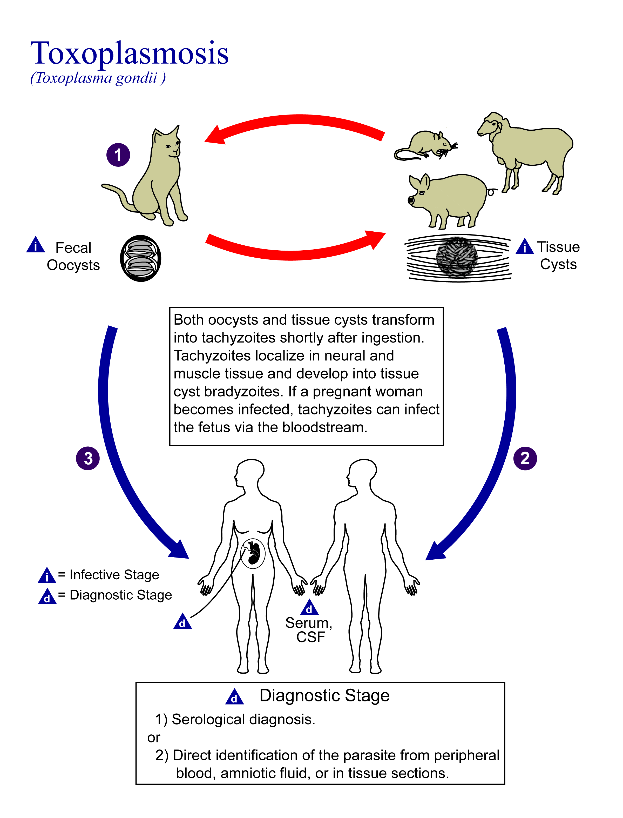
Toxoplasmosis is a parasitic disease caused by ''Toxoplasma gondii'', an apicomplexan. Infections with toxoplasmosis are associated with a variety of neuropsychiatric and behavioral conditions. Occasionally, people may have a few weeks or months of mild, flu-like illness such as muscle aches and tender lymph nodes. In a small number of people, eye problems may develop. In those with a weak immune system, severe symptoms such as seizures and poor coordination may occur. If a person becomes infected during pregnancy, a condition known as congenital toxoplasmosis may affect the child. Toxoplasmosis is usually spread by eating poorly cooked food that contains cysts, exposure to infected cat feces, and from an infected woman to their baby during pregnancy. Rarely, the disease may be spread by blood transfusion. It is not otherwise spread between people. The parasite is known to reproduce sexually only in the cat family. However, it can infect most types of warm-blooded animals, in ... [...More Info...] [...Related Items...] OR: [Wikipedia] [Google] [Baidu] |
Trichinosis
Trichinosis, also known as trichinellosis, is a parasitic disease caused by roundworms of the ''Trichinella'' type. During the initial infection, invasion of the intestines can result in diarrhea, abdominal pain, and vomiting. Migration of larvae to muscle, which occurs about a week after being infected, can cause swelling of the face, inflammation of the whites of the eyes, fever, muscle pains, and a rash. Minor infection may be without symptoms. Complications may include inflammation of heart muscle, central nervous system involvement, and inflammation of the lungs. Trichinosis is mainly spread when undercooked meat containing ''Trichinella'' cysts is eaten. In North America this is most often bear, but infection can also occur from pork, boar, and dog meat. Several species of ''Trichinella'' can cause disease, with ''T. spiralis'' being the most common. After the infected meat has been eaten, the larvae are released from their cysts in the stomach. They then invade the ... [...More Info...] [...Related Items...] OR: [Wikipedia] [Google] [Baidu] |
Electromyogram
Electromyography (EMG) is a technique for evaluating and recording the electrical activity produced by skeletal muscles. EMG is performed using an instrument called an electromyograph to produce a record called an electromyogram. An electromyograph detects the electric potential generated by muscle cells when these cells are electrically or neurologically activated. The signals can be analyzed to detect abnormalities, activation level, or recruitment order, or to analyze the biomechanics of human or animal movement. Needle EMG is an electrodiagnostic medicine technique commonly used by neurologists. Surface EMG is a non-medical procedure used to assess muscle activation by several professionals, including physiotherapists, kinesiologists and biomedical engineers. In Computer Science, EMG is also used as middleware in gesture recognition towards allowing the input of physical action to a computer as a form of human-computer interaction. Clinical uses EMG testing has a variety of ... [...More Info...] [...Related Items...] OR: [Wikipedia] [Google] [Baidu] |
Becker's Muscular Dystrophy
Becker muscular dystrophy is an X-linked recessive inherited disorder characterized by slowly progressing muscle weakness of the legs and pelvis. It is a type of dystrophinopathy. This is caused by mutations in the dystrophin gene, which encodes the protein dystrophin. Becker muscular dystrophy is related to Duchenne muscular dystrophy in that both result from a mutation in the dystrophin gene, but has a milder course. Signs and symptoms Some symptoms consistent with Becker muscular dystrophy are: Individuals with this disorder typically experience progressive muscle weakness of the leg and pelvis muscles, which is associated with a loss of muscle mass (wasting). Muscle weakness also occurs in the arms, neck, and other areas, but not as noticeably severe as in the lower half of the body. Calf muscles initially enlarge during the ages of 5-15 (an attempt by the body to compensate for loss of muscle strength), but the enlarged muscle tissue is eventually replaced by fat and connect ... [...More Info...] [...Related Items...] OR: [Wikipedia] [Google] [Baidu] |
Duchenne Muscular Dystrophy
Duchenne muscular dystrophy (DMD) is a severe type of muscular dystrophy that primarily affects boys. Muscle weakness usually begins around the age of four, and worsens quickly. Muscle loss typically occurs first in the thighs and pelvis followed by the arms. This can result in trouble standing up. Most are unable to walk by the age of 12. Affected muscles may look larger due to increased fat content. Scoliosis is also common. Some may have intellectual disability. Females with a single copy of the defective gene may show mild symptoms. The disorder is X-linked recessive. About two thirds of cases are inherited from a person's mother, while one third of cases are due to a new mutation. It is caused by a mutation in the gene for the protein dystrophin. Dystrophin is important to maintain the muscle fiber's cell membrane. Genetic testing can often make the diagnosis at birth. Those affected also have a high level of creatine kinase in their blood. Although there is no know ... [...More Info...] [...Related Items...] OR: [Wikipedia] [Google] [Baidu] |
Muscular Dystrophy
Muscular dystrophies (MD) are a genetically and clinically heterogeneous group of rare neuromuscular diseases that cause progressive weakness and breakdown of skeletal muscles over time. The disorders differ as to which muscles are primarily affected, the degree of weakness, how fast they worsen, and when symptoms begin. Some types are also associated with problems in other organs. Over 30 different disorders are classified as muscular dystrophies. Of those, Duchenne muscular dystrophy (DMD) accounts for approximately 50% of cases and affects males beginning around the age of four. Other relatively common muscular dystrophies include Becker muscular dystrophy, facioscapulohumeral muscular dystrophy, and myotonic dystrophy, whereas limb–girdle muscular dystrophy and congenital muscular dystrophy are themselves groups of several – usually ultrarare – genetic disorders. Muscular dystrophies are caused by mutations in genes, usually those involved in making muscle proteins. ... [...More Info...] [...Related Items...] OR: [Wikipedia] [Google] [Baidu] |


