|
Mucous Cell
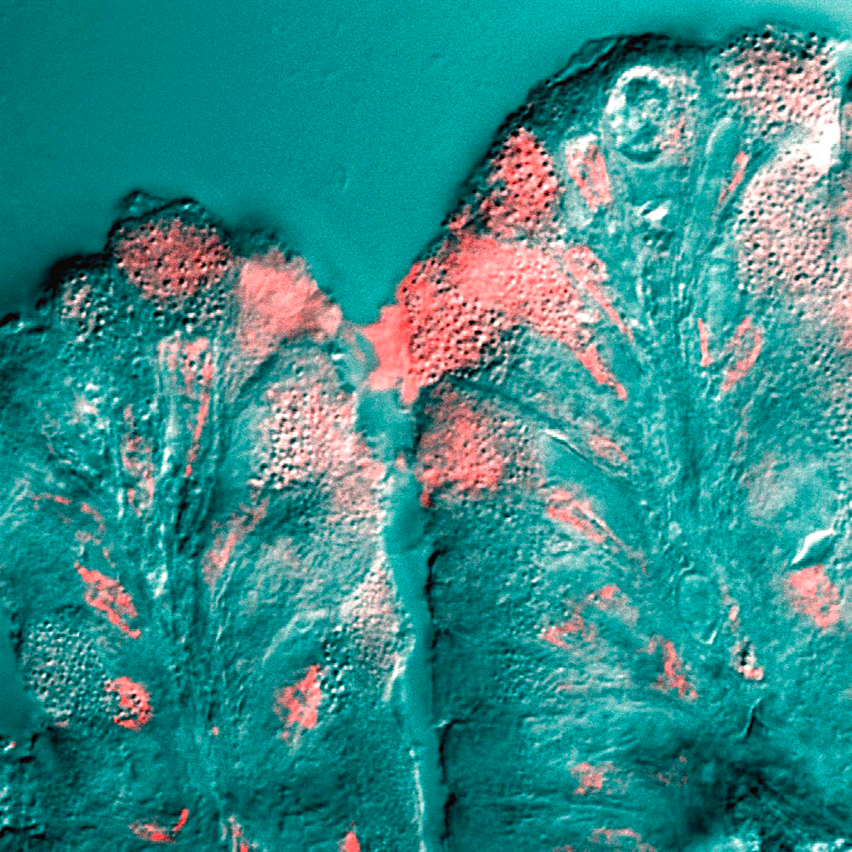
Mucous gland, also known as muciparous glands, are found in several different parts of the body, and they typically stain lighter than serous glands during standard histological preparation. Most are multicellular, but goblet cells are single-celled glands. Mucous salivary glands The mucous salivary glands are similar in structure to the buccal and labial glands. They are found especially at the back part behind the vallate papillae, but are also present at the apex and marginal parts. In this connection the anterior lingual glands require special notice. They are situated on the under surface of the apex of the tongue, one on either side of the frenulum, where they are covered by a fascicle of muscular fibers derived from the styloglossus and inferior longitudinal muscles. They produce a glycoprotein, mucin that absorbs water to form a sticky secretion called mucus. They are from 12 to 25 mm. long, and about 8 mm. broad, and each opens by three or four ducts on ... [...More Info...] [...Related Items...] OR: [Wikipedia] [Google] [Baidu] |
Foliate Papilla
Lingual papillae (singular papilla) are small structures on the upper surface of the tongue that give it its characteristic rough texture. The four types of papillae on the human tongue have different structures and are accordingly classified as circumvallate (or vallate), fungiform, filiform, and foliate. All except the filiform papillae are associated with taste buds. Structure In living subjects, lingual papillae are more readily seen when the tongue is dry. There are four types of papillae present on the tongue: Filiform papillae Filiform papillae are the most numerous of the lingual papillae. They are fine, small, cone-shaped papillae covering most of the dorsum of the tongue. They are responsible for giving the tongue its texture and are responsible for the sensation of touch. Unlike the other kinds of papillae, filiform papillae do not contain taste buds. They cover most of the front two-thirds of the tongue's surface. They appear as very small, conical or cylindrical s ... [...More Info...] [...Related Items...] OR: [Wikipedia] [Google] [Baidu] |
Frenulum Of Tongue
The frenulum of tongue or tongue web (also lingual frenulum or frenulum linguæ; also fraenulum) is a small fold of mucous membrane extending from the floor of the mouth to the midline of the underside of the tongue. Development The tongue starts to develop at about four weeks. The tongue originates from the first, second, and third pharyngeal arches which induces the migration of muscles from the occipital myotomes. A U-shaped sulcus develops in front of and on both sides of the oral part of the tongue. This allows the tongue to be free and highly mobile, except at the region of the lingual frenulum, where it remains attached. Disturbances during this stage cause tongue tie or ankyloglossia. During the sixth week of gestation, the medial nasal processes approach each other to form a single globular process that in time gives rise to the nasal tip, columella, prolabium, frenulum of the upper lip, and the primary palate. As the tongue continues to develop, frenulum cells undergo ... [...More Info...] [...Related Items...] OR: [Wikipedia] [Google] [Baidu] |
Gland
In animals, a gland is a group of cells in an animal's body that synthesizes substances (such as hormones) for release into the bloodstream (endocrine gland) or into cavities inside the body or its outer surface (exocrine gland). Structure Development Every gland is formed by an ingrowth from an epithelial surface. This ingrowth may in the beginning possess a tubular structure, but in other instances glands may start as a solid column of cells which subsequently becomes tubulated. As growth proceeds, the column of cells may split or give off offshoots, in which case a compound gland is formed. In many glands, the number of branches is limited, in others (salivary, pancreas) a very large structure is finally formed by repeated growth and sub-division. As a rule, the branches do not unite with one another, but in one instance, the liver, this does occur when a reticulated compound gland is produced. In compound glands the more typical or secretory epithelium is found forming t ... [...More Info...] [...Related Items...] OR: [Wikipedia] [Google] [Baidu] |
Mucus
Mucus ( ) is a slippery aqueous secretion produced by, and covering, mucous membranes. It is typically produced from cells found in mucous glands, although it may also originate from mixed glands, which contain both serous and mucous cells. It is a viscous colloid containing inorganic salts, antimicrobial enzymes (such as lysozymes), immunoglobulins (especially IgA), and glycoproteins such as lactoferrin and mucins, which are produced by goblet cells in the mucous membranes and submucosal glands. Mucus serves to protect epithelial cells in the linings of the respiratory, digestive, and urogenital systems, and structures in the visual and auditory systems from pathogenic fungi, bacteria and viruses. Most of the mucus in the body is produced in the gastrointestinal tract. Amphibians, fish, snails, slugs, and some other invertebrates also produce external mucus from their epidermis as protection against pathogens, and to help in movement and is also produced in fish ... [...More Info...] [...Related Items...] OR: [Wikipedia] [Google] [Baidu] |
Weber's Glands
The Weber's glands are muciparous glands on the side of the tongue. They are a minor salivary gland in the peritonsillar space. The glands are named after German anatomist Moritz Ignaz Weber Moritz is the German equivalent of the name Maurice. It may refer to: People Given name * Saint Maurice, also called Saint Moritz, the leader of the legendary Roman Theban Legion in the 3rd century * Prince Moritz of Hesse (2007), the son of .... They clear the peritonsillar space of debris. References Glands Tongue Anatomy named for one who described it {{anatomy-stub ... [...More Info...] [...Related Items...] OR: [Wikipedia] [Google] [Baidu] |
Mucin
Mucins () are a family of high molecular weight, heavily glycosylated proteins (glycoconjugates) produced by epithelial tissues in most animals. Mucins' key characteristic is their ability to form gels; therefore they are a key component in most gel-like secretions, serving functions from lubrication to cell signalling to forming chemical barriers. They often take an inhibitory role. Some mucins are associated with controlling mineralization, including nacre formation in mollusks, calcification in echinoderms and bone formation in vertebrates. They bind to pathogens as part of the immune system. Overexpression of the mucin proteins, especially MUC1, is associated with many types of cancer. Although some mucins are membrane-bound due to the presence of a hydrophobic membrane-spanning domain that favors retention in the plasma membrane, most mucins are secreted as principal components of mucus by mucous membranes or are secreted to become a component of saliva. Genes Human mucins ... [...More Info...] [...Related Items...] OR: [Wikipedia] [Google] [Baidu] |
Glycoprotein
Glycoproteins are proteins which contain oligosaccharide chains covalently attached to amino acid side-chains. The carbohydrate is attached to the protein in a cotranslational or posttranslational modification. This process is known as glycosylation. Secreted extracellular proteins are often glycosylated. In proteins that have segments extending extracellularly, the extracellular segments are also often glycosylated. Glycoproteins are also often important integral membrane proteins, where they play a role in cell–cell interactions. It is important to distinguish endoplasmic reticulum-based glycosylation of the secretory system from reversible cytosolic-nuclear glycosylation. Glycoproteins of the cytosol and nucleus can be modified through the reversible addition of a single GlcNAc residue that is considered reciprocal to phosphorylation and the functions of these are likely to be an additional regulatory mechanism that controls phosphorylation-based signalling. In contra ... [...More Info...] [...Related Items...] OR: [Wikipedia] [Google] [Baidu] |
Inferior Longitudinal Muscle Of Tongue
The inferior longitudinal muscle of tongue is an intrinsic muscle of the tongue. It is situated on the under surface of the tongue between the genioglossus and hyoglossus. It helps to move the tongue. Structure The inferior longitudinal muscle of the tongue is an intrinsic muscle of the tongue. It extends from the root to the apex of the tongue. Behind, some of its fibers are connected with the body of the hyoid bone. In front, it blends with the fibers of styloglossus. Nerve supply The inferior longitudinal muscle of the tongue is supplied by the hypoglossal nerve (CN XII). Function The inferior longitudinal muscle of the tongue moves the tongue. Movement of material through the pharynx The pharynx (plural: pharynges) is the part of the throat behind the mouth and nasal cavity, and above the oesophagus and trachea (the tubes going down to the stomach and the lungs). It is found in vertebrates and invertebrates, though its st ... is dependent upon the coordinated a ... [...More Info...] [...Related Items...] OR: [Wikipedia] [Google] [Baidu] |
Styloglossus
The styloglossus, the shortest and smallest of the three styloid muscles, arises from the anterior and lateral surfaces of the styloid process near its apex, and from the stylomandibular ligament. Passing inferiorly and anteriorly between the internal and external carotid arteries, it divides upon the side of the tongue near its dorsal surface, blending with the fibers of the longitudinalis inferior in front of the hyoglossus; the other, oblique, overlaps the Hyoglossus and decussates with its fibers. Innervation The styloglossus is innervated by the hypoglossal nerve (CN XII) like all muscles of the tongue except palatoglossus which is innervated by the pharyngeal plexus of vagus nerve The pharyngeal plexus is a network of nerve fibers innervating most of the palate and pharynx. (The larynx, which is innervated by the superior and recurrent laryngeal nerves from vagus nerve (CN X), is not included.) It is located on the sur ... (CN X). Action The styloglossus draws up ... [...More Info...] [...Related Items...] OR: [Wikipedia] [Google] [Baidu] |
Muscle Fascicle
A muscle fascicle is a bundle of skeletal muscle fibers surrounded by perimysium, a type of connective tissue. Structure Muscle cells are grouped into muscle fascicles by enveloping perimysium connective tissue. Fascicles are bundled together by epimysium connective tissue. Muscle fascicles typically only contain one type of muscle cell (either type I fibres or type II fibres), but can contain a mixture of both types. Function In the heart specialized cardiac muscle cells transmit electrical impulses from the atrioventricular node (AV node) to the Purkinje fibers – fascicles, also referred to as bundle branches. These start as a single fascicle of fibers at the AV node called the bundle of His that then splits into three bundle branches: the right fascicular branch, left anterior fascicular branch, and left posterior fascicular branch. Clinical significance Myositis may cause thickening of the muscle fascicles. This may be detected with ultrasound scans. Mu ... [...More Info...] [...Related Items...] OR: [Wikipedia] [Google] [Baidu] |
Apex Of The Tongue
The tongue is a muscular organ in the mouth of a typical tetrapod. It manipulates food for mastication and swallowing as part of the digestive process, and is the primary organ of taste. The tongue's upper surface (dorsum) is covered by taste buds housed in numerous lingual papillae. It is sensitive and kept moist by saliva and is richly supplied with nerves and blood vessels. The tongue also serves as a natural means of cleaning the teeth. A major function of the tongue is the enabling of speech in humans and vocalization in other animals. The human tongue is divided into two parts, an oral part at the front and a pharyngeal part at the back. The left and right sides are also separated along most of its length by a vertical section of fibrous tissue (the lingual septum) that results in a groove, the median sulcus, on the tongue's surface. There are two groups of muscles of the tongue. The four intrinsic muscles alter the shape of the tongue and are not attached to bone. The f ... [...More Info...] [...Related Items...] OR: [Wikipedia] [Google] [Baidu] |
Submaxillary Gland
The paired submandibular glands (historically known as submaxillary glands) are major salivary glands located beneath the floor of the mouth. They each weigh about 15 grams and contribute some 60–67% of unstimulated saliva secretion; on stimulation their contribution decreases in proportion as the parotid secretion rises to 50%. The average length of the normal human submandibular salivary gland is approximately 27mm, while the average width is approximately 14.3mm. Structure Lying superior to the digastric muscles, each submandibular gland is divided into superficial and deep lobes, which are separated by the mylohyoid muscle: * The superficial lobe comprises most of the gland, with the mylohyoid muscle runs under it * The deep lobe is the smaller part Secretions are delivered into the submandibular duct on the deep portion after which they hook around the posterior edge of the mylohyoid muscle and proceed on the superior surface laterally. The excretory ducts are then crosse ... [...More Info...] [...Related Items...] OR: [Wikipedia] [Google] [Baidu] |


