|
Mouth Infections
Mouth infections, also known as oral infections, are a group of infections that occur around the oral cavity. They include dental infection, dental abscess, and Ludwig's angina. Mouth infections typically originate from Tooth decay, dental caries at the root of Molar (tooth), molars and premolars that spread to adjacent structures. In otherwise healthy patients, removing the offending tooth to allow drainage will usually resolve the infection. In cases that spread to adjacent structures or in immunocompromised patients (cancer, diabetes, transplant immunosuppression), surgical drainage and systemic antibiotics may be required in addition to tooth extraction. Since bacteria that normally reside in the oral cavity cause mouth infections, proper dental hygiene can prevent most cases of infection. As such, mouth infections are more common in populations with poor access to dental care (homeless, uninsured, etc.) or populations with health-related behaviors that damage one's teeth and oral ... [...More Info...] [...Related Items...] OR: [Wikipedia] [Google] [Baidu] |
Oral Cavity
In animal anatomy, the mouth, also known as the oral cavity, or in Latin cavum oris, is the opening through which many animals take in food and issue vocal sounds. It is also the cavity lying at the upper end of the alimentary canal, bounded on the outside by the lips and inside by the pharynx. In tetrapods, it contains the tongue and, except for some like birds, teeth. This cavity is also known as the buccal cavity, from the Latin ''bucca'' ("cheek"). Some animal phyla, including arthropods, molluscs and chordates, have a complete digestive system, with a mouth at one end and an anus at the other. Which end forms first in ontogeny is a criterion used to classify bilaterian animals into protostomes and deuterostomes. Development In the first multicellular animals, there was probably no mouth or gut and food particles were engulfed by the cells on the exterior surface by a process known as endocytosis. The particles became enclosed in vacuoles into which enzymes were secre ... [...More Info...] [...Related Items...] OR: [Wikipedia] [Google] [Baidu] |
Prevertebral Space
The prevertebral space is a space in the neck. On one side it is bounded by the prevertebral fascia. On the other side, some sources define it as bounded by the vertebral bodies, and others define it as bounded by the longus colli. It includes the prevertebral muscles (longus colli and longus capitis), vertebral artery, vertebral vein, scalene muscles, phrenic nerve and part of the brachial plexus. In trauma, an increased thickness of the prevertebral space is a sign of injury, and can be measured with medical imaging Medical imaging is the technique and process of imaging the interior of a body for clinical analysis and medical intervention, as well as visual representation of the function of some organs or tissues (physiology). Medical imaging seeks to rev .... Clinical significance On plain radiography, prevertebral space should be less than 6 mm at C3 vertebral level in children; while in adults, the space should be less than 6 mm at C2 level and less than 22 mm at C6 ... [...More Info...] [...Related Items...] OR: [Wikipedia] [Google] [Baidu] |
Obligate Anaerobe
Obligate anaerobes are microorganisms killed by normal atmospheric concentrations of oxygen (20.95% O2). Oxygen tolerance varies between species, with some species capable of surviving in up to 8% oxygen, while others lose viability in environments with an oxygen concentration greater than 0.5%. Oxygen sensitivity The oxygen sensitivity of obligate anaerobes has been attributed to a combination of factors including oxidative stress and enzyme production. Oxygen can also damage obligate anaerobes in ways not involving oxidative stress. Because molecular oxygen contains two unpaired electrons in the highest occupied molecular orbital, it is readily reduced to superoxide () and hydrogen peroxide () within cells. A reaction between these two products results in the formation of a free hydroxyl radical (OH.). Superoxide, hydrogen peroxide, and hydroxyl radicals are a class of compounds known as reactive oxygen species (ROS), highly reactant products that are damaging to microbes, in ... [...More Info...] [...Related Items...] OR: [Wikipedia] [Google] [Baidu] |
Gram-negative Bacteria
Gram-negative bacteria are bacteria that do not retain the crystal violet stain used in the Gram staining method of bacterial differentiation. They are characterized by their cell envelopes, which are composed of a thin peptidoglycan cell wall sandwiched between an inner cytoplasmic cell membrane and a bacterial outer membrane. Gram-negative bacteria are found in virtually all environments on Earth that support life. The gram-negative bacteria include the model organism ''Escherichia coli'', as well as many pathogenic bacteria, such as ''Pseudomonas aeruginosa'', '' Chlamydia trachomatis'', and ''Yersinia pestis''. They are a significant medical challenge as their outer membrane protects them from many antibiotics (including penicillin), detergents that would normally damage the inner cell membrane, and lysozyme, an antimicrobial enzyme produced by animals that forms part of the innate immune system. Additionally, the outer leaflet of this membrane comprises a complex lipopol ... [...More Info...] [...Related Items...] OR: [Wikipedia] [Google] [Baidu] |
Gram-positive Bacteria
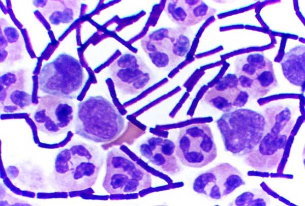
In bacteriology, gram-positive bacteria are bacteria that give a positive result in the Gram stain test, which is traditionally used to quickly classify bacteria into two broad categories according to their type of cell wall. Gram-positive bacteria take up the crystal violet stain used in the test, and then appear to be purple-coloured when seen through an optical microscope. This is because the thick peptidoglycan layer in the bacterial cell wall retains the stain after it is washed away from the rest of the sample, in the decolorization stage of the test. Conversely, gram-negative bacteria cannot retain the violet stain after the decolorization step; alcohol used in this stage degrades the outer membrane of gram-negative cells, making the cell wall more porous and incapable of retaining the crystal violet stain. Their peptidoglycan layer is much thinner and sandwiched between an inner cell membrane and a bacterial outer membrane, causing them to take up the counterstain (sa ... [...More Info...] [...Related Items...] OR: [Wikipedia] [Google] [Baidu] |
Candidiasis
Candidiasis is a fungal infection due to any type of '' Candida'' (a type of yeast). When it affects the mouth, in some countries it is commonly called thrush. Signs and symptoms include white patches on the tongue or other areas of the mouth and throat. Other symptoms may include soreness and problems swallowing. When it affects the vagina, it may be referred to as a yeast infection or thrush. Signs and symptoms include genital itching, burning, and sometimes a white "cottage cheese-like" discharge from the vagina. Yeast infections of the penis are less common and typically present with an itchy rash. Very rarely, yeast infections may become invasive, spreading to other parts of the body. This may result in fevers along with other symptoms depending on the parts involved. More than 20 types of ''Candida'' can cause infection with ''Candida albicans'' being the most common. Infections of the mouth are most common among children less than one month old, the elderly, and those ... [...More Info...] [...Related Items...] OR: [Wikipedia] [Google] [Baidu] |
Pharyngitis
Pharyngitis is inflammation of the back of the throat, known as the pharynx. It typically results in a sore throat and fever. Other symptoms may include a runny nose, cough, headache, difficulty swallowing, swollen lymph nodes, and a hoarse voice. Symptoms usually last 3–5 days, but can be longer depending on cause. Complications can include sinusitis and acute otitis media. Pharyngitis is a type of upper respiratory tract infection. Most cases are caused by a viral infection. Strep throat, a bacterial infection, is the cause in about 25% of children and 10% of adults. Uncommon causes include other bacteria such as ''gonococcus'', fungi, irritants such as smoke, allergies, and gastroesophageal reflux disease. Specific testing is not recommended in people who have clear symptoms of a viral infection, such as a cold. Otherwise, a rapid antigen detection test or throat swab is recommended. PCR testing is becoming commonly used as it is as good as taking a throat swab but gives a ... [...More Info...] [...Related Items...] OR: [Wikipedia] [Google] [Baidu] |
Cervical Vertebrae
In tetrapods, cervical vertebrae (singular: vertebra) are the vertebrae of the neck, immediately below the skull. Truncal vertebrae (divided into thoracic and lumbar vertebrae in mammals) lie caudal (toward the tail) of cervical vertebrae. In sauropsid species, the cervical vertebrae bear cervical ribs. In lizards and saurischian dinosaurs, the cervical ribs are large; in birds, they are small and completely fused to the vertebrae. The vertebral transverse processes of mammals are homologous to the cervical ribs of other amniotes. Most mammals have seven cervical vertebrae, with the only three known exceptions being the manatee with six, the two-toed sloth with five or six, and the three-toed sloth with nine. In humans, cervical vertebrae are the smallest of the true vertebrae and can be readily distinguished from those of the thoracic or lumbar regions by the presence of a foramen (hole) in each transverse process, through which the vertebral artery, vertebral veins, an ... [...More Info...] [...Related Items...] OR: [Wikipedia] [Google] [Baidu] |
Necrotizing Fasciitis
Necrotizing fasciitis (NF), also known as flesh-eating disease, is a bacterial infection that results in the death of parts of the body's soft tissue. It is a severe disease of sudden onset that spreads rapidly. Symptoms usually include red or purple skin in the affected area, severe pain, fever, and vomiting. The most commonly affected areas are the limbs and perineum. Typically, the infection enters the body through a break in the skin such as a cut or burn. Risk factors include poor immune function such as from diabetes or cancer, obesity, alcoholism, intravenous drug use, and peripheral artery disease. It does not typically spread between people. The disease is classified into four types, depending on the infecting organism. Between 55 and 80% of cases involve more than one type of bacteria. Methicillin-resistant ''Staphylococcus aureus'' (MRSA) is involved in up to a third of cases. Medical imaging is often helpful to confirm the diagnosis. Necrotizing fasciitis may b ... [...More Info...] [...Related Items...] OR: [Wikipedia] [Google] [Baidu] |
Mediastinitis
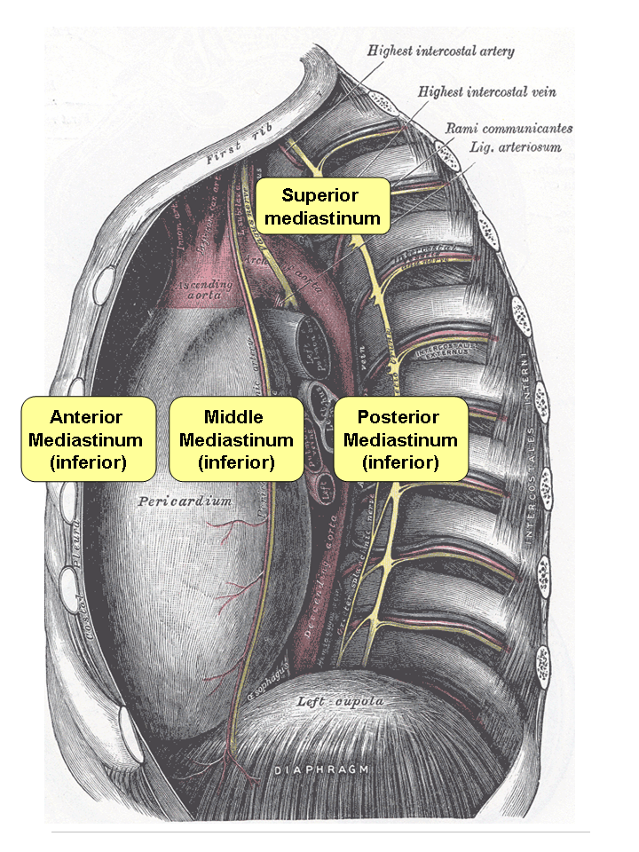
Mediastinitis is inflammation of the tissues in the mid-chest, or mediastinum. It can be either Acute (medical), acute or Chronic (medical), chronic. It is thought to be due to four different etiologies: * direct contamination * hematogenous or Lymphatic system, lymphatic spread * extension of infection from the neck or Retroperitoneal space, retroperitoneum * extension from the lung or pleura Acute mediastinitis is usually caused by bacteria and is most often due to perforation of the esophagus. As the infection can progress rapidly, this is considered a serious condition. Chronic Sclerosis (medicine), sclerosing (or fibrosis, fibrosing) mediastinitis, while potentially serious, is caused by a long-standing inflammation of the mediastinum, leading to growth of acellular collagen and fibrous tissue within the chest and around the central vessels and airways. It has a different cause, treatment, and prognosis than acute infectious mediastinitis. Space infections: Pretracheal space ... [...More Info...] [...Related Items...] OR: [Wikipedia] [Google] [Baidu] |
Deep Cervical Fascia
The deep cervical fascia (or fascia colli in older texts) lies under cover of the platysma, and invests the muscles of the neck; it also forms sheaths for the carotid vessels, and for the structures situated in front of the vertebral column. Its attachment to the hyoid bone prevents the formation of a dewlap. The investing portion of the fascia is attached behind to the ligamentum nuchæ and to the spinous process of the seventh cervical vertebra. The ''alar fascia'' is a portion of the ''deep cervical fascia''. Divisions The deep cervical fascia is often divided into a superficial, middle, and deep layer. The superficial layer is also known as the investing layer of deep cervical fascia. It envelops the trapezius, sternocleidomastoid, and muscles of facial expression. It also contains the submandibular and parotid salivary gland as well as the muscles of mastication (the masseter, pterygoid, and temporalis muscles). The middle layer is also known as the pretracheal fascia. I ... [...More Info...] [...Related Items...] OR: [Wikipedia] [Google] [Baidu] |
Cranial Nerves
Cranial nerves are the nerves that emerge directly from the brain (including the brainstem), of which there are conventionally considered twelve pairs. Cranial nerves relay information between the brain and parts of the body, primarily to and from regions of the head and neck, including the special senses of vision, taste, smell, and hearing. The cranial nerves emerge from the central nervous system above the level of the first vertebra of the vertebral column. Each cranial nerve is paired and is present on both sides. There are conventionally twelve pairs of cranial nerves, which are described with Roman numerals I–XII. Some considered there to be thirteen pairs of cranial nerves, including cranial nerve zero. The numbering of the cranial nerves is based on the order in which they emerge from the brain and brainstem, from front to back. The terminal nerves (0), olfactory nerves (I) and optic nerves (II) emerge from the cerebrum, and the remaining ten pairs arise from the ... [...More Info...] [...Related Items...] OR: [Wikipedia] [Google] [Baidu] |


.jpg)


