|
Mitral
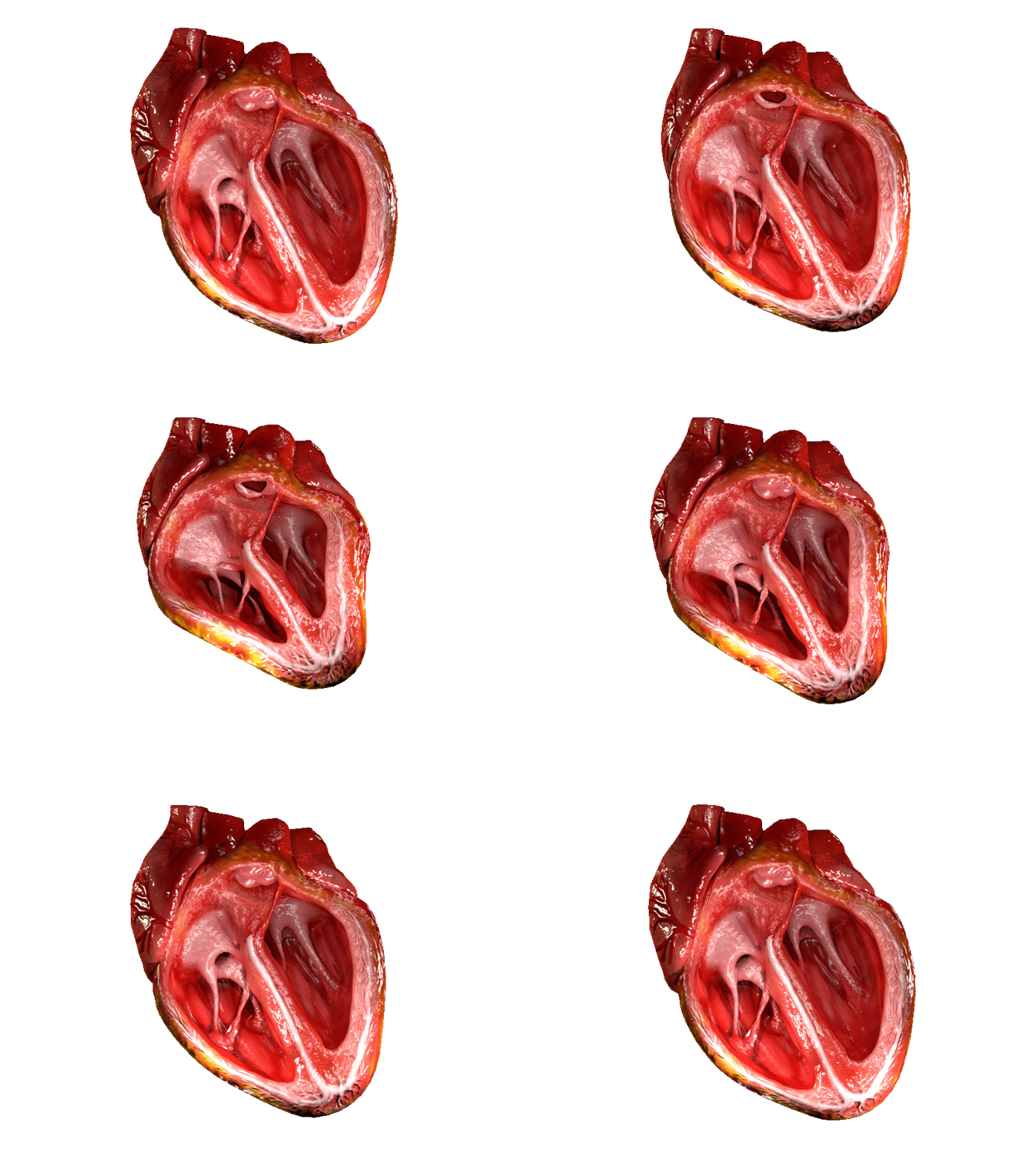
The mitral valve (), also known as the bicuspid valve or left atrioventricular valve, is one of the four heart valves. It has two cusps or flaps and lies between the left atrium and the left ventricle of the heart. The heart valves are all one-way valves allowing blood flow in just one direction. The mitral valve and the tricuspid valve are known as the atrioventricular valves because they lie between the atria and the ventricles. In normal conditions, blood flows through an open mitral valve during diastole with contraction of the left atrium, and the mitral valve closes during systole with contraction of the left ventricle. The valve opens and closes because of pressure differences, opening when there is greater pressure in the left atrium than ventricle and closing when there is greater pressure in the left ventricle than atrium. In abnormal conditions, blood may flow backward through the valve ( mitral regurgitation) or the mitral valve may be narrowed (mitral stenosis). Rhe ... [...More Info...] [...Related Items...] OR: [Wikipedia] [Google] [Baidu] |
Mitral Valve Prolapse
Mitral valve prolapse (MVP) is a valvular heart disease characterized by the displacement of an abnormally thickened mitral valve leaflet into the left atrium during systole. It is the primary form of myxomatous degeneration of the valve. There are various types of MVP, broadly classified as classic and nonclassic. In severe cases of classic MVP, complications include mitral regurgitation, infective endocarditis, congestive heart failure, and, in rare circumstances, cardiac arrest. The diagnosis of MVP depends upon echocardiography, which uses ultrasound to visualize the mitral valve. MVP is the most common valvular abnormality and is estimated to affect 2–3% of the population and 1 in 40 people might have it. The condition was first described by John Brereton Barlow in 1966. It was subsequently termed ''mitral valve prolapse'' by J. Michael Criley. Although mid-systolic click (sound of prolapsing mitral leaflet) and systolic murmur have been noticed earlier with stethoscope ... [...More Info...] [...Related Items...] OR: [Wikipedia] [Google] [Baidu] |
Mitral Regurgitation
Mitral regurgitation (MR), also known as mitral insufficiency or mitral incompetence, is a form of valvular heart disease in which the mitral valve is insufficient and does not close properly when the heart pumps out blood.Mitral valve regurgitation at Mount Sinai Hospital It is the abnormal leaking of blood backwards – regurgitation from the , through the mitral valve, into the |
Mitral Stenosis
Mitral stenosis is a valvular heart disease characterized by the narrowing of the opening of the mitral valve of the heart. It is almost always caused by rheumatic valvular heart disease. Normally, the mitral valve is about 5 cm2 during diastole. Any decrease in area below 2 cm2 causes mitral stenosis. Early diagnosis of mitral stenosis in pregnancy is very important as the heart cannot tolerate increased cardiac output demand as in the case of exercise and pregnancy. Atrial fibrillation is a common complication of resulting left atrial enlargement, which can lead to systemic thromboembolic complications like stroke. Signs and symptoms Signs and symptoms of mitral stenosis include the following: * Heart failure symptoms, such as dyspnea on exertion, orthopnea and paroxysmal nocturnal dyspnea (PND) * Palpitations * Chest pain * Hemoptysis * Thromboembolism in later stages when the left atrial volume is increased (i.e., dilation). The latter leads to increase risk of ... [...More Info...] [...Related Items...] OR: [Wikipedia] [Google] [Baidu] |
Mitral Valve
The mitral valve (), also known as the bicuspid valve or left atrioventricular valve, is one of the four heart valves. It has two cusps or flaps and lies between the left atrium and the left ventricle of the heart. The heart valves are all one-way valves allowing blood flow in just one direction. The mitral valve and the tricuspid valve are known as the atrioventricular valves because they lie between the atria and the ventricles. In normal conditions, blood flows through an open mitral valve during diastole with contraction of the left atrium, and the mitral valve closes during systole with contraction of the left ventricle. The valve opens and closes because of pressure differences, opening when there is greater pressure in the left atrium than ventricle and closing when there is greater pressure in the left ventricle than atrium. In abnormal conditions, blood may flow backward through the valve ( mitral regurgitation) or the mitral valve may be narrowed (mitral stenosis). Rh ... [...More Info...] [...Related Items...] OR: [Wikipedia] [Google] [Baidu] |
Heart Valve
A heart valve is a one-way valve that allows blood to flow in one direction through the chambers of the heart. Four valves are usually present in a mammalian heart and together they determine the pathway of blood flow through the heart. A heart valve opens or closes according to differential blood pressure on each side. The four valves in the mammalian heart are two atrioventricular valves separating the upper atria from the lower ventricles – the mitral valve in the left heart, and the tricuspid valve in the right heart. The other two valves are at the entrance to the arteries leaving the heart these are the semilunar valves – the aortic valve at the aorta, and the pulmonary valve at the pulmonary artery. The heart also has a coronary sinus valve, and an inferior vena cava valve, not discussed here. Structure The heart valves and the chambers are lined with endocardium. Heart valves separate the atria from the ventricles, or the ventricles from a blood vessel. Heart ... [...More Info...] [...Related Items...] OR: [Wikipedia] [Google] [Baidu] |
Cusps Of Heart Valves
A heart valve is a one-way valve that allows blood to flow in one direction through the chambers of the heart. Four valves are usually present in a mammalian heart and together they determine the pathway of blood flow through the heart. A heart valve opens or closes according to differential blood pressure on each side. The four valves in the mammalian heart are two atrioventricular valves separating the upper atria from the lower ventricles – the mitral valve in the left heart, and the tricuspid valve in the right heart. The other two valves are at the entrance to the arteries leaving the heart these are the semilunar valves – the aortic valve at the aorta, and the pulmonary valve at the pulmonary artery. The heart also has a coronary sinus valve, and an inferior vena cava valve, not discussed here. Structure The heart valves and the chambers are lined with endocardium. Heart valves separate the atria from the ventricles, or the ventricles from a blood vessel. Heart va ... [...More Info...] [...Related Items...] OR: [Wikipedia] [Google] [Baidu] |
Heart Valve
A heart valve is a one-way valve that allows blood to flow in one direction through the chambers of the heart. Four valves are usually present in a mammalian heart and together they determine the pathway of blood flow through the heart. A heart valve opens or closes according to differential blood pressure on each side. The four valves in the mammalian heart are two atrioventricular valves separating the upper atria from the lower ventricles – the mitral valve in the left heart, and the tricuspid valve in the right heart. The other two valves are at the entrance to the arteries leaving the heart these are the semilunar valves – the aortic valve at the aorta, and the pulmonary valve at the pulmonary artery. The heart also has a coronary sinus valve, and an inferior vena cava valve, not discussed here. Structure The heart valves and the chambers are lined with endocardium. Heart valves separate the atria from the ventricles, or the ventricles from a blood vessel. Heart ... [...More Info...] [...Related Items...] OR: [Wikipedia] [Google] [Baidu] |
Heart Left Atrial Appendage Tee View
The heart is a muscular organ in most animals. This organ pumps blood through the blood vessels of the circulatory system. The pumped blood carries oxygen and nutrients to the body, while carrying metabolic waste such as carbon dioxide to the lungs. In humans, the heart is approximately the size of a closed fist and is located between the lungs, in the middle compartment of the chest. In humans, other mammals, and birds, the heart is divided into four chambers: upper left and right atria and lower left and right ventricles. Commonly the right atrium and ventricle are referred together as the right heart and their left counterparts as the left heart. Fish, in contrast, have two chambers, an atrium and a ventricle, while most reptiles have three chambers. In a healthy heart blood flows one way through the heart due to heart valves, which prevent backflow. The heart is enclosed in a protective sac, the pericardium, which also contains a small amount of fluid. The wall of t ... [...More Info...] [...Related Items...] OR: [Wikipedia] [Google] [Baidu] |
Heart
The heart is a muscular organ in most animals. This organ pumps blood through the blood vessels of the circulatory system. The pumped blood carries oxygen and nutrients to the body, while carrying metabolic waste such as carbon dioxide to the lungs. In humans, the heart is approximately the size of a closed fist and is located between the lungs, in the middle compartment of the chest. In humans, other mammals, and birds, the heart is divided into four chambers: upper left and right atria and lower left and right ventricles. Commonly the right atrium and ventricle are referred together as the right heart and their left counterparts as the left heart. Fish, in contrast, have two chambers, an atrium and a ventricle, while most reptiles have three chambers. In a healthy heart blood flows one way through the heart due to heart valves, which prevent backflow. The heart is enclosed in a protective sac, the pericardium, which also contains a small amount of fluid. The wall ... [...More Info...] [...Related Items...] OR: [Wikipedia] [Google] [Baidu] |
Chordae Tendineae
The chordae tendineae (tendinous cords), colloquially known as the heart strings, are inelastic cords of fibrous connective tissue that connect the papillary muscles to the tricuspid valve and the mitral valve in the heart. Structure The chordae tendineae connect the atrioventricular valves (tricuspid and mitral), to the papillary muscles within the ventricles. Multiple chordae tendineae attach to each leaflet or cusp of the valves. Chordae tendineae contain elastin in a delicate structure notably at their periphery. Tendon of Todaro The ''tendon of Todaro'' is a continuation of the Eustachian valve of the inferior vena cava and the valve of the coronary sinus. Along with the opening of the coronary sinus and the septal cusp of the tricuspid valve, it makes up Koch's triangle. The apex of Koch's triangle is the location of the atrioventricular node. Function During atrial systole, blood flows from the atria to the ventricles down the pressure gradient. Chordae tendineae are ... [...More Info...] [...Related Items...] OR: [Wikipedia] [Google] [Baidu] |
Regurgitation (circulation)
Regurgitation is blood flow in the opposite direction from normal, as the backward flowing of blood into the heart or between heart chambers. It is the circulatory equivalent of backflow in engineered systems. It is sometimes called reflux. Regurgitation in or near the heart is often caused by valvular insufficiency (insufficient function, with incomplete closure, of the heart valves); for example, aortic valve insufficiency causes regurgitation through that valve, called aortic regurgitation, and the terms ''aortic insufficiency'' and ''aortic regurgitation'' are so closely linked as usually to be treated as metonymically interchangeable. The various types of heart valve regurgitation via insufficiency are as follows: # Aortic regurgitation: the backflow of blood from the aorta into the left ventricle, owing to insufficiency of the aortic semilunar valve; it may be chronic or acute. # Mitral regurgitation: the backflow of blood from the left ventricle into the left atrium, owi ... [...More Info...] [...Related Items...] OR: [Wikipedia] [Google] [Baidu] |
Systole (medicine)
Systole ( ) is the part of the cardiac cycle during which some chambers of the heart contract after refilling with blood. The term originates, via New Latin, from Ancient Greek (''sustolē''), from (''sustéllein'' 'to contract'; from ''sun'' 'together' + ''stéllein'' 'to send'), and is similar to the use of the English term ''to squeeze''. The mammalian heart has four chambers: the left atrium above the left ventricle (lighter pink, see graphic), which two are connected through the mitral (or bicuspid) valve; and the right atrium above the right ventricle (lighter blue), connected through the tricuspid valve. The atria are the receiving blood chambers for the circulation of blood and the ventricles are the discharging chambers. In late ventricular diastole, the atrial chambers contract and send blood to the larger, lower ventricle chambers. This flow fills the ventricles with blood, and the resulting pressure closes the valves to the atria. The ventricles now perform i ... [...More Info...] [...Related Items...] OR: [Wikipedia] [Google] [Baidu] |


