HOME
*


picture info

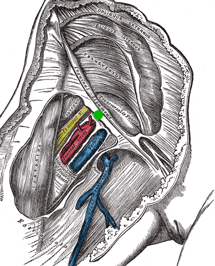
Mid-inguinal Point
The mid-inguinal point (MIP) is located on the inguinal ligament, halfway between the anterior superior iliac spine (ASIS) and the pubic symphysis. It is not to be confused with the midpoint of the inguinal ligament itself, which is located halfway between the anterior superior iliac spine and the pubic tubercle. Significance The external iliac artery becomes the femoral artery The femoral artery is a large artery in the thigh and the main arterial supply to the thigh and leg. The femoral artery gives off the deep femoral artery or profunda femoris artery and descends along the anteromedial part of the thigh in the f ... when it passes deep to the inguinal ligament, at the mid-inguinal point. As such, the point is along the superior boundary of the femoral triangle. References {{Authority control Abdomen Ligaments ...
[...More Info...]      
[...Related Items...]     OR:     [Wikipedia]   [Google]   [Baidu]  


Inguinal Ligament
The inguinal ligament (), also known as Poupart's ligament or groin ligament, is a band running from the pubic tubercle to the anterior superior iliac spine. It forms the base of the inguinal canal through which an indirect inguinal hernia may develop. Structure The inguinal ligament runs from the anterior superior iliac crest of the ilium to the pubic tubercle of the pubic bone. It is formed by the external abdominal oblique aponeurosis and is continuous with the fascia lata of the thigh. There is some dispute over the attachments. Structures that pass deep to the inguinal ligament include: *Psoas major, iliacus, pectineus * Femoral nerve, artery, and vein * Lateral cutaneous nerve of thigh *Lymphatics Function The ligament serves to contain soft tissues as they course anteriorly from the trunk to the lower extremity. This structure demarcates the superior border of the femoral triangle. It demarcates the inferior border of the inguinal triangle. The midpoint of ...
[...More Info...]      
[...Related Items...]     OR:     [Wikipedia]   [Google]   [Baidu]  


Anterior Superior Iliac Spine
The anterior superior iliac spine (abbreviated: ASIS) is a bony projection of the iliac bone, and an important landmark of surface anatomy. It refers to the anterior extremity of the iliac crest of the pelvis. It provides attachment for the inguinal ligament, and the sartorius muscle. The tensor fasciae latae muscle attaches to the lateral aspect of the superior anterior iliac spine, and also about 5  cm away at the iliac tubercle. Structure The anterior superior iliac spine refers to the anterior extremity of the iliac crest of the pelvis. This is a key surface landmark, and easily palpated. It provides attachment for the inguinal ligament, the sartorius muscle, and the tensor fasciae latae muscle. A variety of structures lie close to the anterior superior iliac spine, including the subcostal nerve, the femoral artery (which passes between it and the pubic symphysis), and the iliohypogastric nerve. Clinical significance The anterior superior iliac spine prov ...
[...More Info...]      
[...Related Items...]     OR:     [Wikipedia]   [Google]   [Baidu]  


picture info

Pubic Symphysis
The pubic symphysis is a secondary cartilaginous joint between the left and right superior rami of the pubis of the hip bones. It is in front of and below the urinary bladder. In males, the suspensory ligament of the penis attaches to the pubic symphysis. In females, the pubic symphysis is close to the clitoris. In most adults it can be moved roughly 2 mm and with 1 degree rotation. This increases for women at the time of childbirth. The name comes from the Greek word ''symphysis'', meaning 'growing together'. Structure The pubic symphysis is a nonsynovial amphiarthrodial joint. The width of the pubic symphysis at the front is 3–5 mm greater than its width at the back. This joint is connected by fibrocartilage and may contain a fluid-filled cavity; the center is avascular, possibly due to the nature of the compressive forces passing through this joint, which may lead to harmful vascular disease. The ends of both pubic bones are covered by a thin layer of hyalin ...
[...More Info...]      
[...Related Items...]     OR:     [Wikipedia]   [Google]   [Baidu]  


Pubic Tubercle
The pubic tubercle is a prominent tubercle on the superior ramus of the pubis bone of the pelvis. Structure The pubic tubercle is a prominent forward-projecting tubercle on the upper border of the medial portion of the superior ramus of the pubis bone. The inguinal ligament attaches to it. Part of the abdominal external oblique muscle inserts onto it. The inferior epigastric artery passes between the pubic tubercle and the anterior superior iliac spine. The pubic spine is a rough ridge that extends from the pubic tubercle to the upper border of the pubic symphysis. Clinical significance The pubic tubercle may be palpated. It serves as a landmark for local anaesthetic of the genital branch of the genitofemoral nerve, which lies slightly lateral to the pubic tubercle. This may also be used for the obturator nerve The obturator nerve in human anatomy arises from the ventral divisions of the second, third, and fourth lumbar nerves in the lumbar plexus; the branch from the ...
[...More Info...]      
[...Related Items...]     OR:     [Wikipedia]   [Google]   [Baidu]  


External Iliac Artery
The external iliac arteries are two major arteries which bifurcate off the common iliac arteries anterior to the sacroiliac joint of the pelvis. Structure The external iliac artery arises from the bifurcation of the common iliac artery. They proceed anterior and inferior along the medial border of the psoas major muscles. They exit the pelvic girdle posterior and inferior to the inguinal ligament. This occurs about one third laterally from the insertion point of the inguinal ligament on the pubic tubercle. At this point they are referred to as the femoral arteries. Branches Function The external iliac artery provides the main blood supply to the legs. It passes down along the brim of the pelvis and gives off two large branches - the "inferior epigastric artery" and a "deep circumflex artery." These vessels supply blood to the muscles and skin in the lower abdominal wall. The external iliac artery passes beneath the inguinal ligament in the lower part of the abdomen ...
[...More Info...]      
[...Related Items...]     OR:     [Wikipedia]   [Google]   [Baidu]  


picture info

Femoral Artery
The femoral artery is a large artery in the thigh and the main arterial supply to the thigh and leg. The femoral artery gives off the deep femoral artery or profunda femoris artery and descends along the anteromedial part of the thigh in the femoral triangle. It enters and passes through the adductor canal, and becomes the popliteal artery as it passes through the adductor hiatus in the adductor magnus near the junction of the middle and distal thirds of the thigh. Structure The femoral artery enters the thigh from behind the inguinal ligament as the continuation of the external iliac artery. Here, it lies midway between the anterior superior iliac spine and the symphysis pubis (Mid-inguinal point). Segments In clinical parlance, the femoral artery has the following segments: *The common femoral artery (CFA) is the segment of the femoral artery between the inferior margin of the inguinal ligament and the branching point of the deep femoral artery/profunda femoris artery ...
[...More Info...]      
[...Related Items...]     OR:     [Wikipedia]   [Google]   [Baidu]  


Femoral Triangle
The femoral triangle (or Scarpa's triangle) is an anatomical region of the upper third of the thigh. It is a subfascial space which appears as a triangular depression below the inguinal ligament when the thigh is flexed, abducted and laterally rotated. Structure The femoral triangle is bounded: * superiorly (also known as the base) by the inguinal ligament. * medially by the medial border of the adductor longus muscle. (Some people consider the femoral triangle to be smaller hence the medial border being at the lateral border of the adductor longus muscle.) * laterally by the medial border of the sartorius muscle. The apex of the triangle is continuous with the adductor canal. The roof is formed by the skin, superficial fascia, and deep fascia (fascia lata). The superficial fascia contains the superficial inguinal lymph nodes, femoral branch of the genitofemoral nerve, branches of the ilioinguinal nerve, superficial branches of the femoral artery with accompanying veins, and up ...
[...More Info...]      
[...Related Items...]     OR:     [Wikipedia]   [Google]   [Baidu]  


picture info

Abdomen
The abdomen (colloquially called the belly, tummy, midriff, tucky or stomach) is the part of the body between the thorax (chest) and pelvis, in humans and in other vertebrates. The abdomen is the front part of the abdominal segment of the torso. The area occupied by the abdomen is called the abdominal cavity. In arthropods it is the posterior tagma of the body; it follows the thorax or cephalothorax. In humans, the abdomen stretches from the thorax at the thoracic diaphragm to the pelvis at the pelvic brim. The pelvic brim stretches from the lumbosacral joint (the intervertebral disc between L5 and S1) to the pubic symphysis and is the edge of the pelvic inlet. The space above this inlet and under the thoracic diaphragm is termed the abdominal cavity. The boundary of the abdominal cavity is the abdominal wall in the front and the peritoneal surface at the rear. In vertebrates, the abdomen is a large body cavity enclosed by the abdominal muscles, at front and to ...
[...More Info...]      
[...Related Items...]     OR:     [Wikipedia]   [Google]   [Baidu]