|
Micrometastases
A micrometastasis is a small collection of cancer cells that has been shed from the original tumor and spread to another part of the body through the lymphovascular system. Micrometastases are too few, in size and quantity, to be picked up in a screening or diagnostic test, and therefore cannot be seen with imaging tests such as a mammogram, MRI, ultrasound, PET, or CT scans. These migrant cancer cells may group together to form a second tumor, which is so small that it can only be seen under a microscope. Approximately ninety percent of people who die from cancer die from metastatic disease, since these cells are so challenging to detect. It is important for these cancer cells to be treated immediately after discovery, in order to prevent the relapse (regrowth of the cancer) and the likely death of the patient. Detection of micrometastatic cells The major concern with micrometastases is that the only way to determine if they are present in distant tissue is to remove cells from wh ... [...More Info...] [...Related Items...] OR: [Wikipedia] [Google] [Baidu] |
H&E Stain
Hematoxylin and eosin stain ( or haematoxylin and eosin stain or hematoxylin-eosin stain; often abbreviated as H&E stain or HE stain) is one of the principal tissue stains used in histology. It is the most widely used stain in medical diagnosis and is often the gold standard. For example, when a pathologist looks at a biopsy of a suspected cancer, the histological section is likely to be stained with H&E. H&E is the combination of two histological stains: hematoxylin and eosin. The hematoxylin stains cell nuclei a purplish blue, and eosin stains the extracellular matrix and cytoplasm pink, with other structures taking on different shades, hues, and combinations of these colors. Hence a pathologist can easily differentiate between the nuclear and cytoplasmic parts of a cell, and additionally, the overall patterns of coloration from the stain show the general layout and distribution of cells and provides a general overview of a tissue sample's structure. Thus, pattern recogniti ... [...More Info...] [...Related Items...] OR: [Wikipedia] [Google] [Baidu] |
Sentinel Lymph Nodes
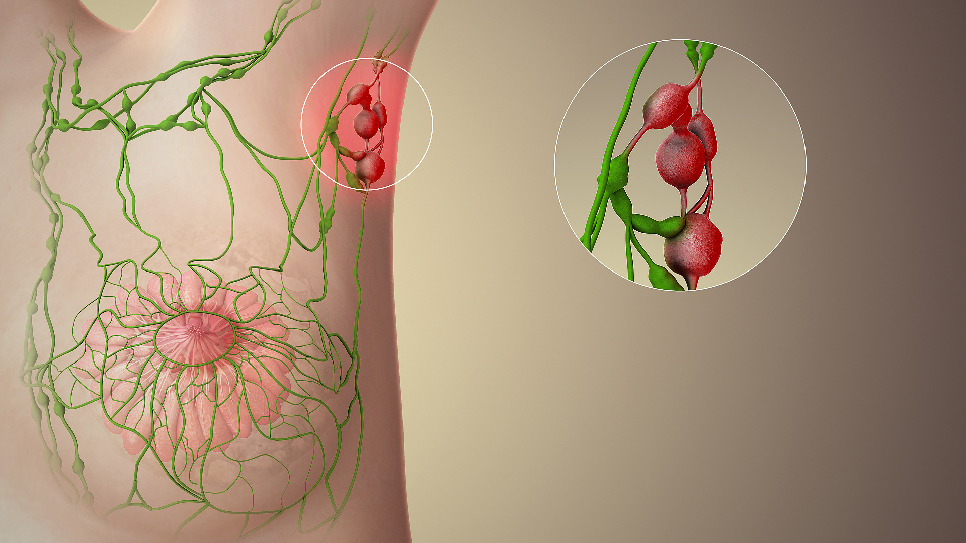
The sentinel lymph node is the hypothetical first lymph node or group of nodes draining a cancer. In case of established cancerous dissemination it is postulated that the sentinel lymph nodes are the target organs primarily reached by metastasizing cancer cells from the tumor. The sentinel node procedure (also termed sentinel lymph node biopsy or SLNB) is the identification, removal and analysis of the sentinel lymph nodes of a particular tumour. Physiology The spread of some forms of cancer usually follows an orderly progression, spreading first to regional lymph nodes, then the next echelon of lymph nodes, and so on, since the flow of lymph is directional, meaning that some cancers spread in a predictable fashion from where the cancer started. In these cases, if the cancer spreads it will spread first to lymph nodes (lymph glands) close to the tumor before it spreads to other parts of the body. The concept of sentinel lymph node surgery is to determine if the cancer has spread ... [...More Info...] [...Related Items...] OR: [Wikipedia] [Google] [Baidu] |

