|
Meningioma
Meningioma, also known as meningeal tumor, is typically a slow-growing tumor that forms from the meninges, the membranous layers surrounding the brain and spinal cord. Symptoms depend on the location and occur as a result of the tumor pressing on nearby tissue. Many cases never produce symptoms. Occasionally seizures, dementia, trouble talking, vision problems, one sided weakness, or loss of bladder control may occur. Risk factors include exposure to ionizing radiation such as during radiation therapy, a family history of the condition, and neurofibromatosis type 2. As of 2014 they do not appear to be related to cell phone use. They appear to be able to form from a number of different types of cells including arachnoid cells. Diagnosis is typically by medical imaging. If there are no symptoms, periodic observation may be all that is required. Most cases that result in symptoms can be cured by surgery. Following complete removal fewer than 20% recur. If surgery is not possibl ... [...More Info...] [...Related Items...] OR: [Wikipedia] [Google] [Baidu] |
Meningioma Seen At Autopsy
Meningioma, also known as meningeal tumor, is typically a slow-growing tumor that forms from the meninges, the membranous layers surrounding the central nervous system, brain and spinal cord. Symptoms depend on the location and occur as a result of the tumor pressing on nearby tissue. Many cases never produce symptoms. Occasionally seizures, dementia, trouble talking, vision problems, one sided weakness, or urinary incontinence, loss of bladder control may occur. Risk factors include exposure to ionizing radiation such as during radiation therapy, a family history of the condition, and neurofibromatosis type 2. As of 2014 they do not appear to be related to cell phone use. They appear to be able to form from a number of different types of cell (biology), cells including Arachnoid mater, arachnoid cells. Diagnosis is typically by medical imaging. If there are no symptoms, periodic observation may be all that is required. Most cases that result in symptoms can be cured by surgery ... [...More Info...] [...Related Items...] OR: [Wikipedia] [Google] [Baidu] |
Neurofibromatosis Type 2
Neurofibromatosis type II (also known as MISME syndrome – multiple inherited schwannomas, meningiomas, and ependymomas) is a genetic condition that may be inherited or may arise spontaneously, and causes benign tumors of the brain, spinal cord, and peripheral nerves. The types of tumors frequently associated with NF2 include vestibular schwannomas, meningiomas, and ependymomas. The main manifestation of the condition is the development of bilateral benign brain tumors in the nerve sheath of the cranial nerve VIII, which is the "auditory-vestibular nerve" that transmits sensory information from the inner ear to the brain. Besides, other benign brain and spinal tumors occur. Symptoms depend on the presence, localisation and growth of the tumor(s), in which multiple cranial nerves can be involved. Many people with this condition also experience vision problems. Neurofibromatosis type II (NF2 ''or'' NF II) is caused by mutations of the "Merlin" gene, which seems to influence the fo ... [...More Info...] [...Related Items...] OR: [Wikipedia] [Google] [Baidu] |
Brain Tumors
A brain tumor occurs when abnormal cells form within the brain. There are two main types of tumors: malignant tumors and benign (non-cancerous) tumors. These can be further classified as primary tumors, which start within the brain, and secondary tumors, which most commonly have spread from tumors located outside the brain, known as brain metastasis tumors. All types of brain tumors may produce symptoms that vary depending on the size of the tumor and the part of the brain that is involved. Where symptoms exist, they may include headaches, seizures, problems with vision, vomiting and mental changes. Other symptoms may include difficulty walking, speaking, with sensations, or unconsciousness. The cause of most brain tumors is unknown. Uncommon risk factors include exposure to vinyl chloride, Epstein–Barr virus, ionizing radiation, and inherited syndromes such as neurofibromatosis, tuberous sclerosis, and von Hippel-Lindau Disease. Studies on mobile phone exposure have not s ... [...More Info...] [...Related Items...] OR: [Wikipedia] [Google] [Baidu] |
Haemangiopericytoma
A hemangiopericytoma is a type of soft-tissue sarcoma that originates in the pericytes in the walls of capillaries. When inside the nervous system, although not strictly a meningioma tumor, it is a meningeal tumor with a special aggressive behavior. It was first characterized in 1942. Signs and symptoms Symptoms of hemangiopericytoma vary greatly depending on both tumor stage and affected organs. Most patients report pain and mass-related symptoms, while others also report vascular disease-related symptoms, and some have no symptoms until late in the disease process. Hemangiopericytomas are most commonly found in the meninges, lower extremities, retroperitoneum, pelvis, lungs, and pleura. Histopathology Hemangiopericytomas are tumors that are derived from specialized spindle shaped cells called pericytes, which line capillaries. Hemangiopericytoma located in the cerebral cavity is an aggressive tumor of the mesenchyme with oval nuclei with scant cytoplasm. "There is dense inter ... [...More Info...] [...Related Items...] OR: [Wikipedia] [Google] [Baidu] |
Neuro-oncology
Neuro-oncology is the study of brain neoplasms, brain and Spinal neoplasms, spinal cord neoplasms, many of which are (at least eventually) very dangerous and life-threatening (astrocytoma, glioma, glioblastoma multiforme, ependymoma, pontine glioma, and brain stem tumors are among the many examples of these). Among the malignant brain cancers, gliomas of the brainstem and pons, glioblastoma multiforme, and Grading (tumors), high-grade (highly anaplastic) astrocytoma/oligodendroglioma are among the worst. In these cases, untreated survival usually amounts to only a few months, and survival with current radiation and chemotherapy treatments may extend that time from around a year to a year and a half, possibly two or more, depending on the patient's condition, immune function, treatments used, and the specific type of malignant brain neoplasm. Surgery may in some cases be curative, but, as a general rule, malignant brain cancers tend to regenerate and emerge from remission (medicine) ... [...More Info...] [...Related Items...] OR: [Wikipedia] [Google] [Baidu] |
Felix Plater
Felix Platter (also Plater ; ; Latinized: Platerus; 28 October 1536 – 28 July 1614) was a Swiss physician, well known for his classification of psychiatric diseases, and was also the first to describe an intracranial tumour (a meningioma). Biography Felix Platter was the son of Lutheran humanist, schoolmaster and printer, Thomas Platter, and the half-brother of Thomas Platter the Younger. In 1552, and only fifteen years old, Platter travelled by pony from Basel to the University of Montpellier to start a course of study under Guillaume Rondelet. He earned his medical doctorate from Montpellier in 1557. Once arrived, he lodged in the house of Laurent Catalan, the town pharmacist and a Maran or Christian Jew. Platter occasionally sent packages of fruits and seeds to his father. His studies took place in an atmosphere of terror and religious persecution. Rondelet taught his students the technique of pressing, drying and mounting botanical specimens on paper, a process practised by ... [...More Info...] [...Related Items...] OR: [Wikipedia] [Google] [Baidu] |
Radiosurgery
Radiosurgery is surgery using radiation, that is, the destruction of precisely selected areas of tissue using ionizing radiation rather than excision with a blade. Like other forms of radiation therapy (also called radiotherapy), it is usually used to treat cancer. Radiosurgery was originally defined by the Swedish neurosurgeon Lars Leksell as "a single high dose fraction of radiation, stereotactically directed to an intracranial region of interest". In stereotactic radiosurgery (SRS), the word "stereotactic" refers to a three-dimensional coordinate system that enables accurate correlation of a virtual target seen in the patient's diagnostic images with the actual target position in the patient. Stereotactic radiosurgery may also be called stereotactic body radiation therapy (SBRT) or stereotactic ablative radiotherapy (SABR) when used outside the central nervous system (CNS). History Stereotactic radiosurgery was first developed in 1949 by the Swedish neurosurgeon Lars Leksell ... [...More Info...] [...Related Items...] OR: [Wikipedia] [Google] [Baidu] |
Arachnoid Mater
The arachnoid mater (or simply arachnoid) is one of the three meninges, the protective membranes that cover the brain and spinal cord. It is so named because of its resemblance to a spider web. The arachnoid mater is a derivative of the neural crest mesoectoderm in the embryo. Structure It is interposed between the two other meninges, the more superficial and much thicker dura mater and the deeper pia mater, from which it is separated by the subarachnoid space. The delicate arachnoid layer is not attached to the inside of the dura but against it and surrounds the brain and spinal cord. It does not line the brain down into its sulci (folds), as does the pia mater, with the exception of the longitudinal fissure, which divides the left and right cerebral hemispheres. Cerebrospinal fluid (CSF) flows under the arachnoid in the subarachnoid space, within a meshwork of trabeculae which span between the arachnoid and the pia. The arachnoid mater makes arachnoid villi, small protrusions ... [...More Info...] [...Related Items...] OR: [Wikipedia] [Google] [Baidu] |
Meninges
In anatomy, the meninges (, ''singular:'' meninx ( or ), ) are the three membranes that envelop the brain and spinal cord. In mammals, the meninges are the dura mater, the arachnoid mater, and the pia mater. Cerebrospinal fluid is located in the subarachnoid space between the arachnoid mater and the pia mater. The primary function of the meninges is to protect the central nervous system. Structure Dura mater The dura mater ( la, tough mother) (also rarely called ''meninx fibrosa'' or ''pachymeninx'') is a thick, durable membrane, closest to the Human skull, skull and vertebrae. The dura mater, the outermost part, is a loosely arranged, fibroelastic layer of cells, characterized by multiple interdigitating cell processes, no extracellular collagen, and significant extracellular spaces. The middle region is a mostly fibrous portion. It consists of two layers: the endosteal layer, which lies closest to the Calvaria (skull), skull, and the inner meningeal layer, which lies closer ... [...More Info...] [...Related Items...] OR: [Wikipedia] [Google] [Baidu] |
Autopsy
An autopsy (post-mortem examination, obduction, necropsy, or autopsia cadaverum) is a surgical procedure that consists of a thorough examination of a corpse by dissection to determine the cause, mode, and manner of death or to evaluate any disease or injury that may be present for research or educational purposes. (The term "necropsy" is generally reserved for non-human animals). Autopsies are usually performed by a specialized medical doctor called a pathologist. In most cases, a medical examiner or coroner can determine the cause of death. However, only a small portion of deaths require an autopsy to be performed, under certain circumstances. Purposes of performance Autopsies are performed for either legal or medical purposes. Autopsies can be performed when any of the following information is desired: * Determine if death was natural or unnatural * Injury source and extent on the corpse * Manner of death must be determined * Post mortem interval * Determining the deceas ... [...More Info...] [...Related Items...] OR: [Wikipedia] [Google] [Baidu] |
Symptom
Signs and symptoms are the observed or detectable signs, and experienced symptoms of an illness, injury, or condition. A sign for example may be a higher or lower temperature than normal, raised or lowered blood pressure or an abnormality showing on a medical scan. A symptom is something out of the ordinary that is experienced by an individual such as feeling feverish, a headache or other pain or pains in the body. Signs and symptoms Signs A medical sign is an objective observable indication of a disease, injury, or abnormal physiological state that may be detected during a physical examination, examining the patient history, or diagnostic procedure. These signs are visible or otherwise detectable such as a rash or bruise. Medical signs, along with symptoms, assist in formulating diagnostic hypothesis. Examples of signs include elevated blood pressure, nail clubbing of the fingernails or toenails, staggering gait, and arcus senilis and arcus juvenilis of the eyes. Indicati ... [...More Info...] [...Related Items...] OR: [Wikipedia] [Google] [Baidu] |
Focal Seizure
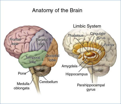
Focal seizures (also called partial seizures and localized seizures) are seizures which affect initially only one hemisphere of the brain. The brain is divided into two hemispheres, each consisting of four lobes – the frontal, temporal, parietal and occipital lobes. A focal seizure is generated in and affects just one part of the brain – a whole hemisphere or part of a lobe. Symptoms will vary according to where the seizure occurs. When seizures occur in the frontal lobe the patient may experience a wave-like sensation in the head. When seizures occur in the temporal lobe, a feeling of déjà vu may be experienced. When seizures are localized to the parietal lobe, a numbness or tingling may occur. With seizures occurring in the occipital lobe, visual disturbances or hallucinations have been reported. , Epilepsy S ... [...More Info...] [...Related Items...] OR: [Wikipedia] [Google] [Baidu] |


