|
Melanopsin
Melanopsin is a type of photopigment belonging to a larger family of light-sensitive retinal proteins called opsins and encoded by the gene ''Opn4''. In the mammalian retina, there are two additional categories of opsins, both involved in the formation of visual images: rhodopsin and photopsin (types I, II, and III) in the rod and cone photoreceptor cells, respectively. In humans, melanopsin is found in intrinsically photosensitive retinal ganglion cells (ipRGCs). It is also found in the iris of mice and primates. Melanopsin is also found in rats, amphioxus, and other chordates. ipRGCs are photoreceptor cells which are particularly sensitive to the absorption of short-wavelength (blue) visible light and communicate information directly to the area of the brain called the suprachiasmatic nucleus (SCN), also known as the central "body clock", in mammals. Melanopsin plays an important non-image-forming role in the setting of circadian rhythms as well as other functions. Mutations in ... [...More Info...] [...Related Items...] OR: [Wikipedia] [Google] [Baidu] |
Melanopsin In Retina
Melanopsin is a type of photopigment belonging to a larger family of light-sensitive retinal proteins called opsins and encoded by the gene ''Opn4''. In the mammalian retina, there are two additional categories of opsins, both involved in the formation of visual images: rhodopsin and photopsin (types I, II, and III) in the rod and cone photoreceptor cells, respectively. In humans, melanopsin is found in intrinsically photosensitive retinal ganglion cells (ipRGCs). It is also found in the iris of mice and primates. Melanopsin is also found in rats, amphioxus, and other chordates. ipRGCs are photoreceptor cells which are particularly sensitive to the absorption of short-wavelength (blue) visible light and communicate information directly to the area of the brain called the suprachiasmatic nucleus (SCN), also known as the central "body clock", in mammals. Melanopsin plays an important non-image-forming role in the setting of circadian rhythms as well as other functions. Mutations in ... [...More Info...] [...Related Items...] OR: [Wikipedia] [Google] [Baidu] |
Ignacio Provencio
Ignacio Provencio (born 29 June 1965) is an American neuroscientist and the discoverer of melanopsin, an opsin found in specialized photosensitive ganglion cells of the mammalian retina. Provencio served as the program committee chair of the Society for Research on Biological Rhythms from 2008 to 2010. Biography Provencio was born in Bitburg, Germany and attended Lebanon Catholic High School in Lebanon, PA. During his undergraduate career at Swarthmore College, Provencio became interested in neuroscience while studying crayfish, cockroaches, and fireflies under Jon Copeland. From 1987 to 1989 he worked as a lab technician in Steve Reppert's research laboratory at Massachusetts General Hospital, where he was introduced to the field of circadian biology. He graduated in 1987 from Swarthmore College with a B.A. in Biology and went on to earn his Ph.D. from the University of Virginia, a university with a strong network of circadian biologists, in 1996. During his postdoctoral tr ... [...More Info...] [...Related Items...] OR: [Wikipedia] [Google] [Baidu] |
Intrinsically Photosensitive Retinal Ganglion Cells
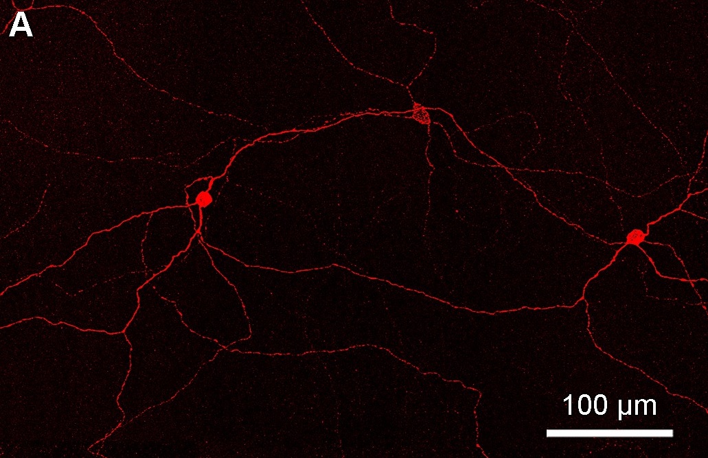
Intrinsically photosensitive retinal ganglion cells (ipRGCs), also called photosensitive retinal ganglion cells (pRGC), or melanopsin-containing retinal ganglion cells (mRGCs), are a type of neuron in the retina of the mammalian eye. The presence of (something like) ipRGCs was first suspected in 1927 when rodless, coneless mice still responded to a light stimulus through pupil constriction, This implied that rods and cones are not the only light-sensitive neurons in the retina. Yet research on these cells did not advance until the 1980s. Recent research has shown that these retinal ganglion cells, unlike other retinal ganglion cells, are intrinsically photosensitive due to the presence of melanopsin, a light-sensitive protein. Therefore they constitute a third class of photoreceptors, in addition to rod and cone cells. Overview Compared to the rods and cones, the ipRGCs respond more sluggishly and signal the presence of light over the long term. They represent a very small ... [...More Info...] [...Related Items...] OR: [Wikipedia] [Google] [Baidu] |
Photoreceptor Cell
A photoreceptor cell is a specialized type of neuroepithelial cell found in the retina that is capable of visual phototransduction. The great biological importance of photoreceptors is that they convert light (visible electromagnetic radiation) into signals that can stimulate biological processes. To be more specific, photoreceptor proteins in the cell absorb photons, triggering a change in the cell's membrane potential. There are currently three known types of photoreceptor cells in mammalian eyes: rods, cones, and intrinsically photosensitive retinal ganglion cells. The two classic photoreceptor cells are rods and cones, each contributing information used by the visual system to form an image of the environment, sight. Rods primarily mediate scotopic vision (dim conditions) whereas cones primarily mediate to photopic vision (bright conditions), but the processes in each that supports phototransduction is similar. A third class of mammalian photoreceptor cell was discovered ... [...More Info...] [...Related Items...] OR: [Wikipedia] [Google] [Baidu] |
Opsin
Animal opsins are G-protein-coupled receptors and a group of proteins made light-sensitive via a chromophore, typically retinal. When bound to retinal, opsins become Retinylidene proteins, but are usually still called opsins regardless. Most prominently, they are found in photoreceptor cells of the retina. Five classical groups of opsins are involved in Visual perception, vision, mediating the conversion of a photon of light into an electrochemical signal, the first step in the Visual phototransduction, visual transduction cascade. Another opsin found in the mammalian retina, melanopsin, is involved in circadian rhythms and Pupillary light reflex, pupillary reflex but not in vision. Humans have in total nine opsins. Beside vision and light perception, opsins may also sense temperature, sound, or chemicals. Structure and function Animal opsins detect light and are the molecules that allow us to see. Opsins are G-protein-coupled receptors (GPCRs), which are chemoreceptors and hav ... [...More Info...] [...Related Items...] OR: [Wikipedia] [Google] [Baidu] |
Samer Hattar
Samer Hattar ( ar, سامر حتر) is a chronobiologist and a leader in the field of non-image forming photoreception. He is the Chief of the Section on Light and Circadian Rhythms at the National Institute of Mental Health, part of the National Institutes of Health. He was previously an associate professor in the Department of Neuroscience and the Department of Biology at Johns Hopkins University in Baltimore, MD. He is best known for his investigation into the role of melanopsin and intrinsically photosensitive retinal ganglion cells (ipRGC) in the entrainment of circadian rhythms. Life Samer Hattar was born in Amman, Jordan to a Jordanian father and a Lebanese mother. Raised in a Christian family, he planned on becoming a priest. He studied at Terra Sancta High School, a Catholic high school in Amman, from 1978 to 1988. He earned good grades in his classes and fell in love with biology when introduced to Mendel's pea plant experiments. This passion inspired him to pursue a ... [...More Info...] [...Related Items...] OR: [Wikipedia] [Google] [Baidu] |
Amphioxus
The lancelets ( or ), also known as amphioxi (singular: amphioxus ), consist of some 30 to 35 species of "fish-like" benthic filter feeding chordates in the order Amphioxiformes. They are the modern representatives of the subphylum Cephalochordata. Lancelets closely resemble 530-million-year-old ''Pikaia'', fossils of which are known from the Burgess Shale. Zoologists are interested in them because they provide evolutionary insight into the origins of vertebrates. Lancelets contain many organs and organ systems that are closely related to those of modern fish, but in more primitive form. Therefore, they provide a number of examples of possible evolutionary exaptation. For example, the gill-slits of lancelets are used for feeding only, and not for respiration. The circulatory system carries food throughout their body, but does not have red blood cells or hemoglobin for transporting oxygen. Lancelet genomes hold clues about the early evolution of vertebrates: by comparing genes from ... [...More Info...] [...Related Items...] OR: [Wikipedia] [Google] [Baidu] |
Retinal Ganglion Cell
A retinal ganglion cell (RGC) is a type of neuron located near the inner surface (the ganglion cell layer) of the retina of the human eye, eye. It receives visual information from photoreceptor cell, photoreceptors via two intermediate neuron types: Bipolar cell of the retina, bipolar cells and retina amacrine cells. Retina amacrine cells, particularly narrow field cells, are important for creating functional subunits within the ganglion cell layer and making it so that ganglion cells can observe a small dot moving a small distance. Retinal ganglion cells collectively transmit image-forming and non-image forming visual information from the retina in the form of action potential to several regions in the thalamus, hypothalamus, and mesencephalon, or midbrain. Retinal ganglion cells vary significantly in terms of their size, connections, and responses to visual stimulation but they all share the defining property of having a long axon that extends into the brain. These axons form th ... [...More Info...] [...Related Items...] OR: [Wikipedia] [Google] [Baidu] |
Retinal Ganglion Cells
A retinal ganglion cell (RGC) is a type of neuron located near the inner surface (the ganglion cell layer) of the retina of the eye. It receives visual information from photoreceptors via two intermediate neuron types: bipolar cells and retina amacrine cells. Retina amacrine cells, particularly narrow field cells, are important for creating functional subunits within the ganglion cell layer and making it so that ganglion cells can observe a small dot moving a small distance. Retinal ganglion cells collectively transmit image-forming and non-image forming visual information from the retina in the form of action potential to several regions in the thalamus, hypothalamus, and mesencephalon, or midbrain. Retinal ganglion cells vary significantly in terms of their size, connections, and responses to visual stimulation but they all share the defining property of having a long axon that extends into the brain. These axons form the optic nerve, optic chiasm, and optic tract. A small pe ... [...More Info...] [...Related Items...] OR: [Wikipedia] [Google] [Baidu] |
Photopigment
Photopigments are unstable pigments that undergo a chemical change when they absorb light. The term is generally applied to the non-protein chromophore moiety of photosensitive chromoproteins, such as the pigments involved in photosynthesis and photoreception. In medical terminology, "photopigment" commonly refers to the photoreceptor proteins of the retina. Photosynthetic pigments Photosynthetic pigments convert light into biochemical energy. Examples for photosynthetic pigments are chlorophyll, carotenoids and phycobilins. These pigments enter a high-energy state upon absorbing a photon which they can release in the form of chemical energy. This can occur via light-driven pumping of ions across a biological membrane (e.g. in the case of the proton pump bacteriorhodopsin) or via excitation and transfer of electrons released by photolysis (e.g. in the photosystems of the thylakoid membranes of plant chloroplasts). In chloroplasts, the light-driven electron transfer chain in t ... [...More Info...] [...Related Items...] OR: [Wikipedia] [Google] [Baidu] |
Cone Cell
Cone cells, or cones, are photoreceptor cells in the retinas of vertebrate eyes including the human eye. They respond differently to light of different wavelengths, and the combination of their responses is responsible for color vision. Cones function best in relatively bright light, called the photopic region, as opposed to rod cells, which work better in dim light, or the scotopic region. Cone cells are densely packed in the fovea centralis, a 0.3 mm diameter rod-free area with very thin, densely packed cones which quickly reduce in number towards the periphery of the retina. Conversely, they are absent from the optic disc, contributing to the blind spot. There are about six to seven million cones in a human eye (vs ~92 million rods), with the highest concentration being towards the macula. Cones are less sensitive to light than the rod cells in the retina (which support vision at low light levels), but allow the perception of color. They are also able to perceive ... [...More Info...] [...Related Items...] OR: [Wikipedia] [Google] [Baidu] |


